59
Author of the article: Marina Dmitrievna
2017.07.17
28 004
Symptoms
When there is pain in the heart area, most people are wary, because a person’s full life depends on the work of this organ. Many people go to the hospital to see a cardiologist. However, electrocardiography does not always answer all their questions. Can a person’s heart hurt if the cardiogram is good? Why does my heart hurt for more than one month?
In what cases is an electrocardiogram prescribed?
If a person has the symptoms described below, the cardiologist will refer him to an electrocardiogram:
- legs swell;
- fainting conditions;
- there is shortness of breath;
- chest pain, back pain, neck pain.
An ECG is mandatory for pregnant women for examination, for people preparing for surgery, or for a medical examination.
ECG results are also required if you travel to a sanatorium or if you need permission for any sports activities.
For prevention and if a person has no complaints, doctors recommend taking an electrocardiogram once a year. Often this can help diagnose cardiac pathologies that are asymptomatic.
What does an electrocardiogram determine?
- Heart rate - heart rate.
- Rhythms of heart contractions.
- Heart attack.
- Arrhythmias.
- Ventricular hypertrophy.
- Ischemic and cardystrophic changes.
The most disappointing and serious diagnosis on the electrocardiogram is myocardial infarction. In the diagnosis of heart attacks, the ECG plays an important and even the main role. Using a cardiogram, the zone of necrosis, the localization and depth of lesions in the heart area are revealed. Also, when decoding the cardiogram tape, you can recognize and distinguish acute myocardial infarction from an aneurysm and past scars. Therefore, when undergoing a medical examination, it is necessary to do a cardiogram, because it is very important for the doctor to know what the ECG will show.
Most often, a heart attack is associated directly with the heart. But it is not so. A heart attack can occur in any organ. Pulmonary infarction occurs (when lung tissue partially or completely dies if the arteries are blocked).
There is a cerebral infarction (otherwise known as ischemic stroke) - the death of brain tissue, which can be caused by thrombosis or rupture of brain vessels. With a cerebral infarction, functions such as speech, physical movement, and sensation may be completely lost or lost.
When a person has a heart attack, living tissue in their body dies or becomes necrosis. The body loses tissue or a section of an organ, as well as the functions performed by this organ.
Myocardial infarction is the death or ischemic necrosis of areas or areas of the heart muscle itself due to complete or partial loss of blood supply. Heart muscle cells begin to die approximately 20-30 minutes after blood flow stops. If a person has a myocardial infarction, blood circulation is disrupted. One or more blood vessels fail. Most often, heart attacks occur due to blockage of blood vessels by blood clots (atherosclerotic plaques). The area of distribution of the infarction depends on the severity of the dysfunction of the organ, for example, extensive myocardial infarction or microinfarction. Therefore, you should not immediately despair if the ECG shows a heart attack.
This becomes a threat to the functioning of the entire cardiovascular system of the body and threatens life. In the modern period, heart attacks are the main cause of death among the population of developed countries.
Will an ECG show heart failure – Treatment of hypertension
OUR READERS RECOMMEND!
Our readers successfully use ReCardio to treat hypertension. Seeing how popular this product is, we decided to bring it to your attention. Read more here...
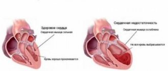
Chronic heart failure is a syndrome in which progressive structural and functional changes in the heart lead to the inability of the heart to release oxygenated blood in the amount required by all tissues and organs.
Acute and chronic heart failure leads the world in terms of mortality. This is more than the mortality rate for cancer pathologies and infections.
Where does chronic heart failure come from?
Chronic cardiovascular failure is the most common type of circulatory failure.
Chronic heart failure is caused either by factors directly damaging the myocardium (infections, injuries, drugs or chemical cardiotoxic drugs), or by factors leading to functional cardiac overload. Such an overload occurs either with a larger volume of inflowing blood, or with an increase in vascular resistance (progression of valve defects, cardiosclerosis, vasoconstriction due to hypertension).
Characterized by a progressive decrease in the tolerance of the cardiovascular system to stress, fluid retention, and a decrease in the duration and quality of life.
Various attempts have been made to classify chronic heart failure. Here are the most popular and therefore the most convenient.
Classification according to N.D. Strazhesko (by stages)
I is the initial, easiest. Heart failure is only revealed by significant physical activity.
Those who are not picky about their health can attribute the shortness of breath, palpitations and fatigue to age and workload.
In the absence of load and any tension, all manifestations disappear on their own. Instrumental methods will also not show any abnormalities in the patient at rest.
Stage II has 2 periods. In period A, the phenomena of circulatory failure appear at rest, but moderately. There is already a decrease in stress tolerance. Fatigue sets in quickly. Shortness of breath may also appear when getting out of bed; the heart rate is always increased. Hemodynamics are disrupted in both circulation circles.
Period B is characterized by significantly pronounced symptoms of heart failure without any load. Shortness of breath becomes such that it begins to interfere with the normal course of life. Significant swelling appears in the legs, and the heart rate increases even more.
III final stage, in which significant, irreversible damage to organs appears.
Classification taking into account the severity of the main symptoms
Chronic heart failure 1st degree - compensated. Complaints of fatigue, sleep disturbances, breathing problems after increased exercise.
2nd degree – subcompensated (moderate). At grade 2A, shortness of breath with moderate exertion and rapid heartbeat are characteristic. Grade 2B is much more severe. Shortness of breath at rest, the liver is enlarged, swelling appears, bluishness of the skin, heart rate up to 100 per minute, chest pain is possible.
Chronic failure of the 3rd degree is irreversible. The condition becomes significantly worse: shortness of breath does not stop, swelling spreads throughout the body. Cough may produce bloody sputum.
It is more popular in the USA. It identifies 4 functional classes, focused on the level of possible physical activity.
Class I unites those who, having heart disease, feel well and do not need to limit their previous activity.
Class II includes those who need moderate limitation of their activity.
Class III unites those who are unable to limit themselves in physical activity due to cardiac pathology.
Class IV includes those who experience severe discomfort with the slightest exertion.
The pathogenesis of chronic heart failure consists in the inability of the heart, as a pump, given the existing resistance of the vessels, to pump into them the necessary blood that enters the heart through the veins. From this starvation and the inability to remove all toxic metabolic products with the blood, all organ systems suffer to varying degrees.
Chronic heart failure is compensated for a long time.
For example, the heart increases the number of its contractions to compensate for the insufficient output volume, vasoconstriction occurs (reflex vasoconstriction) in the periphery to avoid a lack of blood supply to the main organs (brain, heart), the myocardium hypertrophies (the muscle mass of the heart increases for increased work) and others . Then the person feels practically healthy.
But then one day the mechanisms become exhausted. As a result, all tissues and organs suffer from a lack of oxygenated blood, and, consequently, oxygen, and an excess of cell metabolism products. And then complaints appear.
As a result of the fact that the blood flow does not correspond to what is necessary for the organs, the chemical processes of the tissues are disrupted. As a result, irreversible dystrophic changes in organs are triggered.
The kidneys react to the narrowing of their blood vessels and disruption of microcirculation by retention of sodium and fluid in the body. As a result, the constancy of the internal environment is disrupted and swelling increases.
Due to insufficient pumping function of the heart, preload increases and prolonged stagnation of blood in the organs occurs. The lungs and liver are especially affected. Tissue fibrosis develops in them, and the number of cells capable of functioning normally decreases. These syndromes form the basis of the pathogenesis of chronic heart failure.
Manifestations of chronic circulatory failure
Clinical symptoms of chronic heart failure vary for each patient. One of the first manifestations is usually shortness of breath. Tachycardia, swelling, cough, and cyanosis of the skin occur.
Edema in bedridden patients appears in the area of the sacrum and sides of the abdomen. In patients without limited mobility, swelling begins in the ankles and spreads higher. With decompensation, hydrothorax, hydropericardium, and ascites develop.
Skin cyanosis increases.
Chronic heart failure is diagnosed after a careful clinical examination. The diagnosis is confirmed by ECG, x-ray examination of the heart and lungs. Echocardiography (ultrasound examination) of the heart is indicative.
Directions of treatment
Treatment is more symptomatic. Its goal is to reduce the manifestations of deficiency and prolong a person’s life. Treatment of chronic heart failure consists of treating the underlying cause that led to CHF.
A low-salt diet, a special regime of physical activity, and regular examinations by a cardiologist are required.
Medications used include ACE inhibitors, beta-adrenergic receptor blockers, diuretics, cardiac glycosides, and aldosterone antagonists. Drugs are prescribed exclusively by a cardiologist, who takes into account the severity of the patient’s condition and concomitant diseases.
Today, abstracts, articles, and dissertations are written on the topic of chronic heart failure. But it still leaves questions to which there are still no clear answers that do not raise doubts.
For example, they are still arguing about which mechanism of edema is leading, who gets sick more often - men or women. Perhaps someday they will be answered.
And maybe they will find a medicine that will make this disease reversible.
Watch a video animation about heart failure:
Source: https://lechenie-gipertoniya.ru/serdechnaya-nedostatochnost/pokazhet-li-ekg-serdechnuyu-nedostatochnost/
Causes of heart attack
- Atherosclerosis.
- Rheumatism.
- Congenital heart defect.
- Diabetes.
- Smoking, obesity.
- Arterial hypertension.
- Vasculitis.
- Increased blood viscosity (thrombosis).
- Previous heart attacks.
- Severe coronary artery spasms (for example, when taking cocaine).
- Age-related changes.
An ECG can also identify other diseases, such as tachycardia, arrhythmia, and ischemic disorders.
Arrhythmia
What to do if the ECG shows arrhythmia?
Arrhythmia can be characterized by numerous changes in the contraction of the heartbeat.
Arrhythmia is a condition in which there is a disturbance in the heart rhythm and heart rate. More often, this pathology is marked by an irregular heartbeat; The patient has either a rapid or slow heartbeat. An increase is observed when inhaling, and a decrease is observed when exhaling.
Diseases detected with its help
Thanks to the ECG, it is possible to diagnose many abnormalities in cardiac activity. The main ones are:
- Hypertrophy of departments.
This problem occurs due to hemodynamic disturbances. Deviations in the movement of blood through the vessels cause overload of the organ chambers, causing the atria or ventricles to increase in size.This problem can be identified by the following signs:
- Changes in the electrical axis of the heart.
- Increase in R wave amplitude.
- Changing the position of the transition zone.
Increasing the excitation vector.
When there are no attacks of the disease, there may be no signs of it on the ECG.
This disease exhibits the following features:
- The location of the ST segment is below the isoline.
Changes in T wave mapping.
In the presence of this pathology, disturbances in the formation of the impulse occur. Because of this, disruptions in the pulse rhythm occur.
It appears on the ECG as follows:
- There are fluctuations in the P-Q and QT display.
Deviations from the norm in the interval between R-waves.
This is a type of arrhythmia in which the heart rate increases.
Its signs on the cardiogram:
- The gap between the R-teeth is less than normal.
The PQ area decreases.
This is another type of arrhythmia in which the heart rate decreases. Signs:
- The gap between R and R is increased.
There is an increase in the QT region.
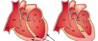
In this case, the myocardium increases due to changes in the muscle layers or pathologies in the development of the organ during the prenatal period.
During extrasystole, a focus is formed in the heart that is capable of creating an electrical impulse, which disrupts the rhythm of the sinus node.
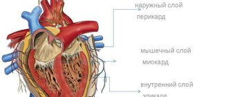
This disease is characterized by inflammation of the layers of the pericardial sac.
Other diseases that can be detected using a cardiogram include ischemic heart disease, myocardial infarction, myocarditis, heart failure, etc.
Angina pectoris
If the patient experiences attacks of pain under the sternum or to the left of it in the area of the left arm, which can last a few seconds or can last up to 20 minutes, then the ECG will show angina.
The pain usually intensifies with lifting weights, heavy physical activity, or going out into the cold and may disappear with rest. Such pain decreases within 3-5 minutes when taking nitroglycerin. The patient's skin turns pale and the pulse becomes uneven, which causes interruptions in the functioning of the heart.
Angina pectoris is one of the forms of coronary heart disease. It is often quite difficult to diagnose angina pectoris, because such abnormalities can also manifest themselves in other cardiac pathologies. Angina pectoris can further lead to heart attacks and strokes.
Tachycardia
Many people are very worried when they find out that the ECG showed tachycardia.
Tachycardia is an increase in heart rate at rest. Heart rhythms during tachycardia can reach 100-150 beats per minute. This pathology can also occur in people, regardless of age, when lifting heavy objects or during increased physical activity, as well as during strong psycho-emotional arousal.
Still, tachycardia is considered not a disease, but a symptom. But it is no less dangerous. If the heart begins to beat too quickly, then it cannot have time to fill with blood, which subsequently leads to a decrease in blood output and a lack of oxygen in the body, as well as the heart muscle itself. If tachycardia lasts more than a month, it can lead to further disruption of the heart muscle and an increase in heart size.
Will a cardiogram show heart failure - Treatment of hypertension
Blood pressure is an important parameter for the functioning of the cardiovascular and circulatory systems. By measuring indicators, the condition of the heart muscle, blood vessels and arteries is assessed.
The systolic (upper) value is recorded at the moment of blood ejection from the heart muscle and depends on the strength and speed of its contraction. Normally, the designation on the tonometer varies from 101 to 139 mmHg.
Diastolic pressure (lower) is recorded during the period of myocardial relaxation. The value varies depending on the level of vascular resistance and the total volume of circulating fluid in the human body.
Let's find out what DM and DD mean, why they can increase and what reasons lead to pathological deviation? How is arterial hypertension diagnosed and treated?
When the atrium contracts (systole), blood is pushed out of the myocardium. However, the natural physiological process can be “interfered” with increased blood viscosity and narrowing of the lumen of the vascular walls, which leads to impaired circulation in the body.
Due to hypoxia - a deficiency of oxygen, as well as nutrients or an excess of toxic components formed during the metabolic process, the functionality of internal organs and systems is disrupted.
The systolic parameter can fluctuate under the influence of various causes and provoking factors. The dominant reason for the increase is the formation of atherosclerotic plaques, since the metabolism of proteins and carbohydrates in the body is disrupted.
Deposits of harmful cholesterol lead to the growth of connective tissue, insoluble calcium salts accumulate in it, the gaps between the vessels become smaller, and they become clogged.
DM often increases due to excess weight. In obese men and women, especially the elderly, hypertrophied adipose tissue compresses blood vessels, which leads to obstructed blood flow and increased blood pressure.
The top figure may decrease due to the following reasons:
- Osteochondrosis of the cervical spine.
- Neuroses, diseases of the autonomic system.
- Impaired functionality of the gastrointestinal tract.
- Internal or external bleeding.
- Deficiency of vitamins and minerals.
- Inadequate therapy with antihypertensive drugs.
- Brain stroke.
In an absolutely healthy person, blood pressure can increase, and this is not a pathology. For example, excessive physical activity leads to a short-term jump in values.
From 5 to 20 years of age, DM increases, until the age of 40 it stabilizes, after which it increases again.
Bottom digit
The next phase after systole involves contraction of the ventricles and relaxation of the atrium. The overall cardiac cycle ends with a short pause lasting about 0.4 seconds. After the blood enters the peripheral vessels, the atrium and ventricles are filled.
DD shows the state of the vessels in the periphery, since at the moment the value is recorded on the tonometer, the heart is in a relaxed state. That is, it does not participate in the formation of this meaning.
The value will depend on the speed of blood flow. If the designation grows, this indicates obstacles, that is, impaired blood circulation.
It is noted that an increase in the lower indicator to 100-110 indicates pathologies of the cardiovascular system. Statistics indicate a high mortality rate due to these disorders.
Normally, the renal parameter is 80 mmHg. Its limit is 89 mm. When registering from 90 and above, pathology is diagnosed.
Reduced DD is a consequence of the following reasons:
- Diseases of the cardiovascular system (congenital and acquired).
- Poisoning of the body.
- Incorrect treatment of hypertension.
- Chronic stress, shock.
- Inflammatory processes.
- Infectious diseases.
- Hormonal imbalance.
Diastolic pressure parameters deviate from normal due to kidney disease, deficiency of minerals and vitamins. A low value may indicate tumor formation.
Women aged 25-45 years experience low blood pressure. It is noted that hypotension is diagnosed in them more often than in men, this is due to physiology, characteristics of the hormonal system, and a certain difference in blood circulation.
With age, the rate of renal pressure increases. For an elderly person, the norm is considered to be 90 mm.
Pathological and normal blood pressure
The acceptable limits for lower and upper blood pressure vary depending on the age group of the person. For teenagers, the norm is 129 to 69. For middle-aged adults, the ideal is 120/80 mm.
If an elderly person’s systolic value increases to 140 mm, this indicates a high risk of developing complications from the cardiovascular system. When the tonometer shows 140 to 90 or higher, they speak of arterial hypertension of the 1st degree.
During treatment, the main goal is to achieve a value of 135/70-85. If there is a history of atherosclerotic changes in the blood vessels, then the indicators need to be reduced slowly and gradually, since there is a significant risk of heart attack or stroke.
When hypertension is caused by impaired renal function, the target level is 120-130/85 mmHg.
The lower limits of blood pressure in a healthy person are 101/65. If it decreases even more, the patient will experience signs of circulatory disorders, general health will worsen - headache, weakness, nausea, etc.
Normal DM and DD in women are slightly lower than in men. After 60 years of age, rates become equal for both sexes.
The ratio of SD and DD
Pulse pressure (PP) represents the difference between two parameters; it has its own norm, which varies from 30 to 40 mmHg. The difference determines the patency of arteries and veins, blood vessels, their elasticity, the presence or absence of inflammation in a certain area.
If the PD is small and deviates from the corresponding norm, then this signals serious disorders in the body. These include left ventricular stroke, heart failure, myocarditis, cardiosclerosis, etc.
When the difference increases, this is a more dangerous condition, since the blood vessels, heart, brain and kidneys, working with double and triple load, quickly wear out. As a rule, such a difference is detected in hypertensive patients.
Other factors of high PD:
- Temperature.
- Severe stress, shock.
- Heart block.
- Inflammatory processes in the inner lining of the heart.
- Anemia.
To normalize the difference, drug therapy is used. Drugs are prescribed to strengthen the vascular walls. The patient is additionally recommended to exercise for hypertension.
When the pulse increases against the background of a large difference, Phenobarbital, Motherwort, Persen, Relanium are prescribed to stabilize it.
Diagnosis and treatment
Blood pressure should normally correspond to the values of 120-139/80-89. If the numbers on the tonometer increase, there is a suspicion of the development of hypertension. There are several degrees of the disease depending on the recorded blood parameters.
During a visit to the doctor, the first thing to do is measure your blood pressure and pulse. To make an accurate diagnosis, it is recommended to undergo ABPM - daily monitoring of arterial parameters.
The study will provide average values per day and show the dynamics of the lability of indicators during the day and night.
Diagnostic measures include general urine and blood analysis, cardiogram, arteriography, ultrasound, computed tomography and other instrumental diagnostic methods.
The treatment regimen depends on the blood pressure level. At 140 to 90, it is recommended to exclude factors that can increase blood pressure parameters. These include alcohol, smoking, unhealthy diet, sedentary lifestyle, etc.
If non-drug methods do not give the desired result, the following groups of medications are prescribed:
- Beta blockers.
- Diuretics.
- ACE inhibitors.
- Calcium antagonists.
The main condition of therapy is strict adherence to all doctor’s recommendations, lifestyle changes and daily monitoring of parameters.
Blood pressure is one of the dominant indicators of health. The emotional background, performance, and life expectancy depend on its meaning. If diabetes and DD are unstable, it is necessary to visit a cardiologist, nephrologist and gastroenterologist.
The best modern remedy for hypertension and high blood pressure. 100% guarantee of pressure control and excellent prevention!
TO THE DOCTOR
how can I call you?:
Email (not published)
Subject of the question:
Question:
Last questions for specialists:
- Do IVs help with hypertension?
- If you take Eleutherococcus, does it lower or increase your blood pressure?
- Is it possible to treat hypertension with fasting?
- How much pressure should be reduced in a person?
How is this disease diagnosed?
The presence of the disease can be determined by carrying out simple procedures. When a person complains to a doctor about certain ailments, he is sent for an examination of possible ECG signs. Using a special device, the heart rhythm is measured. The cardiogram will immediately show whether there is tachycardia or not.
In rare cases, such an examination does not reveal pathology. To be completely sure, experts advise undergoing an ultrasound, which shows the functioning of the heart as a whole. At this stage, it is possible to detect areas in which arrhythmias appear.
How do doctors fight the disease?
It is not uncommon for doctors to prescribe antiarrhythmic medications. In other cases, long-term treatment with removal of the cardioverter-defibrillator. And, for example, tachycardia with paroxysms (pirouettes) dictates mandatory outpatient treatment.
Treatment of ventricular tachycardia of the unsustained pirouette type may not be considered an emergency option. Only if the attacks lengthened did the frequency of the organ beat increase. Then the doctor prescribes an antiarrhythmic drug.
The duty of the attending physician is to prevent the death of the patient. The second most important is the elimination of arrhythmia. Implantation of a cardioverter-defibrillator is the optimal solution in serious situations.
Long-term treatment is inappropriate if the disease appears as a result of a recent myocardial infarction. In other cases, patients require hospitalization.
What complications are possible?
- An acute form of decreased blood circulation in the brain followed by loss of consciousness with silent ventricular tachycardia. This complication often plunges the patient into a prolonged coma.
- Heart failure with characteristic pulmonary edema. If the disease is not treated in a timely manner, the complication will become regular and even fatal.
- Stopping blood circulation.
The optimal solution here is to restart the organ using defibrillation and implantation of a pacemaker.