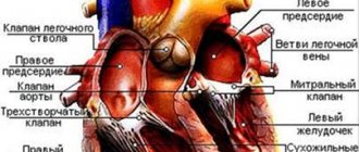
The cardiac muscle (myocardium) in the structure of the human heart is located in the middle layer between the endocardium and epicardium. It is this that ensures the uninterrupted operation of “distilling” oxygenated blood into all organs and systems of the body.
Any weakness affects blood flow and requires compensatory restructuring and coordinated functioning of the blood supply system. Insufficient ability to adapt causes a critical decrease in the performance of the heart muscle and its diseases. The endurance of the myocardium is ensured by its anatomical structure and endowed capabilities.
Features of muscle tissue are:
- striated striations formed by myofibrils of cardiomyocyte cells;
- the presence of two types of fibers: thin (actin) and thick (myosin), connected by cross bridges;
- the connection of myofibrils into bundles of different lengths and directions, which makes it possible to distinguish three layers (superficial, internal and middle).
The structure of the heart muscle is different from skeletal and smooth muscle muscles, which provide movement and protection of internal organs
The morphological features of the structure provide a complex mechanism of heart contraction.
Treatment and diagnosis
A cardiologist can determine why the heart muscle hurts. For diagnosis, he does an ultrasound and cardiogram. If there are no pathologies, the patient is sent for a thorough examination of the abdominal cavity and an x-ray of the spine. He also needs to consult with an endocrinologist and neurologist. If a serious pathology is detected, the specialist selects individual treatment. It is based on the type and stage of the disease, the characteristics of the body.
Modern medicine is making great strides in the treatment of heart diseases. Various and highly effective treatment methods are used. It is even possible to get rid of many pathologies in order to return to a full life.
In the treatment of cardiac pathologies the following are used:
- Medications;
- Surgical intervention - atherectomy, heart muscle transplant, valve replacement, installation of pacemakers, coronary artery bypass grafting, etc.;
- Physiotherapy;
- All kinds of alternative medicine methods.
Many cardiologists also use innovative methods such as:
- External synchronized counterpulsation;
- Regenerative Medicine;
- Shock wave therapy.
How does the heart contract?
Contractility is one of the properties of the myocardium, which consists in creating rhythmic movements of the atria and ventricles, allowing blood to be pumped into the vessels. The chambers of the heart constantly pass through 2 phases:
- Systole - is caused by the combination of actin and myosin under the influence of ATP energy and the release of potassium ions from the cells, while thin fibers slide over thick ones and the bundles decrease in length. The possibility of wave-like movements has been proven.
- Diastole - relaxation and separation of actin and myosin occurs, restoration of expended energy due to the synthesis of enzymes, hormones, and vitamins obtained through the “bridges”.
It has been established that the force of contraction is provided by calcium entering the myocytes.
The entire cycle of heart contraction, including systole, diastole and the general pause after them, with a normal rhythm fits into 0.8 seconds. Begins with atrial systole, the ventricles are filled with blood. Then the atria “rest”, moving into the diastole phase, and the ventricles contract (systole). Calculating the time of “work” and “rest” of the heart muscle showed that per day the state of contraction accounts for 9 hours 24 minutes, and the state of relaxation accounts for 14 hours 36 minutes.
The sequence of contractions, ensuring the physiological characteristics and needs of the body during stress and anxiety depend on the connection of the myocardium with the nervous and endocrine systems, the ability to receive and “decipher” signals, and actively adapt to human living conditions.
The spread of excitation from the sinus node can be traced by intervals and ECG waves
List of heart diseases
The normal functioning of the heart and vascular system can be disrupted by a large number of different diseases. Several umbrella categories are often used, from which different types of diseases branch.
Atherosclerotic disease
While many disease processes can affect blood vessels, the term "cardiovascular disease" generally covers disorders of the blood vessels (arteries) that are associated with either atherosclerosis, hypertension, or heart disease.
Atherosclerosis is a disease in which plaque made up of fat, calcium, cholesterol and other substances builds up and hardens in the arteries, impairing blood flow.
There are different types of atherosclerotic diseases, including coronary artery disease, carotid artery disease, and peripheral artery disease.
Coronary heart disease (CHD), common in most parts of the world, can lead to heart attacks (myocardial infarction) and is the most common type of heart disease. In coronary artery disease, atherosclerotic plaques form in the lining of the coronary arteries, hardening and narrowing the arteries.
Atherosclerosis and high blood pressure (hypertension or hypertension) can lead not only to coronary artery disease, but also to carotid artery disease, which affects the carotid arteries on both sides of the neck, and peripheral artery disease, which can affect almost any other artery in organism.
Cardiac mechanisms that provide contraction
The properties of the heart muscle have the following purposes:
- support myofibril contraction;
- ensure the correct rhythm for optimal filling of the cavities of the heart;
- maintain the ability to push blood through any extreme conditions for the body.
For this, the myocardium has the following abilities.
Excitability - the ability of myocytes to respond to any incoming pathogens. Cells protect themselves from above-threshold stimulation by a state of refractoriness (loss of the ability to excite). In a normal contraction cycle, a distinction is made between absolute and relative refractoriness.
- During the period of absolute refractoriness, for 200 to 300 msec, the myocardium does not respond even to extremely strong stimuli.
- When relative, it is able to respond only to sufficiently strong signals.
This property prevents the heart muscle from “distracting” the contraction mechanism during the systole phase
Conductivity is the property of receiving and transmitting impulses to different parts of the heart. It is provided by a special type of myocytes that have processes very similar to neurons in the brain.
Automaticity - the ability to create its own action potential inside the myocardium and cause contractions even when isolated from the body. This property allows resuscitation in emergency cases and maintains blood supply to the brain. The importance of the located network of cells and their accumulation in nodes during transplantation of a donor heart.
Pacemaker cells (pacemakers) become the main ones if the processes of repolarization and depolarization in the main nodes are weakened. They suppress “other people’s” excitability and impulses, and try to take on a leadership role. Localized in all parts of the heart. The possibilities are restrained by the sufficient strength of the sinus node.
Features of heart pain in men
In both sexes, heart pain is similar, and the differences in this symptom are dictated not so much by gender as by individual characteristics. However, the pain sensitivity threshold is different for men and women. The pain threshold of men is influenced by the degree of activity of the antinociceptive system.
Neurotransmitters-endorphins, which belong to the group of endogenous opiates, dampen pain. Since men have certain characterological characteristics and are more prone to systematic physical activity, they release endorphins more regularly and in greater quantities than women.
The threshold is also positively influenced by the hormone testosterone, which by definition is several times higher in men. Such gender-specific differences in the perception of pain signals lead to late visits to the doctor, which delays the start of adequate therapeutic measures. Of course, it is not just one symptom that is treated, but the disease itself as a whole.
The earlier treatment begins, the more effective it will be. It is noteworthy that regular alcohol intake also increases the pain threshold, which increases the risk of coronary death, because the heart begins to hurt weakly and often after the onset of irreversible ischemic changes.
Typical chest pain refers to the clinical manifestation of ordinary angina. The heart hurts behind the sternum and radiates to the upper limb, collarbone, neck, lower jaw on the left. To identify the painful area, patients point to the mid-left area of the chest. These symptoms are very similar to myocardial infarction.
There are situations when a man focuses on areas of irradiation and describes them first, which is already regarded as atypical angina. Sometimes the individual characteristics of the innervation of internal organs determine the fact that it hurts at the level of the stomach, in the subcostal area. The nature of this symptom in this case is more diffuse, bursting, pressing.
And sometimes vegetative symptoms come to the fore, when the heart hurts, but not so much. With dextracardia, the pain is more on the right side.
In most cases, the pain syndrome is not fundamentally different from men, however, women react to pain in the heart earlier and more actively, which is associated with their physiological high sensitivity and, accordingly, a lower pain threshold.
Women are less likely to use substances that increase the pain threshold and are less likely to engage in activities that require physical exertion, which determines their lower levels of endorphins.
Their testosterone is several times lower than that of healthy men, which contributes to the formation of the pain sensitivity threshold. Most often, women experience the following symptoms:
- ordinary cough;
- general weakness and pallor;
- elevated temperature;
- high or low blood pressure;
- general swelling;
- dizziness in crowded places and motion sickness in any transport;
- frequent shortness of breath;
- periodic vomiting and nausea;
- pain in the neck or shoulder blades, reminiscent of ordinary osteochondrosis;
- pain in the chest area;
- strong and frequent heartbeats.
Typically, cough is a symptom of viral diseases, flu, colds and bronchitis. However, it is worth paying special attention to such a minor symptom if expectorant medications do not help. A dry cough that suddenly appears in a patient while lying down is a reasonable cause for alarm.
As for general weakness of the body and pallor, such symptoms are signs of disorders and disorders of the nervous system. Such symptoms are characteristic of neuroses of the heart muscle, although they are often associated with other diseases.
The importance of biochemical processes in the myocardium
The viability of cardiomyocytes is ensured by the supply of nutrients, oxygen and the synthesis of energy in the form of adenosine triphosphoric acid.
All biochemical reactions occur maximally during systole. The processes are called aerobic because they are possible only with a sufficient amount of oxygen. The left ventricle consumes 2 ml of oxygen per minute per 100 g of mass.
To produce energy, the following are used in the blood:
- glucose,
- lactic acid,
- ketone bodies,
- fatty acid,
- pyruvic and amino acids,
- enzymes,
- B vitamins,
- hormones.
If the heart rate increases (physical activity, anxiety), the need for oxygen increases 40–50 times, and the consumption of biochemical components also increases significantly.
What compensatory mechanisms does the heart muscle have?
A person does not develop pathology as long as the compensation mechanisms work well. Regulation is carried out by the neuroendocrine system.
The sympathetic nerve delivers signals to the myocardium about the need for increased contractions. This is achieved by more intense metabolism and increased ATP synthesis.
A similar effect occurs with increased synthesis of catecholamines (adrenaline, norepinephrine). In such cases, increased work of the myocardium requires an increased supply of oxygen.
If atherosclerotic narrowing of the coronary vessels does not allow the heart muscle to be supplied in the required volume, then the mediator acetylcholine is released. It protects the myocardium and helps maintain contractile activity in conditions of oxygen deficiency.
The vagus nerve helps reduce the frequency of contractions during sleep, during rest periods, and preserve oxygen reserves.
It is important to consider reflex adaptation mechanisms.
Tachycardia is caused by congestive stretching of the mouths of the vena cava.
Reflex slowing of the rhythm is possible with aortic stenosis. In this case, increased pressure in the cavity of the left ventricle irritates the endings of the vagus nerve, contributing to bradycardia and hypotension.
The duration of diastole increases. Favorable conditions are created for the functioning of the heart. Therefore, aortic stenosis is considered a well-compensated defect. It allows patients to live to an old age.
Features of the clinical picture and course of the disease in various infections
The manifestations of infectious inflammation of the myocardium are influenced by the strength of the pathogen, the state of the patient’s immunity, and individual compensatory capabilities.
With diphtheria, both the “working” muscles of the myocardium and the conduction pathways are affected. Toxins cause cell necrosis and hemorrhages under the endocardium. The clinic develops in the second week of the disease. The more pronounced the bradycardia, the deeper and more widespread the lesion. The myocardium “activates” the protective mechanism of automatism. At the same time, nerves and blood vessels are affected.
Typhus “selects” small vessels, including the coronary arteries, and begins as vasculitis with inflammation in the surrounding tissues. Primary disturbances in cerebral vascular tone and a sharp drop in blood pressure. Myocarditis occurs secondary.
Typhoid fever is rarely accompanied by myocarditis in the third week of the disease. The myocardium changes along the path of fatty degeneration and dystrophy. Classic symptoms occur after a decrease in temperature and are post-typhoid in nature.
With scarlet fever, the morphology of tissue changes has a different form: from fatty degeneration to the formation of specific granulomas. The transverse striation of myofibrils disappears. Clinically, it occurs in accordance with the general manifestations.
Influenza - heart damage occurs after a decrease in temperature and intoxication.
Some researchers believe that the flu can be equated to diphtheria myocarditis in terms of its effect on the heart.
Considering the tendency of influenza viruses to damage the heart with severe atherosclerosis of the coronary vessels in older people, it is recommended to adhere to longer bed rest in treatment.
During lobar pneumonia, focal inflammation develops against the background of impaired blood flow to the right ventricle, insufficient filling of the left ventricle and coronary vessels. The process in the lungs complicates the outflow and saturation of oxygen. Therefore, circulatory failure develops much faster.
How to treat hypertrophy?
Typically, prolonged increased load causes hypertrophy. The thickness of the left ventricular wall increases by more than 15 mm. An important point in the formation mechanism is the lag in the growth of capillaries deep into the muscle. In a healthy heart, the number of capillaries per mm2 of cardiac muscle tissue is about 4000, and with hypertrophy the figure decreases to 2400.
Therefore, the condition is considered compensatory up to a certain point, but with significant thickening of the wall it leads to pathology. It usually develops in the part of the heart that must work hard to push blood through a narrowed hole or overcome a vascular obstruction.
Hypertrophied muscle is able to maintain blood flow for a long time in case of heart defects.
The muscle of the right ventricle is less developed; it works against a pressure of 15–25 mm Hg. Art. Therefore, compensation for mitral stenosis and cor pulmonale does not last long. But right ventricular hypertrophy is of great importance in acute myocardial infarction, cardiac aneurysm in the left ventricular area, and relieves overload. The significant capabilities of the right sections in training during physical exercises have been proven.
Thickening of the left ventricle compensates for aortic valve defects and mitral insufficiency
Features of rheumatic myocarditis
Rheumatic myocarditis stands apart among all myocardial inflammations. Although caused by infection, an autoimmune response has been proven to play a role.
It has characteristic differences from other types:
- almost always accompanied by endocarditis with damage to the heart valves;
- associated with a primary streptococcal infection, symptoms appear after a latent period 2 weeks after a sore throat, acute nephritis;
- most often affects children and adolescents;
- the morphological substrate is specific granulomas (Ashof-Talalaev nodules);
- the location of granulomas is most often found in the posterior wall of the left atrium and in the papillary muscle of the left ventricle;
- accompanied by damage to other organs and tissues (joints, nervous system);
- has a chronic course.
Idiopathic Abramov-Fiedler myocarditis etiology remains unclear. It has a very severe acute course. Rapid development of decompensation with pronounced expansion of the cavities of the heart. The patient dies from heart failure or rhythm disturbances.
Can the heart adapt to work in hypoxic conditions?
An important property of adaptation to work without sufficient oxygen supply is the anaerobic (oxygen-free) process of energy synthesis. A very rare occurrence for human organs. Turns on only in emergency situations. Allows the heart muscle to continue contracting. Negative consequences are the accumulation of breakdown products and overwork of muscle fibrils. One cardiac cycle is not enough for energy resynthesis.
However, another mechanism is involved: tissue hypoxia reflexively causes the adrenal glands to produce more aldosterone. This hormone:
- increases the amount of circulating blood;
- stimulates an increase in the content of red blood cells and hemoglobin;
- increases venous flow to the right atrium.
This means that it allows the body and myocardium to adapt to the lack of oxygen.
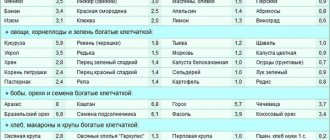
Using natural recipes
It's no secret that to find out how to strengthen the heart muscle, you need to look at folk remedies.
Basically, among folk methods for strengthening the heart, there are the following recommendations regarding nutrition. This can be done at home to strengthen the heart.
To strengthen the heart muscle, it is advisable to follow these tips:
- use honey instead of sugar in food to strengthen;
- drink more tea with mint and valerian to strengthen. This will bring a calming effect to the body;
- make more juices from vegetables to strengthen. Carrot juice will be an excellent neutralizer for arrhythmia;
- more nuts and dried fruits as heart-healthy snacks.
In this simple way you can feed and strengthen the human body with vitamins and beneficial microelements.
Also, folk remedies for strengthening the heart may not include medicinal decoctions that are prepared from various herbs. To prepare a medicinal decoction to strengthen the heart, you need to take calendula, viburnum berries, and lingonberry leaves. Mix this set thoroughly and brew it in a thermos. You must wait at least 12 hours, and then drink a quarter glass every three hours to strengthen the body.
This folk remedy imperceptibly at first glance reduces the risk of myocardial damage.
How myocardial pathology occurs, mechanisms of clinical manifestations
Myocardial diseases develop under the influence of various causes, but appear only when adaptation mechanisms fail.
Long-term loss of muscle energy, the impossibility of independent synthesis in the absence of components (especially oxygen, vitamins, glucose, amino acids) lead to a thinning of the actomyosin layer, breaking the connections between myofibrils, replacing them with fibrous tissue.
This disease is called dystrophy. It accompanies:
We recommend reading: What is hibernating myocardium
- anemia,
- avitaminosis,
- endocrine disorders,
- intoxications.
Arises as a consequence:
- hypertension,
- coronary atherosclerosis,
- myocarditis.
Patients experience the following symptoms:
- weakness,
- arrhythmia,
- shortness of breath during physical exertion,
- heartbeat.
At a young age, the most common cause may be thyrotoxicosis and diabetes mellitus. In this case, there are no obvious symptoms of an enlarged thyroid gland.
The inflammation of the heart muscle is called myocarditis. It accompanies both infectious diseases of children and adults, as well as those unrelated to infection (allergic, idiopathic).
It develops in focal and diffuse forms. Proliferation of inflammatory elements affects myofibrils, interrupts pathways, and changes the activity of nodes and individual cells.
We advise you to learn more about inflammatory diseases of the myocardium from this article
As a result, the patient develops heart failure (usually right ventricular failure). Clinical manifestations consist of:
- pain in the heart area;
- rhythm interruptions;
- shortness of breath;
- dilation and pulsation of the neck veins.
Atrioventricular blocks of varying degrees are recorded on the ECG.
The most well-known disease caused by impaired blood flow to the heart muscle is myocardial ischemia. It proceeds in the form:
- angina attacks,
- acute heart attack,
- chronic coronary insufficiency,
- sudden death.
The main morphological substrate for this pathology are areas of the heart muscle depleted of nutrients and oxygen. Depending on the degree of damage, cardiomyocytes change and undergo necrosis.
All forms of ischemia are accompanied by paroxysmal pain. They are figuratively called “the cry of the starving myocardium.” The course and outcome of the disease depends on:
- speed of assistance;
- restoration of blood circulation due to collaterals;
- the ability of muscle cells to adapt to hypoxia;
- formation of a strong scar.
A controversial drug included in the doping list for providing additional energy to the heart muscle
Reasons for development
Inflammation of the heart muscle (myocarditis) is focal or diffuse inflammation of the myocardium, covering the entire heart muscle. Mild forms of myocarditis are more common. However, in severe cases, scars may remain in places where there were foci of inflammation, which causes a disruption in the propagation of impulses through the conduction system of the heart and the occurrence of cardiac arrhythmias. Insufficiency of heart function appears, the heart muscle thickens.
Inflammation of the myocardium causes various diseases. These are usually inflammatory diseases that affect the heart. With active rheumatism, myocarditis is possible. The main cause of myocarditis is bacterial and viral infections, such as tonsillitis, diphtheria, scarlet fever or influenza, as well as toxoplasmosis and trichinosis. If you have one of these diseases, you must strictly adhere to bed rest. In addition, myocarditis can occur with allergic diseases and diseases of the body's immune system. However, usually the cause of myocarditis remains unknown. It may depend on the epidemiological situation of the region; for example, in Europe and North America, myocarditis is more often caused by viruses. Many viral infections are thought to affect the heart muscle, but this does not always result in clear clinical symptoms.