The heart is one of the main vital organs of a person. The organ is located in the chest cavity, and is shifted to the left side. The exact location and mass of the muscle depends on several factors. These include a person’s physique, gender differences, age characteristics and chest shape. The average weight of the organ for women is approximately 250 g, for men the figures are slightly higher, reaching 300 g. The heart is a muscle, and therefore can increase during various workouts. Therefore, in athletes and people actively involved in physical labor, the muscle grows and increases in volume.
Features of the structure of the heart muscle
The heart is a group of muscle fibers that have a hollow space inside. There are four cavities inside the organ:
- Right atrium;
- Left atrium;
- Right ventricle;
- Left ventricle.
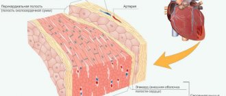
The structure of the heart muscle includes three types of wall layers. The first includes the inner endothelial layer together with the valves, called the endocardium. Next is the middle layer - the myocardium. The next outer layer is the epicardium, which is covered with single-layer epithelium. The entire heart is covered from the outside with a pericardial sac. The structural features also include the serous fluid located between the pericardium and epicardium. This fluid is necessary to reduce tissue friction during contraction of muscle fibers during beating.
The structure of the heart muscle also includes valves. The first is located on the left side between the ventricle and atrium. In medicine, this valve is classified as mitral or bicuspid. On the right side there is a second valve called the tricuspid valve. The semilunar valves are located at the mouth of the aorta. With their help, the flow of blood back to the heart, into the ventricle, is blocked. The middle layer of the heart is formed with the help of muscle cells - cardiomyocytes. In the area of the atria, the myocardium is thinner, in the ventricles it becomes denser, especially in the left ventricle. The myocardium is a transversely striated muscle, which in turn has a number of distinctive features. Cardiomyocytes adhere very tightly to each other and form a single tissue structure - syncytium. It is thanks to this structural feature of the heart muscle that very rapid excitation and simultaneous contraction of the entire heart is ensured.
Myocardial properties and functional load
Due to the structural features of the myocardium and its cells, this muscle group has a number of characteristic features:
- automation (the function generates various impulses directed from external influences);
- conductivity (the ability of an organ to transmit excitation);
- excitability (heart cells are excited by impulse signals emanating from the organ’s conduction system);
- contractility (the ability to contract as a result of receiving an impulse).
Impulse signals arise in the pacemaker located in the right atrium at the mouth of the vena cava. The structure and properties of the heart muscle are mainly aimed at contracting the organ, providing blood supply to all tissues in the body with oxygen and nutrients. The contractile function must work correctly and clearly. If normal functioning is disrupted, various health problems arise not only for the organ itself, but also for the entire human body.
Chapter 5. PHYSIOLOGICAL PROPERTIES OF CARDIAC MUSCLE
Basic questions : The structure of the human heart, the systemic and pulmonary circulation. Morpho-functional features and physiological properties of the heart muscle. Conduction system of the heart. Gradient of heart automaticity. Action potential of working cardiomyocytes. The relationship between the phases of excitation and periods of excitability of working cardiomyocytes. Extrasystoles and their types.
Cardiac cycle. Phases of the cardiac cycle and their characteristics.
The human heart is a hollow muscular organ divided into four chambers - two atria
and two
ventricles
.
The work of the heart is a continuous cyclic alternation of systole - contraction and diastole - relaxation. The frequency of contractions and relaxations of the atria and ventricles ensures the pumping of blood into the great vessels of the systemic and pulmonary circulation.
The atria communicate with the ventricles through the atrioventricular openings
.
In the left half of the heart they are closed by a bicuspid valve
(
mitral
), and in the right -
by a tricuspid valve
(Fig. 20
)
. These valves can only allow blood to flow in one direction - from the atria to the ventricles.
Fig.20. The structure of the human heart
The aorta arises from the left ventricle
, from which
the systemic circulation
(Fig. 21).
Arterial blood entering the vessels of the large circle supplies all organs and tissues of the body with oxygen and nutrients. The large circle ends in the right atrium, where venous blood flowing from organs and tissues flows through the superior
and
inferior vena cava
. From here the blood passes through the atrioventricular opening into the right ventricle.
The pulmonary artery arises from the right ventricle
, which is the beginning of
the pulmonary circulation
. The blood vessels of the small circle, passing through the lungs, ensure the enrichment of the blood with oxygen and the removal of carbon dioxide from it. The pulmonary circulation ends in the left atrium.
The openings from which the aorta and pulmonary artery begin are covered by the semilunar valves
–
aortic
and
pulmonary
(Fig. 20). They pass blood in only one direction - from the ventricles to the great vessels.
Fig.21. Diagram of the systemic and pulmonary circulation
Cardiac muscle - myocardium,
which is a collection of muscle cells, has six physiological properties:
1) irritability;
2) excitability;
3) conductivity;
4) contractility;
5) lability;
6) automation.
Irritability is a common property of all living tissues (plant and animal) to respond to irritation by changing metabolism and energy, as well as by changing the processes of reproduction, growth and differentiation of cells.
Some living tissues have the ability to generate bioelectric potentials. Such highly specialized living tissues (nervous, muscle and glandular) are called excitable .
At rest, cells in excitable tissues are polarized
, their cytoplasm is negatively charged relative to the intercellular fluid.
This relative potential difference existing at rest between the inner and outer sides of the cell membrane is called the resting membrane potential (RMP).
Polarization is due to ion asymmetry
and the unequal
permeability
of the semi-permeable cell membrane for various ions.
The membrane of an excitable cell contains pores called ion channels
. Depending on the ions passed through, sodium, potassium, calcium and chloride ion channels are distinguished.
The cytoplasm of nerve cells contains 50 times more K+ ions and 10 times less Na+ ions than the extracellular fluid. The consequence of such ionic asymmetry is concentration, electrical and electrochemical gradients
.
A concentration gradient is a force that causes a substance to move from an area of higher concentration to a lower one.
Electric gradient is a force that ensures the movement of charged particles from the electric pole of the same name to the opposite one.
The sum or difference of the concentration and electrical gradient constitutes the electrochemical gradient .
The main contribution to the formation of the resting membrane potential is made by K+ ions. This is due to the fact that at rest the permeability of the cell membrane for K+ ions is tens of times greater than for Na+ ions.
In the resting state, K+ ions passively leave the cell along a concentration gradient (Fig. 22).
Fig.22. Scheme of ionic mechanisms of MPP
CG – concentration gradient; ECG – electrochemical gradient.
The passive movement of positively charged K+ particles from the cell causes an increase in the electronegativity of the cytoplasm. A transmembrane potential difference arises
– the difference in electrical potential on both sides of the cell membrane.
Due to the low permeability at rest, only a small number of Na+ ions can diffuse passively along the electrochemical gradient into the cytoplasm. Entering the cell in small quantities, positively charged Na+ ions somewhat reduce the electronegativity of the cytoplasm and the transmembrane potential difference, and hence the value of the resting membrane potential.
An additional contribution to the formation of muscle cell MPP is made by Cl- ions. At rest, Cl- ions passively, along a concentration gradient, penetrate into the cell from the intercellular fluid. Entering the cell, negatively charged Cl- ions increase the electronegativity of the cytoplasm, and therefore lead to an increase in the level of transmembrane potential difference, i.e. the value of the resting membrane potential.
Under the influence of stimuli, highly specialized cells move from a state of rest to a state of activity, which manifests itself in the form of local or spreading excitation.
Excitation is a bioelectrical process that occurs in living excitable tissue as a result of the action of a stimulus and is characterized by depolarization of the cell membrane in the form of the generation of a spreading action potential (AP) or a local (local), non-propagating response (LO).
The main reason for depolarization under the influence of a stimulus is an increase in the permeability of the cell membrane to Na+ ions. This leads to an increase in the flow of Na+ ions entering the cell. Positively charged Na+ ions passively enter the cytoplasm along an electrochemical gradient, where they partially neutralize negative intracellular anions. The electronegativity of the cytoplasm, and therefore the transmembrane potential difference, decreases.
The type of excitation (LO or AP) depends on the strength of the stimulus.
An irritant is a factor of the external or internal environment that acts on tissue and has sufficient strength, duration and rate of increase in strength over time to excite it.
Stimuli of minimal strength that are still capable of causing the generation of spreading excitation when acting on tissue are called threshold . Stimuli of lesser strength than the threshold, which are not capable of causing the generation of spreading excitation, are called subthreshold . Stimuli of greater strength than the threshold, capable of causing the generation of spreading excitation, are classified as suprathreshold .
The ability of excitable tissue to respond to irritation by the process of excitation is called excitability .
A measure of excitability is the excitability threshold - the minimum strength of the stimulus that causes the generation of a propagating PD when exposed to tissue. There is an inverse relationship between the threshold of excitability and excitability: the lower the threshold, the higher the excitability and vice versa. Myocardial excitability is less than that of skeletal muscle, but greater than that of smooth muscle.
Spreading excitation causes a specific reaction of the excitable tissue. The specific ability of muscle tissue to respond to spreading excitation by the process of contraction is contractility
.
Contractility is a specific property of muscle tissue to respond to stimulation by the process of contraction.
The myocardium is a functional syncytium
– a union of muscle fiber cells, each of which is connected to the others using
nexuses - slot-like contacts equipped with insertion disks with low electrical resistance.
Having arisen in any part of the myocardium, excitation spreads freely along the syncytium and, passing from one muscle cell to another through nexuses, covers the entire muscle as a whole, causing its contraction. Thanks to the nexuses, the heart muscle functions as a single whole and contracts according to the “all or nothing” law: the heart responds to the action of threshold and suprathreshold stimuli causing spreading excitation with a maximum contraction of all working muscle fibers (“all”), and to subthreshold stimuli the contractile reaction of the myocardium does not arise (“nothing”). There are two types of cells in the heart muscle: workers
(
typical
)
cardiomyocytes
and
atypical
(
conducting
)
cardiomyocytes
.
The main task of atypical muscle cells of the heart is spontaneous (spontaneous) generation of excitation. They form the conduction system of the heart
which provides:
1) rhythmic spontaneous generation of excitation;
2) conducting excitation;
3) sequence of contractions of the atria and ventricles;
4) synchronicity of involvement of myocardial muscle cells in the process of excitation and contraction.
Automaticity of the heart is the ability of atypical cardiomyocytes to spontaneously generate AP under the influence of metabolic processes occurring in these cells.
Atypical muscle cells of the conduction system of the human heart form two main nodes of automation
(Fig.23
)
:
1) sinoatrial node (sinoatrial node);
2) atrioventricular node (atrioventricular node).
Fig.23. Conduction system of the heart
The sinoatrial node is located at the point where the vena cava enters the right atrium. It is the main, leading center of heart automation - the pacemaker
(
pacemaker
) of the first order.
The sinoatrial node sets the rate of excitation, and therefore contractions, of other parts of the heart. The cells of the sinoatrial node are called pacemaker P-cells
, and the action potential that they spontaneously generate is called
the pacemaker potential
(Fig. 24).
The resting membrane potential (RMP) of pacemaker β-cells during cardiac diastole does not exceed -60 mV. It is characterized by instability: under the influence of metabolic processes, slow diastolic depolarization periodically occurs - a gradual decrease in the transmembrane potential difference. This is due to a spontaneous decrease in the permeability of P-cells for K+ ions that leave the cell and an increase in permeability for Ca2+ and Na+ ions entering it.
When a critical level is reached (about -50 mV), the rate of depolarization of the P-cell membrane increases - slow diastolic depolarization turns into fast depolarization .
After complete depolarization, when the transmembrane potential difference becomes zero, the flow of positively charged ions entering the cell (Ca2+ and Na+) still exceeds the flow of positively charged particles leaving it (K+). reversion occurs (up to +10 mV) - recharging of the P-cell, which becomes positively charged inside and negatively charged outside.
Then, the permeability of P-cells for K+ ions increases, and for Ca2+ and Na+ ions decreases. Therefore, the reversal phase is replaced first by a phase of fast and then slow repolarization . During the process of slow repolarization, the membrane potential reaches the initial level of -60 mV. After this, the entire cycle of spontaneous excitation is repeated again.
Slow diastolic depolarization is a local, non-propagating excitation called the P-cell prepotential
. Only when a critical level is reached is this prepotential transformed into propagating excitation, which causes myocardial contraction. Therefore, the rate and duration of slow diastolic depolarization (SDD) determine the heart rate. The faster the speed of DMD and the shorter its duration, the higher the heart rate (HR).
Fig.24. Graph of the action potential of pacemaker β-cells of the heart
1) slow diastolic depolarization, 2) fast depolarization, 3) reversal, 4) fast repolarization, 5) slow repolarization.
The speed and duration of DMD can vary under the influence of various biologically active substances. Thus, acetylcholine, a mediator of the parasympathetic nervous system, increases the permeability of β-cells to K+ ions. As a result, depolarization becomes more difficult, the speed decreases, and the duration of DMD increases. This leads to a decrease in heart rate. Under the influence of adrenaline and norepinephrine, the permeability of β-cells to Ca2+ and Na+ ions increases. As a result, depolarization is facilitated, the speed is increased and the duration of DMD is reduced, which leads to an increase in heart rate.
From the sinoatrial node, excitation spreads at a speed of about 1 m/s through the atrial myocardium and through the conduction system of the heart to the atrioventricular node.
The atrioventricular node is a second-order center of automation, which is located in the cardiac septum at the border of the atria and ventricles. In a healthy person, it is excited in the rhythm of the sinoatrial node. Under normal conditions, excitation can pass through it in only one direction - to the ventricles. , an atrioventricular delay in the conduction of excitation occurs
– reduction in the speed of excitation propagation to 0.02-0.05 m/s. This ensures the sequence of contractions of the atria and ventricles.
From the atrioventricular node, excitation spreads at a speed of 4-5 m/s along the bundle and legs of His
to the apex of the heart, and from there it returns along
the Purkinje fibers
to its base. Much more slowly, at a speed of about 1 m/s, excitation diffuses through the nexuses along the fibers of the working ventricular myocardium. However, due to the high speed of excitation propagation along the conduction pathways of the heart, the muscle cells of the myocardium of both ventricles are involved in the contraction process almost simultaneously, i.e. synchronously.
The ability of different nodes of the conduction system to be automatic is expressed differently and is characterized by a gradient of automaticity
. The automaticity gradient decreases from the base to the apex of the heart: in the sinoatrial node, the natural frequency of AP generation is 60-80 impulses/min, in the atrioventricular node - 40-50, in His bundle cells - 30-40, and in Purkinje fibers - about 20 pulses/min.
Under normal natural conditions, the sinoatrial node subordinates all underlying formations of the conduction system of the heart. However, if automatic excitation does not occur in it or its transmission is blocked, the atrioventricular node takes on the role of pacemaker.
If it is impossible to transmit excitation to the ventricles, they begin to contract in the His bundle automatic mode. However, the intrinsic frequency of the His bundle and especially the Purkinje fibers cannot ensure normal blood flow and maintain vital functions for a long period of time. Damage to all potential pacemakers leads to cardiac arrest.
Typical ( working) myocardial cardiomyocytes do not have the ability to automate. However, they are able to excite and contract under the influence of electrical impulses coming to them from the cells of the conduction system of the heart. The main function of typical cardiomyocytes is to ensure the contraction process and the pumping function of the heart.
The biopotentials of typical cardiomyocytes are characterized by:
1) stable level of MPP compared to atypical cardiomyocytes;
2) longer duration compared to PD of skeletal muscles.
The resting membrane potential of working cardiomyocytes is -90 mV (Fig. 25). As a result of stimulation of typical cardiomyocytes with the next electrical impulse that comes from the conduction system of the heart, their slow depolarization - a decrease in the transmembrane potential difference. The reason for depolarization of working cardiomyocytes is an increase in their permeability to Na+ ions.
The phase of slow depolarization of working cardiomyocytes takes a very short period of time (about 0.01 s). When a critical level is reached (about -70 mV), the rate of depolarization increases - slow depolarization turns into fast depolarization .
After complete depolarization, when the potential difference on either side of the cell membrane is zero, the flow of positively charged Na+ ions entering the cell is still increased compared to the resting state. reversion occurs - recharging of a typical cardiomyocyte, which is charged positively inside and negatively outside.
After reaching the peak AP (+30 mV), there is a rapid decrease in the permeability of the membrane of the working cardiomyocyte for Na+ ions entering the cell, and a simultaneous increase in permeability for K+ ions leaving it. The flow of positively charged particles leaving the cell (K+) exceeds the flow of positively charged particles entering it (Na+). repolarization begins - restoration of the resting membrane potential.
However, soon after the onset of repolarization, slow-responding calcium ion channels are activated. The total flow of positively charged particles (Ca2+ and Na+) entering the cell increases again. Therefore, the rate of repolarization slows down significantly, and it enters a plateau , which is characterized by a relatively stable level of membrane potential (near zero).
The calcium ion channels are then inactivated. Therefore, the rate of repolarization increases significantly - the plateau goes into a phase of rapid repolarization , which ends with the achievement of the initial level of polarization of the cell membrane at rest (-90 mV). By this time, the permeability of the membrane of working cardiomyocytes for K+, Na+ and Ca2+ ions ensures the maintenance of the resting membrane potential until the next electrical impulse arrives from the sinoatrial node.
Fig.25. PP schedule of working (typical) cardiomyocytes
1) slow depolarization, 2) fast depolarization, 3) reversal, 4) plateau, 5) fast repolarization.
Certain periods of the excitation cycle (EC) in the cells of the working myocardium correspond to phase changes in excitability (Fig. 26).
Fig.26. Correlation of AP phases with periods of excitability of the working cardiomyocyte
1) primary supernormal excitability, 2) absolute refractoriness, 3) relative refractoriness, 4) exaltation (secondary supernormal excitability).
In the initial resting state (during cardiac diastole), the excitability of typical cardiomyocytes is conventionally taken as 100%. Slow depolarization corresponds to a period of primary supernormal ( increased ) excitability , which lasts about 0.01 s.
The rapid depolarization, reversal, and plateau correspond to the absolute refractory , which lasts approximately 0.27 s (270 ms). At this time, the heart muscle does not respond to either threshold or suprathreshold stimuli.
The initial part of rapid repolarization (to a critical level) corresponds to a period of relative refractoriness (about 0.03 s), during which the myocardium is able to respond with excitation and contraction only to suprathreshold stimuli.
The final part of the rapid repolarization corresponds to a relatively short period (0.03 s) of secondary supernormal excitability ( exaltation ), during which working cardiomyocytes are able to respond with spreading excitation and contraction even to subthreshold stimuli.
The refractory period lasts as long as the contraction of myocardial muscle fibers continues - systole. Long-term refractoriness protects the myocardium from too rapid re-excitation, and therefore eliminates the possibility of a continuous continuous contraction of the heart. Thanks to this, the heart works in the rhythm of single muscle contractions, which ensures the pumping of blood into the main vessels.
Irritation of the myocardium during the period of its relaxation (diastole), when excitability has already been sufficiently restored, causes extrasystole - an extraordinary premature contraction that temporarily disrupts the heart rhythm.
The focus of extraordinary excitation that occurs in the myocardium outside the conduction system of the heart is called ectopic . Extrasystoles, depending on the location of the ectopic focus in relation to the sinoatrial node, are divided into atrial
and
ventricular
.
If ectopic excitation occurs in the atrial myocardium when the refractory period has already ended, but the next automatic impulse in the sinoatrial node has not yet been generated, then an extraordinary contraction of the heart occurs - sinus (atrial) extrasystole . The pause that follows a sinus extrasystole lasts the same time as a normal one.
An extraordinary contraction of the heart that occurs during ventricular diastole or general cardiac pause, when ectopic excitation occurs in the ventricular myocardium, is called ventricular extrasystole . Extraordinary excitation that occurs in the ventricles does not affect the automation of the sinoatrial node. It promptly sends the next impulse, which reaches the ventricles at a time when they are still in a refractory state after the extrasystole. Therefore, the ventricular myocardium does not respond to the next timely electrical impulse coming from the sinoatrial node, which leads to a long compensatory pause
ventricles (felt as interruptions in the functioning of the heart).
Due to the long period of absolute refractoriness, the working myocardium is characterized by low lability.
Lability is the speed with which the tissue, having responded to irritation with excitation, becomes again capable of generating a new PD in response to the next irritation.
Lability is measured by the maximum number of APs in 1 second (1 s = 1000 ms) that the tissue can reproduce without distorting the rhythm of stimulation. It depends on the duration of the absolute refractoriness phase of living tissue during AP generation. The longer the absolute refractory period, the lower the lability.
To calculate lability, 1000 ms must be divided by the duration of absolute refractoriness. Myocardial lability is 1000 ms: 270 ms = 3.7 PD/s (lability of skeletal muscle - 200 PD/s, nervous tissue - 1000 PD/s). Low lability, as well as long-term refractoriness, ensures myocardial contraction in the mode of a single muscle contraction, which is necessary for the pumping function of the heart.
The work of the heart is a continuous cyclic alternation of periods of contraction ( systole ) and relaxation ( diastole ).
The cardiac cycle is a sequential, periodically repeated, strictly coordinated alternation of contractions and relaxations of the atria and ventricles.
The cardiac cycle is divided into three phases (Fig. 27): I - atrial systole, ventricular diastole
(0.1 s), II -
ventricular systole, atrial diastole
(0.33 s), III -
general pause
(0.37 s).
Fig.27. Phases of the cardiac cycle
During atrial systole, muscle bundles of the myocardium compress the openings of the vena cava flowing into the right atrium. Blood flows along a pressure gradient (about 7 mm Hg) through the open atrioventricular valves into the ventricles, supplementing the volume of blood filling the ventricles during the general pause of the heart.
Then ventricular systole begins. It is divided into several periods and phases:
1) period of contraction (0.08 s): a) phase of asynchronous contraction (0.05 s),
b) isometric contraction phase (0.03 s);
2) expulsion period (0.25 s): a) rapid expulsion phase (0.12 s),
b) slow expulsion phase (0.13 s).
Ventricular systole begins with asynchronous contraction of myocardial muscle fibers, resulting from uneven distribution of excitation throughout the ventricular myocardium. Therefore, the pressure in the ventricles remains virtually unchanged. It begins to grow only when excitation covers all muscle fibers. When the pressure in the ventricles reaches a value sufficient to close the atrioventricular valves, but not enough to open the semilunar valves, the isometric contraction phase begins.
During isometric contraction, the ventricular cavity is hermetically sealed, since the atrioventricular and semilunar valves are closed. The length of the myocardial fibers does not change, only their mechanical tension increases. At the same time, the pressure in the ventricles rapidly increases to 70-80 mm Hg. Art. in the left and 12-15 mm Hg. Art. in the right. When the pressure in the ventricles exceeds the pressure in the great vessels by 1 mmHg. Art., the semilunar valves open and the period of blood expulsion begins.
During the rapid ejection phase, blood pressure in the ventricular cavity continues to rise, reaching approximately 130 mmHg. Art. in the left and 25-30 mm Hg. Art. in the right. This ensures first fast and then slow flow of blood into the aorta and pulmonary artery.
Then the ventricular myocardium begins to relax, the pressure in them decreases and diastole occurs, which includes:
1) protodiastolic period (0.04 s);
2) period of isometric relaxation (0.08 s);
3) the blood filling period (0.25 s), which is divided into two phases: a) fast filling phase (0.08 s), b) slow filling phase (0.17 s);
4) presystolic period (0.1 s), which coincides with atrial systole.
The period of time from the beginning of ventricular relaxation to the closure of the semilunar valves is called the protodystolic period.
In the main arteries, the pressure decreases much more slowly than in the ventricles, which ensures the closure of the semilunar valves and prevents the reverse flow of blood. After the closure of the semilunar valves, the volume of blood remaining in the ventricles and the length of the myocardial fibers do not change, but the pressure continues to fall, i.e. a period of isometric relaxation begins.
When the pressure in the ventricles becomes slightly less than in the atria, the atrioventricular valves open and blood rushes into the ventricles - the period of filling with blood begins. The pressure in the ventricles during this period changes slightly, and the blood volume increases - first quickly (fast filling phase), then more slowly (slow filling phase). During this time, there is a continuous flow of blood from the main veins through the atria into the ventricles.
Towards the end of the slow filling phase, atrial systole begins again - the presystolic period. By the time the atria contract, the filling of the ventricles with blood is almost complete, and therefore, during atrial systole, the intraventricular blood volume increases by no more than 8%. Then a new cycle of ventricular activity begins.
Major diseases of the organ
Any deviation in the functioning of the heart leads to pathological processes. The most common diseases include the following:
- Myocarditis. This is an inflammatory process localized in the organ. The cause is a bacterial or viral infection. In some cases, myocardial dystrophy develops;
- Cardiomyopathy. The most common cause is alcohol abuse;
- Tachycardia. Manifested by rapid heartbeat, increased pulsation;
- Arrhythmia. Improper contraction of the heart muscle. In some cases it can be completely cured. In others, regular and systematic treatment is required.
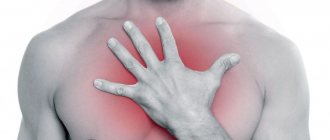
Shortness of breath, lack of oxygen, pain in the chest, under the shoulder blade or hypochondrium indicate various diseases of the organ. In this case, examination by specialists is required. To prevent pathological processes, it is necessary to regularly train the muscle and not abuse bad habits. For preventive purposes, it is recommended to use vitamin complexes, including vitamins C, B6, E, and F. Among medications, such drugs as Asparkam, Riboxin, and Rhodiola rosea have proven themselves well. It is recommended to use all medications only with the permission of the attending physician.
What are the first symptoms of heart problems?
Congestive cardiomyopathy
Congestive cardiomyopathy is a common disease of the cardiovascular system. Men are more often affected.
The disease can be recognized by the characteristic symptoms of heart failure and heart rhythm disturbances: shortness of breath, wheezing, irregular heartbeat, swelling around the ankles, fatigue; in some cases, pain in the heart area and hemoptysis are possible.
Congestive cardiomyopathy is usually treated with medications designed to treat abnormal heart rhythms and heart failure. .
The very first sign of incipient heart problems. Shortness of breath occurs when the heart is still slightly damaged, but can no longer pump enough blood.
Article on the topic
Swelling in the legs
These are signs of vascular disorders. Edema due to heart disease begins to appear in cases where the heart can no longer cope with the increased load and decompensation occurs.
Blue lips
In case of circulatory failure of the heart, a pale or bluish color of the lips is noted. If the lips are completely pale, anemia (anemia) should be excluded.
If you see an obese person in front of you, you are almost guaranteed to suspect cardiovascular disease. Extra pounds are a serious additional burden on the heart.
Bluish-red cheek color may be an indicator of mitral valve dysfunction.
Red bumpy nose
A red, bumpy nose streaked with blood vessels suggests hypertension.
superficial shortness of breath, in which the patient cannot take a full breath; extreme pallor or abnormally red complexion; weakly palpable but frequent pulse; suddenly clouded vision; the appearance of slurred speech; the patient’s inability to respond to speech addressed to him; loss of consciousness.
You should not ignore the feeling of discomfort in the chest, heaviness or pain behind the sternum, pain radiating to the arm, back, under the shoulder blade, throat, jaw, lack of air - these are symptoms of a heart attack.