Cardiac tamponade (CT) is a condition caused by the accumulation of fluid in the pericardial cavity. This provokes an increase in diastolic pressure to critical levels in the intrapericardial cavity, which greatly reduces cardiac output. This condition is very dangerous. It causes pathological disorders of the central and peripheral circulation and metabolic disorders.
As a result, the work of the heart is sharply disrupted, acute failure is formed, which leads to cardiac arrest. The pathological condition, accompanied by negative manifestations, is rapidly developing. Pathogenesis is characterized by unpredictability of the course.
Schematic comparison of normal cardiac conditions and tamponade
Reasons for development
The main reason for the development of tamponade (hydropericardium) is an inflammatory process caused by another disease:
| Diseases | Most common cases |
| Infectious diseases | The occurrence of pathology can be provoked by protracted or advanced diseases:
|
| Malignant tumors | Most often, the occurrence of tamponade is affected by oncology:
|
| Diseases of the endocrine system and connective tissue | It is observed in people with lupus erythematosus. But it also occurs in myxedema and rheumatoid arthritis. |
| Metabolic disorders | Occurs in chronic kidney disease and diabetes. |
Hemotamponade occurs due to mechanical damage or heart disease, leading to disruption of the integrity of the heart muscle.
Its appearance can be caused by complications after operations, chest injuries, myocardial rupture with Beck's triad (in this case, the cause may be either damage to the chest due to trauma or injury, or stress, acquired or congenital diseases):
- Penetrating and non-penetrating wounds;
- Myocardial infarction, congenital malformations, tumors, etc.;
- Dissection or rupture of the aorta/vessels.
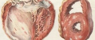
Causes of cardiac tamponade, including blood
Most often, this critical complication occurs with closed or open heart injury, intense bleeding, or hemopericardium. The development of tamponade is caused by:
- operations,
- taking a sample of myocardial tissue,
- camera sensing,
- rupture of the muscle layer during a heart attack,
- installation of a venous catheter,
- dissection of the aortic walls,
- anticoagulant therapy,
- pericarditis due to tuberculosis, bacterial infection,
- tumor process in the heart or lungs,
- uremia,
- lupus erythematosus,
- hypothyroidism
Circulatory disorders are associated not only with the volume of fluid in the pericardium, but also with the possibility of stretching the cardiac sac, so the rapid entry of even a small amount leads to a sharp increase in pressure between the layers and compression of the vena cava inside the pericardium.
The blood flow into the heart cavity decreases, and during diastole the filling of the ventricles decreases. A small volume of blood enters the systemic arteries, and stagnant processes develop in the venous network.
We recommend reading the article about exudative pericarditis. From it you will learn about the disease and its classification, causes and signs of pathology, diagnosis and treatment.
And here is more information about heart biopsy.
Symptoms
The symptoms of tamponade depend on the amount of accumulated fluid - the amount of pressure on the heart, and the level of decrease in cardiac output. But due to the fact that this pathology develops against the background of another disease, it is difficult to identify abnormalities in the early stages - fluid accumulates and makes itself felt only with a serious decrease in the level of cardiac output.
Patients with clinical tamponade experience the following symptoms:
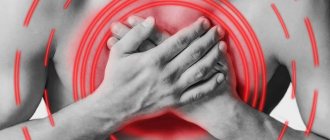
- pain in the chest area;
- shortness of breath and/or weakness;
- strong feeling of anxiety, fear of death;
- shallow breathing;
- cold sweat;
- tachycardia.
Pathophysiological consequences in case of neglect of the disease or its rapid progression (strong increase in pressure inside the heart) lead to the following characteristic symptoms:
- patient's posture (people with pathology often lean forward or lie on their sides and pull their legs to their chest);
- tension or enlargement of the jugular veins;
- wheezing in the lungs;
- pale skin or unnatural blue color;
- coughing.
Cardiac hemotamponade develops rapidly. Symptoms may include:
- a sharp drop in blood pressure;
- increased breathing rate;
- muffled heart sounds;
- frequent but weak heartbeat;
- disappearance of the pulse when inhaling and its appearance when exhaling;
- fainting.
Both diseases can be fatal. At the slightest suspicion of a disease, you should immediately consult a doctor.
Signs of exacerbation
Due to a sharp weakening of the contractility of the heart, blood output during systole decreases. Manifestations of circulatory failure may include:
- increasing difficulty breathing,
- heaviness in the chest
- severe weakness
- rapid pulse,
- fear of death,
- excited state
- cold sweat.
Diagnostic methods
Methods for detecting tamponade and hemotamponade do not differ.
In total, there are four examination options that are used in combination:
| Diagnostic method | Signs of the disease |
| Physical examination of the patient | This is the first thing the doctor does if the patient has signs of tamponade or hemotamponade. These pathologies may be indicated by:
|
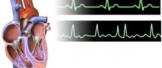
| Electrocardiogram (ECG) | This method reveals signs that may indicate other diseases. However, the ECG shows:
|
| Chest X-ray | X-ray (as well as fluoroscopy) reveals:
|
| Echocardiogram (EchoCG) | Echocardiography is the most informative diagnosis. It is able to detect even minimal liquid emissions. It is used when there is a risk of hemotamponade after surgery and when it is difficult to detect tamponade. |
Diagnostics
Algorithm of medical actions for various diagnostic signs of cardiac tamponade
Based on the presence of the above signs, the doctor can determine the presence of cardiac tamponade. In this case, there is an increased central venous pressure with a decreased arterial pressure. It is characteristic that there is no symptom of left ventricular failure, but the presence of paradoxical pulsus is noted.
It is important to note that the latter is not a clear sign of the pathology in question and can manifest itself when:
- chronic obstructive pulmonary disease;
- exacerbation of persistent bronchial asthma;
- pulmonary embolism;
- right ventricular infarction;
- pericarditis.
The note. Paradoxical pulse does not always appear in patients; for example, it may not be present if local compression of the myocardium by blood clots occurs, with aortic insufficiency, atrial septal defect and severe hypotension.
You can watch the video in this article in more detail about the features of defining the disease. The table below shows methods that help identify the presence of pathology.
Table. Diagnosis of cardiac tamponade:
| Name | A comment |
| As a rule, the clinical picture on the ECG is not specific, so additional studies are required. More obvious changes are recorded only when a large amount of fluid is concentrated in the pericardium. |
| In identifying TC, it is of great value because even the slight presence of fluid in the pericardium can be detected. In addition, it shows: diastolic collapse, disturbances of hemocirculation through the valves. |
| In the photo you can see a thicker cardiac shadow with weak pulsation; venous congestion is not detected in the lung tissue. |
| Shows the presence of stenosis in the vessels, as well as the condition of the coronary arteries and heart. When a catheter is inserted on the right side, it is possible to diagnose TS and find out how much hemodynamics are impaired. |
| The correlation of blood movement through the valve cavities from respiratory excursions is assessed, which complements the overall diagnostic picture. |
The note. Transesophageal echocardiography is prescribed after cardiac surgery, and also when there are certain difficulties in determining the presence of pericardial effusion.
Urgent Care
Emergency care for tamponade or hemotamponade of the heart is carried out only by qualified physicians.
If a person feels unwell at home, on the street or in another place where urgent removal of fluid from the pericardial sac is impossible, the best option would be to follow the following algorithm:
- call an ambulance;
- provide a person with peace and fresh air;
- measure blood pressure;
- If breathing stops, begin resuscitation.
Features of hemodynamics
Circulatory pathologies are primarily determined by the speed of effusion permeable into the pericardium and, to a somewhat lesser extent, by the amount and degree of stretching of cardiac tissue.
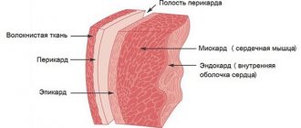
Note. In a healthy person, the outer connective tissue membrane of the heart contains approximately 20–40 ml of fluid, with a pressure of around 0 mm. rt. Art.
The pericardium is characterized by the ability to get used to an increase in fluid volume and pressure to one or even two liters and a slight increase in pressure, but only if the effusion is slow. Otherwise, a rapid increase in intrapericardial pressure is recorded, which creates tension on the heart and the vena cava passing nearby.
A sharp increase in volume, even by 100–300 ml, can already be fatal. Such negative processes interfere with the normal flow of blood into the ventricles, thereby causing insufficient filling of the chambers during diastole, reducing stroke volume and blood ejection.
At the end of diastole, it is considered normal if the pressure in the right ventricle and atrium is five and seven mm Hg. Art., and in the left twelve and fourteen mm Hg. Art., respectively.
Important. TS is formed when the intrapericardial pressure is compared with the ventricular pressure at the end of diastole (EDP).
With the development of pathology, certain mechanisms are formed that are aimed at maintaining normal or acceptable cardiac filling and output.
In this case, compensation will be provided through:
- increased heart rate;
- increased peripheral resistance;
- increase in pressure in the central veins (CVP).
How is the treatment carried out?
After first aid is provided, the patient undergoes pericardiocentesis - under local anesthesia, a needle is inserted into the pericardial sac, after which fluid or part of the blood is pumped out. The procedure is performed only with monitoring of the victim’s condition using echocardiography or radiography.
In the case of tamponade, further treatment depends on the pathology causing the complication.
If, after puncture of the pericardium, fluid continues to be ejected, and the reason for this has not yet been identified, the patient is given a catheter to drain it. Infusion therapy is also used - intravenous administration of nootropic drugs or blood plasma to maintain the patient's fluid balance. For relapses, surgery is required.
Symptoms of cardiac tamponade
The clinical manifestations of cardiac tamponade are quite characteristic, so it is often possible to determine this syndrome without
instrumental research methods.
Symptoms of cardiac tamponade with rapid fluid filling:
- Gradually increasing dyspnea;
- Frequent shallow breathing;
- Blueness of the skin and mucous membranes;
- Fear of dying;
- Palpitations;
- Cold sweating;
- Dizziness;
- Loss of consciousness – if the patient has acute cardiac tamponade;
- The patient's weak condition worsens;
- Pain in the cardiac region is of a pressing nature;
- Enlarged liver;
- Cough;
- Hoarse voice;
- Swallowing disorders.
For reference. Cough with cardiac tamponade develops due to compression of the trachea. The patient's hoarse voice occurs when the recurrent nerve is compressed. Compression of the esophagus causes the inability to swallow. These clinical manifestations appear when a large volume of exudate accumulates.
If the exudate accumulates in the heart sac slowly, the symptoms do not appear so acutely:
- Pain under the right ribs;
- Feeling of nausea, vomiting;
- Enlarged belly, swelling of the legs.
For reference. Patients with cardiac tamponade can be recognized by their characteristic postures: most often they sit, leaning forward, sometimes they can kneel, resting their heads on the pillow. The skin is pale, grayish in color, and sweating is observed. Swelling is visible on the face and neck. The pulse is palpable weakly, the pressure is low.
On examination during percussion, it can be noted that the cardiac boundaries are significantly expanded, the impulse is not detected, and tachycardia is noted. With the slow formation of cardiac tamponade, there is an accumulation of fluid in the abdominal cavity and pain on palpation of the liver.
Forecast
Both with tamponade and hemotamponade, failure to consult a doctor in a timely manner or to detect the disease can result in death.
In case of inflammation, recovery depends on the pathology that provoked the complication. In case of mechanical damage, everything is decided by the speed and correctness of surgical intervention.
Author of the article: Yulia Dmitrieva (Sych) - In 2014, she graduated with honors from Saratov State Medical University named after V. I. Razumovsky. Currently working as a cardiologist at the 8th City Clinical Hospital in the 1st clinic.
Causes
Most often, cardiac tamponade is caused by the following factors:
- hemopericarditis due to damage to the integrity of the sternum and heart;
- rupture of dissecting aortic aneurysm;
- heart rupture due to myocardial infarction;
- hemorrhages during cardiac surgery;
- long-term course of chronic diseases (pericarditis, hemopericarditis, lymphoma, myxedema, systemic lupus erythematosus, cancer of the breast, lungs, etc.);
- renal failure developing during hemodialysis;
- taking anticoagulants;
- irradiation, etc.
Treatment
Cardiac tamponade is an emergency and is a life-threatening situation. Emergency removal of the pericardial transudate is necessary. To do this, the surgeon performs a puncture of the pericardial wall - pericardiocentesis or surgery. To provide hemodynamic support for the cardiovascular system, doctors administer intravenous infusions of blood components and nootropic drugs.
The puncture is performed under the control of echocardiography or fluoroscopy. Constant monitoring of blood pressure, central venous pressure, and heart rate is carried out. The clinical effectiveness of pericardiocentesis is noticeable when aspirating small volumes of effusion (25-30 ml). After the fluid has been removed, the doctor may introduce an antibiotic, hormonal drug, or sclerosing agent into the pericardial cavity, if indicated. To prevent re-accumulation of fluid, drainage is installed. The next stage of treatment is therapy for the condition that led to the development of the complication.
Attention! If the risk of recurrence of the complication is high, surgical treatment of cardiac tamponade is preferable. Performed for life-saving reasons, for example, rupture of the heart or aorta.
Types of operations:
- Pericardiotomy - an opening in the pericardial wall to drain the cavity. At the same time, an inspection of the inner surface of the walls is carried out to identify injury or neoplasm.
- Subtotal pericardectomy is a radical method of treating tamponade. Used for chronic exudative pericarditis, the presence of scars or calcifications.
Causes and symptoms of cardiac tamponade
most common causes of cardiac tamponade
- Damage to the integrity of the heart and/or sternum (open wound, blunt trauma, etc.);
- Hemorrhage due to cardiac surgery;
- Dissecting aortic aneurysm, that is, its rupture;
- Heart rupture due to myocardial infarction;
- Long-term and chronic diseases (acute viral, idiopathic or post-radiation pericarditis, hemopericardium, tuberculosis, lymphoma, lung cancer, breast cancer, etc.);
- Chronic or acute renal failure during hemodialysis;
- Anticoagulant therapy;
- Radiation damage, etc.
tamponade and pericarditis due to trauma
Signs of cardiac tamponade are the consequences of a sharp drop in cardiac output, decreased pumping function and systemic venous stasis. So, the most obvious symptoms of cardiac tamponade include:
- Chest discomfort;
- Increasing shortness of breath;
- Increased anxiety, “fear of death”;
- Sudden weakness;
- Pale skin and profuse sweating;
- Drop in blood pressure;
- Venous hypertension;
- Low mobility of the heart, accompanied by dull heart sounds.
The last three symptoms form the so-called “classical Beck triad,” that is, the classic symptomatic picture of cardiac tamponade. However, it manifests itself in cases of pronounced pathology (heart injury, etc.). In most cases, the disease progresses progressively, and the symptoms are in many ways similar to those of heart failure:
- Weakness, lethargy, general malaise and loss of appetite;
- Pain under the ribs on the right;
- Shortness of breath, forcing the patient to take a sitting position to facilitate breathing - the so-called orthopnea;
- Pathological enlargement of the liver (hepatomegaly);
- Accumulation of fluid in the peritoneal cavity (ascites);
- Increased pressure in the jugular veins and their swelling.
Sometimes cardiac tamponade may not manifest itself at all for a long time, which ultimately threatens with such a complication as pericarditis - inflammation of the serous membrane of the heart. That is why the presence in a person of even some of the above symptoms of tamponade (especially for blood pressure and pressure) should already cause caution.
When “clothes” don’t fit or how tamponade develops
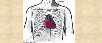
Cardiac tamponade
Let's figuratively imagine what happens to the heart during tamponade... How would you feel wearing clothes 2 sizes smaller? You would feel cramped. You would not be able to carry out the daily load, at first you would make every effort to complete the work, and then, tired, you would remove it, tear it, unable to withstand the discomfort... The heart feels the same way during tamponade, since in this condition there is an excessive accumulation of fluid in the pericardial cavity .
When this cavity fills with fluid, the pressure in it increases, the heart becomes compressed, its ability to contract decreases, the venous flow to it decreases, and cardiac output decreases. Cardiac tamponade is a life-threatening complication of pathological conditions of the cardiovascular system. How quickly this condition progresses depends on the rate of fluid accumulation and its volume in the pericardial cavity.
Pressure in the pericardial cavity
If fluid accumulation occurs quickly, then even 230-240 ml of fluid is enough for the development of tamponade; with slow fluid accumulation, the pericardium has time to stretch, adapt, and tamponade occurs with a fluid volume exceeding 2 liters, and sometimes even 3 liters can fit in the heart sac... Tamponade can develop rapidly, dramatically, literally in a couple of hours, or even within a few minutes, in some cases it develops slowly, and then we are talking about subacute compression of the heart.
But regardless of the rate of compression to which the heart muscle is subjected, the mechanisms of its development are the same: intrapericardial pressure increases rapidly, fluid compression causes a decrease in the volume of the left ventricle and other cardiac chambers, there is an increase in diastolic and a fall in systolic pressure of the ventricles, a sharp decrease in the volume of stroke, cardiac ejection As a result of the pathological changes in hemodynamics, the blood supply to organs and tissues is catastrophically reduced, the blood flow to the brain, as well as coronary blood flow, drops sharply.
So what causes compression of the heart muscle? What diseases and pathological conditions can lead to life-threatening disorders?
Causes of blood accumulation
The reasons are always serious. The development of the condition takes a matter of minutes, less often hours.
Among the factors:
- Extensive infarction with disruption of the anatomical integrity of the heart.
- Penetrating and closed wounds of the chest organs.
- Surgical interventions on cardiac structures, diagnostic measures (including minimally invasive ones). The risk of such a traumatic outcome is always there, although not great.
It is almost impossible to react in time. Against the background of massive bleeding, compression turns out to be an additional lethal factor, reducing the chances of survival to almost zero.
Tests and diagnostics
In addition to an objective examination with percussion and auscultation, the patient is prescribed the following basic research methods:
- ECG . There is a decrease in the voltage of the teeth, and signs characteristic of myocardial infarction .
- R-graphy of the chest organs. The image shows smoothness of the cardiac arcs, an increase in the shadow of the heart, and the absence or decrease of pulsation of the contours.
- EchoCG. There is a gap in the echo signal between the pericardium and the walls of the heart. Examination may reveal a small amount of accumulated blood.
Chronic hemopericardium requires dynamic monitoring based on the results of the examination methods described above.
To clarify the origin of the hemopericardium, additional examination methods are prescribed:
- pericardioscopy;
- biopsy ;
- bacterial examination of pericardial fluid;
- cytological analysis of pericardial fluid;
- angiocardiography;
- cardiac catheterization;
- puncture or diagnostic pericardiocentesis .
Additionally, differential diagnosis with other diseases is carried out:
- myocarditis;
- acute exudative (or hemorrhagic, serous-purulent) pericarditis ;
- pneumopericardium;
- non-inflammatory hydropericardium;
- chylopericardium.
Forms of the disease
Compression of the cardiac cavities and increased intrapericardial pressure lead to difficulty in normal heart contractions, impaired diastolic filling of the ventricles and a significant decrease in cardiac output. As a result, cardiac tamponade can cause acute heart failure, shock and complete cessation of cardiac activity.
The normal volume of fluid in the heart sac does not exceed 20-40 ml. The condition in which the volume of liquid reaches 250 ml may already be critical. Sometimes this volume even reaches 1000 ml or more: this is possible if the effusion increases gradually and the cardiac sac has time to stretch, thus adapting to the growing volume of exudate. These clinical manifestations are characteristic of the chronic form of tamponade.
Acute cardiac tamponade progresses rapidly and its course is unpredictable. Thus, if the integrity of the aorta or heart muscle is damaged, the patient may suddenly lose consciousness and fall into hemorrhagic collapse, which requires urgent surgery to avoid death.
Video: the occurrence of cardiac tamponade (eng)
What is systolic and diastolic pressure: normal, deviations
Systolic blood pressure is the blood pressure in the vessels at the moment the heart contracts. The indicator depends on the strength, speed of myocardial contraction, and its condition. Diastolic blood pressure is the blood pressure in the vessels at the moment the heart relaxes. The difference between the values is called pulse pressure. Normally, its value is 30-40 mmHg. Art., SBP should not be higher than 129 mm Hg. Art., DBP – not higher than 89 mm Hg. Art.
If the obtained figure is higher, arterial hypertension should be excluded. If the reading is 20% below normal or below 90/60 mmHg. Art., then we are talking about arterial hypotension. There are factors that lead to physiological fluctuations in blood pressure. When they are eliminated, the pressure returns to normal levels. If this does not happen, you need to contact a cardiologist.
Emergency treatment
Given the health risks, the first thing you need if cardiac tamponade develops is emergency care.
Primary treatment measures include emergency pericardiocentesis. It is carried out under the control of echocardiography with mandatory monitoring of blood pressure, pulse and temperature.
A feature of the blood obtained during pericardiocentesis is that the fluid does not clot, no matter how long it stands.
Next, depending on the indications, the patient is prescribed antibiotics (their use is mandatory, since there is a risk of introducing infection directly into the vascular bed during cardiocentesis). At the same time, sclerosing therapy is carried out (to speed up the healing of the injury caused by the needle).
If the accumulation of exudate continues, it is recommended to place a catheter in the pericardial cavity, but it should be remembered that when placing it, the risk of wound infection increases significantly.
Prognosis and prevention of cardiac tamponade
Cardiac tamponade is a dangerous clinical syndrome that threatens the life and health of the patient. In the absence of urgent medical care, the disease can be fatal. In the case of accumulation of blood in the pericardial cavity (hemotamponade), the prognosis is quite disappointing - death occurs in approximately half of the patients.
The possibility of complete recovery depends entirely on the cause that led to the formation of cardiac tamponade. Viral, bacterial or fungal infections are usually treated successfully.
Attention. If the cause of the development of cardiac tamponade is a cancerous tumor, then the outcome of the syndrome largely depends on the degree of the oncological process and its prevalence.
Prevention of cardiac tamponade consists of timely treatment of inflammatory processes in the pericardium, high-quality performance of cardiac operations, monitoring the state of the blood coagulation system during treatment with anticoagulants, as well as timely treatment of various diseases that can cause the development of cardiac tamponade.