Pericardiocentesis is a puncture of the pericardium. In most cases, doctors perform the procedure to relieve cardiac tamponade and determine the cause of pericarditis. The Yusupov Hospital has all the conditions for treating patients with diseases of the cardiovascular system:
- cozy rooms of various comfort classes;
- dietary nutrition;
- use of modern equipment from leading global manufacturers;
- attentive attitude of medical staff to the wishes of patients.
The therapy clinic employs professors and doctors of the highest category. Cardiologists take an individual approach to choosing a treatment method for each patient. Doctors use effective and safe drugs registered in the Russian Federation. At a meeting of the expert council, the presence of indications and contraindications for pericardiocentesis is discussed.
Methods for performing pericardial puncture
Pericardial puncture is not performed in patients suffering from coagulopathy and receiving anticoagulant treatment. A relative contraindication to the procedure is a limited-volume effusion and a platelet count in the blood of less than 50×109/L. Pericardial puncture is not performed for dissecting aortic aneurysm, post-infarction myocardial ruptures, or traumatic hemopericardium. In all these cases, leading cardiac surgeons perform surgery at partner clinics.
Before pericardial puncture, chest X-ray and echocardiography are performed. If the puncture is carried out under the control of electrocardiography or blindly, doctors make sure that at least 2 cm of effusion has accumulated between the layers of the pericardium. The purpose of the procedure is explained to the patient and his consent is obtained.
In order for the pericardial effusion to move to the anterior inferior pericardial sinus, the patient is placed in a semi-sitting position. Peripheral venous access is established, electrocardiogram, blood pressure, pulse and oxygen saturation are monitored (measurement of the amount of oxygen bound to hemoglobin cells in the circulatory system). In case complications develop, everything necessary for resuscitation measures is prepared, including a defibrillator.
Pericardiocentesis kit includes:
- introducer needles;
- expander;
- conductor;
- bent radiopaque catheter;
- multi-purpose tube adapter.
The point for puncture of the pericardium when using the Larrey technique corresponds to the apex of the angle between the left costal arch and the base of the xiphoid process on the left. When performing a Marfan puncture, the doctor punctures at a special point located under the xiphoid process.
Indications and contraindications for the procedure
Indications for pericardial puncture in a patient arise when he is diagnosed with pericarditis or hemopericardium. In this case, the procedure reduces the pressure of fluid on the heart and prevents its further entry into the pericardium.
If the patient has an exudative stage of pericarditis, then in such a situation the puncture is performed solely for the purpose of diagnosing pericardial effusion, and the indications for treating the pathology are somewhat different. As for contraindications for puncture, they can arise if the following phenomena are present in the patient’s body:
- bleeding disorder,
- previous coronary bypass surgery,
- insufficient pleural effusion,
- obliteration of the pericardial plane.
Important! Indications for the puncture procedure are established exclusively by one doctor - a cardiac surgeon.
Larrey pericardiocentesis technique
The doctor puts on a sterile gown, mask and gloves. After treating the skin of the chest and upper abdomen with an antiseptic solution, limit the puncture site with sterile material or use a sterile film. The injection site is numbed with a thin needle. To do this, 2-5 ml of a 1% lidocaine solution is injected into the point located between the left costal arch and the xiphoid process. Attach a ten-millimeter syringe with 1% lidocaine to a long needle measuring 16-18 G or to a long intravenous “catheter on a needle”.
In the first variant of needle insertion, the needle is injected at an angle of 30° to the skin in the frontal plane and directed forward along the axis of the body. The needle should go flush with the costal edge. When using the second option, the needles are directed to the patient's left shoulder. The needle is positioned at a shallower angle relative to the heart. This makes it easier to pass the guidewire and catheter.
After the end of the fluid has reached the edge of the costal arch, 0.5-1.0 ml of lidocaine is carefully injected. Then the needle is advanced 4-5 mm forward. In this case, the syringe plunger is constantly pulled towards itself. Then the cycle is repeated. With this approach in adults, the average distance from the skin to the pericardium is 6-8 cm.
The needle can be inserted under electrocardiogram control. The doctor attaches the chest lead wire of the electrocardiograph or cardioscope to the needle using a clamp. Then turns on the “chest lead” mode on the recording device or connects the distal electrode to the wire of the right hand, and the proximal electrode to the wire of the left hand. The nurse turns on the first lead on the cardioscope or cardiograph. If the needle is in the pericardial cavity, a negative ST wave will appear on the ECG.
The needle can be inserted under ultrasound cardiography control. In this case, it is easier for the doctor to choose the optimal access point. An apical or left parasternal approach is used. Measure the distance to the effusion and mark the direction of the central ultrasound beam. The direction of the needle should be the same.
After receiving the liquid, disconnect the syringe with the remaining lidocaine and make sure that it is not blood that is coming from the needle, but pericardial fluid. Collect 10-15 ml of pericardial effusion into a clean 10-15 ml tube for testing. If the patient's condition is severe due to cardiac tamponade, 50-100 ml of effusion is aspirated through a needle before installing the catheter. A catheter is installed. If the catheter is on the needle, remove the needle. If not, use the standard Seldinger catheterization technique.
After making sure that the liquid is freely aspirated, secure the catheter with a nylon suture or adhesive tape and connect the extension to the drainage container. To avoid acute dilatation of the right ventricle as a result of sudden decompression, fluid from the pericardium is removed in parts, no more than one liter at a time.
Recovery after puncture
The patient remains in the hospital for observation for some time. After discharge, the doctor gives recommendations for a speedy recovery. For the first time after pericardiocentesis, it is strictly forbidden to lift weights, overexert yourself, or have sex. Smoking and alcohol are excluded if possible altogether, at least for 2 weeks.
When you are discharged, you should consult with your doctor about what medications you can take. A painkiller will be prescribed; you need to drink it in a clearly indicated dosage. At the first sign of complications, you should consult a doctor or call an ambulance.
For quick rehabilitation, a healthy diet is recommended; as a rule, diet No. 15 is prescribed. From time to time you need to go for leisurely walks and breathe fresh air. In winter, be sure to dress warmly; in summer, avoid overheating and sunbathing on the beach. Stressful situations are especially undesirable in case of heart disease and should be avoided as much as possible. For those who are especially sensitive, a cardiologist may recommend sedatives. You can return to an active lifestyle after your doctor approves it.
Pericardial puncture according to Marfan
When performing a Marfan puncture, the doctor makes a puncture under the xiphoid process. The needle insertion site is located strictly in the middle of this anatomical landmark, neither on the right nor on the left side of it. The middle position of the needle ensures its penetration into the pericardial cavity in the area of the right ventricle. The sterile needle is advanced smoothly and slowly at an angle of 30-450. The puncture needle is first directed obliquely upward, then slightly behind.
When approaching the heart area, the doctor sometimes feels the pulsation transmitted by the pulse. When the outer pericardial layer is punctured directly, a feeling of overcoming a certain obstacle appears. After entering the pericardial cavity, the existing contents are removed using aspiration movements of the syringe attached to the needle. Often, a catheter is inserted through a needle into the pericardial cavity and fixed for a certain time to ensure drainage of the cavity and removal of exudate. After puncturing and inserting a catheter into patients who are in serious condition, doctors administer medications into the pericardial cavity: hydrocortisone, prednisolone, antibiotics.
Types of diagnostics
Puncture is carried out using several methods:
- Pirogov-Delorme technique
. The needle is inserted at the level between the fourth and fifth ribs on the left side. - Method of pericardial puncture according to Larrey
. The area between the cartilage tissue and the xiphoid process on the left side is pierced. Level - between 8-10 ribs. - Technique of pericardial puncture according to Marfan
. A needle is inserted in the middle of the xiphoid process (usually under it).
The last two types of puncture are considered the most atraumatic. The risks of premature needle displacement and damage to the pleural layers are minimal. And with an accidental puncture of the heart wall, the risks of complications are small, they do not lead to myocardial rupture.
The procedure is divided into emergency and planned. The first type of intervention is required for, and the second for effusion pericarditis.
Complications during pericardial puncture
Pericardial puncture for cardiac tamponade is not an easy procedure, but if the technique is followed correctly, it does not cause any complications in the patient. If the course of the procedure is somewhat disrupted for reasons beyond the doctor’s control, the risk of damage to the tissue of the heart, thoracic or coronary artery increases.
The most serious complication of pericardial puncture is rupture and perforation of the coronary artery or heart muscle. If the needle or catheter has perforated the heart and is in its cavity, the needle is removed and the catheter is secured. In both cases, patients are consulted by a cardiac surgeon. Alternatively, a new pericardial puncture is attempted. If it is successful, surgery can be avoided. In this case, autotransfusion of blood from the pericardium is performed.
There are complications of pericardial puncture such as pneumothorax, air embolism, arrhythmias, as well as abdominal puncture or perforation of abdominal organs. Rarely, purulent pericarditis, pulmonary edema, and internal mammary artery fistula develop.
Make an appointment with a cardiologist by calling the Yusupov Hospital. The doctor will examine the patient and prescribe effective treatment. If cardiac tamponade develops, a pericardial puncture will be performed.
Author
Patient preparation
The puncture is performed in emergency situations or planned. Depending on the situation, the patient is prescribed several types of examination.
In urgent cases, the cardiologist decides what tests are needed before intervention. For a planned procedure the following is prescribed:
- echocardiogram;
- chest x-ray;
- blood tests (required for clotting);
- electrocardiogram.
Patients undergoing systematic treatment should inform their doctor about all medications they are taking. 4-6 hours before the procedure, you are prohibited from eating or drinking. Be sure to tell the doctor about all medications that were taken during the last 24 hours.
Bibliography
- ICD-10 (International Classification of Diseases)
- Yusupov Hospital
- Cherenkov V. G. Clinical oncology. — 3rd ed. - M.: Medical book, 2010. - 434 p. — ISBN 978-5-91894-002-0.
- Shirokorad V.I., Makhson A.N., Yadykov O.A. The state of oncourological care in Moscow // Oncourology. - 2013. - No. 4. - P. 10-13.
- Volosyanko M.I. Traditional and natural methods of preventing and treating cancer, Aquarium, 1994
- John Niederhuber, James Armitage, James Doroshow, Michael Kastan, Joel Tepper Abeloff's Clinical Oncology - 5th Edition, eMEDICAL BOOKS, 2013
Features of the event
Manipulations are carried out in several ways; such techniques are named after their inventors. The Larrey and Marfan methods are most often used; they differ only in the needle insertion points. For the patient himself, it practically does not matter what technique will be used. Whatever type of pericardiocentesis is prescribed, the operation will be almost the same for the patient.
The patient changes into sterile hospital clothing or exposes the upper body. Takes a semi-sitting position on the treatment table, sometimes a pillow is placed under the back. Sedatives are injected into the vein, all other manipulations are carried out after 20 minutes. The chest is treated with antiseptic agents.
After determining the insertion point, the doctor processes the instrument. The puncture needle is thin and will deliver an anesthetic for local anesthesia. First, the anesthetic is injected to numb the skin, then a little deeper, all the way to the pericardium.
The process will be carried out under the control of fluoroscopy or echocardiography. The doctor will slowly insert the needle, take samples, and withdraw the instrument. To remove air or liquids, a catheter is inserted into the needle cavity, through which the excess is drained out.
In case of pericarditis, the cavity can be rinsed and an antibiotic and oxygen injected into it. At the end of the operation, the puncture site is treated with an antiseptic and sometimes sealed with Xeol.
The patient then remains under medical supervision for at least 2 hours. If drainage has been installed, the hospital stay is extended for several days. The biomaterials are sent for analysis, and another examination is carried out: a chest x-ray (to ensure the integrity of the organs), a pulse and blood pressure check.
Diagnostic indications
Diagnostic indications boil down to the need to confirm the presence of fluid or blood in the pericardium, as well as take a sample of exudate for laboratory testing. In such cases, pericardial puncture is performed using the Larrey or Marfan method, since these are the safest and least traumatic methods.
Thus, pericardial puncture allows doctors to simultaneously confirm the disease and alleviate the person’s condition.
From this video you can learn more about pericardial puncture:
Pericardial puncture
Indications. Pericardial puncture is performed for diagnostic and therapeutic purposes. It is carried out only when fluid accumulates in the cavity of the cardiac membrane (hydropericardium, hemopericardium, exudative pericarditis). The presence of effusion should be confirmed by echocardiography and radiography. Pericardial puncture can be emergency (performed for cardiac tamponade) and planned (performed for effusion pericarditis).
Puncture technique. When performing a pericardial puncture, the patient should be in a semi-sitting position with his head tilted back and a pillow placed under the lower back (Marfan position). Regardless of whether the intervention is performed on a patient lying on the bed or on the operating table, this position is mandatory. For pain relief, local infiltration anesthesia with a 0.5% solution of novocaine is used. To perform a puncture, a long needle connected to a syringe is used. The puncture is performed in the deepest part of the pericardium to avoid entering the chest cavity. Pericardial puncture can be performed in several ways.
Method 1. A needle is inserted into the V-VI intercostal space on the left along the midclavicular line or slightly outward from it. The direction of the needle should be strictly perpendicular to the chest wall. They sequentially pass through the skin, subcutaneous tissue, muscles, intrathoracic fascia, parietal pleura and pericardium.
Method 2. The puncture can also be made from an injection into the angle formed by the costal arch and the xiphoid process (Larrey's method), or under the apex of the xiphoid process (Marfan's method). In both cases, the skin is punctured at a right angle in the cranial direction. The skin, subcutaneous tissue, and rectus abdominis muscle with aponeurosis are pierced. This depth, with an average thickness of the abdominal wall, is usually 1.5-2 cm. After puncturing the inner edge of the rectus abdominis muscle (or linea alba), the needle is advanced almost parallel to the chest wall upward and inward. In this way, advancing the needle to a depth of about 2-3 cm, the pericardium is punctured. The approach to the pericardium is determined by the beginning of the needle oscillations in the rhythm of heart contractions. If there is a significant amount of liquid, it is clearly felt as if the needle is falling into the cavity. In the case of purulent fibrous pericarditis, the thickened epicardium rubs against the tip of the needle, as if it were being rhythmically drawn along sandpaper. An electrocardiograph can be used to clarify the position of the needle. If the needle is located in a collection of pericardial fluid, the electrocardiographic curve will not change. As soon as the tip of the needle comes into contact with the epicardium, characteristic changes occur in the form of deformation of the QRS complex, expressed in a pathological Q wave and a decrease in the R wave. For hemopericardium, as a temporary measure in preparing the patient for surgery, a catheter is inserted into the cavity of the heart sac using the Seldinger method for continuous aspiration blood. A similar manipulation is performed with progressive exudative pericarditis. When sucking blood with a syringe during pericardial puncture, it is necessary to immediately decide whether this blood is the contents of the pericardium (hemorrhagic pericarditis). To do this, the sucked liquid must be collected in a test tube or on a piece of white gauze.
Fresh blood from the bloodstream is scarlet in color and differs sharply from stagnant hemolyzed lacquer-like blood.
Complications.
When performing a pericardial puncture, one should be wary of injury to the heart by the puncture needle and damage to the internal mammary artery. When the needle penetrates into the heart cavity, it is necessary to slowly remove the needle, holding the syringe in the suction position, since it is possible that when it comes back, the needle will enter the pericardial cavity.
If this fails, then the intervention is stopped, and the patient needs intensive monitoring. In most cases, there is no bleeding when the heart wall is punctured.
Pericardectomy
The operation is performed for chronic adhesive inflammation of the pericardium, which is often accompanied by compression of the heart and vena cava. The pericardium adheres to the epicardium, and lime deposits occur in this scarred tissue. The heart is, as it were, in a stone bag. The essence of compressive pericarditis is that the heart is unable to expand during diastole, and therefore its diastolic filling decreases to a greater extent.
Technique of the operation. The operation is performed under endotracheal anesthesia. Total pericardiectomy is performed only through a median sternotomy. After spreading the edges of the sternum, the mouths of the great vessels and the chambers of the heart are sequentially isolated. An incision of the pericardium is made in a scarred, hard, but, if possible, non-calcified area to such a depth that the contracting heart can be seen. It is fundamentally important to strictly observe the sequence of isolating the parts of the heart. They begin with the separation of adhesions that compress the outflow tract from the heart. First, the root of the aorta and pulmonary artery are released, and then the lateral wall of the left ventricle, the right ventricle and the right atrium. The operation is completed by releasing the compression of the mouths of the vena cava. The areas that have compressed the pericardial shell are removed.
The peculiarity of this operation is that it is necessary to correctly find the layer between the pericardium and epicardium. After this, the edges of the dissected pericardium are grabbed with clamps and the epicardium is gradually released using a blunt and sharp method. Calcified areas penetrating deeply into the myocardium are not isolated, but are bypassed, leaving them on the epicardium.
These places look like islands protruding from the surface. Calcified areas of the pericardium are bitten with Luer or Liston forceps.
Extreme caution must be used when excision of the pericardium in the area of the coronary vessels, atria and vena cava. The posterior part of the pericardium is usually left in place.
In addition, removal of the pericardium is done with caution to avoid damaging the phrenic nerve. The operation ends with drainage left in the anterior mediastinum to control bleeding and exudative process.
Contents of the topic “Punctures.”: 1. 2. 3. 4.
Pericardial puncture
produced to remove exudate from the pericardial cavity.
Point for puncture of the pericardial cavity
(
Larrey's point
) corresponds to the apex of the angle between the left costal arch (attachment of the cartilage of the VII rib to the sternum) and the base of the xiphoid process on the left. The total length of the needle injection does not exceed 6 cm - 1.5-2 cm for the skin with anesthetic + 3.5-4 cm for passing through the muscles, diaphragm and penetration into the pericardium.
The Marfan point is of secondary importance
for pericardial puncture - when the needle is inserted immediately under the xiphoid process and moves along the strictly midline to the cephalic end. The total length of the needle insertion is 5 cm in thin patients and up to 10 cm in obese patients. The patient's position is the same as for puncture at Larrey's point.
, the Pirogov-Delorme puncture point is important
, when the needle is inserted strictly perpendicular to the chest right at the left edge of the sternum in the area of the IV-V intercostal space. The depth of needle insertion does not exceed 3-4 cm.
Puncture of the pericardium to the right of the sternum symmetrically to the Pirogov-Delorme puncture point is called the Voynich-Syanozhetsky point
.
Kurshman puncture point
has historical significance, as it suggests a puncture point in the V-VI intercostal space 2.5 cm medially from absolute cardiac dullness identified by percussion. Transsternal puncture points of the pericardium - Deso, Laeneca, Riolan - have a similar historical significance - today no one will try to puncture the pericardium through the sternum.
We will examine in detail the favorite puncture point of the pericardium by surgeons - Larrey's point
. After local anesthesia with novocaine, a long needle connected to a syringe is inserted into the puncture point in the cranial direction at an angle of 45° to the surface of the body. The skin, subcutaneous tissue, and rectus abdominis muscle with aponeurosis are pierced.
After puncturing the front wall
the sheaths of the rectus abdominis muscle change the direction of the syringe and needle parallel to the plane of the sternum, after which they move the needle upward by 2-3 cm. The direction of the needle is from bottom to top and somewhat posteriorly. The needle passes through the muscle bundles of the sternal diaphragm and the lower surface of the pericardium. The feeling of pulsation indicates the proximity of the heart.
As the needle advances, the syringe plunger is periodically pulled back to record the moment of puncture of the pericardium.
, after which needle advancement should be stopped to avoid damage to the heart. Fluid from the pericardial cavity is sucked out very slowly so as not to disrupt the functioning of the heart.
Operations for heart wounds.
Heart wounds are accompanied by three main symptoms:
a) intrathoracic bleeding
b) pericardial tamponade
c) cardiac dysfunction.
The right ventricle is most often damaged, adjacent most of its surface to the anterior chest wall.
In case of heart injuries it is necessary:
1. Inject plasma-substituting agents or blood intravenously to replenish the volume of circulating blood
2. Eliminate hemopericardium and eliminate cardiac tamponade by puncture of the pericardium (removal of even 10-15 ml of blood from the pericardial cavity raises blood pressure to 70-80 mm Hg)
3. Perform immediate thoracotomy with suturing of the heart wound.
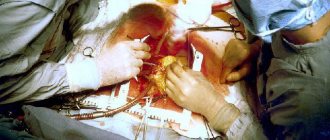
Fig. a – stitches on a heart wound; the thumb covers the wound opening and stops the bleeding. Figure b – sutures on the myocardium without damage to the coronary artery when the heart is wounded near it; U-shaped sutures pass under the coronary artery
Technique for suturing heart wounds:
1. Left anterolateral thoracotomy in the 4th-5th intercostal space (if necessary, the incision is expanded by crossing several more intercostal cartilages)
2. Opening the pericardium anteriorly or behind the phrenic nerve, aspiration of blood and removal of blood clots
3. If a bleeding heart wound is detected, it is sutured. To do this, four fingers of the left hand are placed on the back wall of the heart, fixed and slightly lifted towards the surgeon, while at the same time pressing the wound with the thumb and stopping the bleeding. With the right hand, stitches are applied to the wound with atraumatic needles, and the assistant ties them.
For large lacerations of the heart, a wide circular purse-string or U-shaped suture is applied, for wounds of the atrium - a purse-string suture, if the wound is located near the coronary arteries - U-shaped sutures under the coronary arteries, when cutting through the applied sutures - U-shaped sutures on Teflon pads . For hemostatic purposes, fibrin film and autologous tissue (muscle, pericardium) can also be fixed to the wound. 4. After suturing the bleeding wound, the heart is examined in search of other wounds (especially on the posterior wall).
5. The pericardium is sutured with rare interrupted sutures to ensure adequate outflow of residual blood from the pericardium.
6. Inspection of the pleural cavity, drainage of the pleural sinuses.
7. Suturing the chest wound tightly in layers, leaving drainage in the pleural cavity.
Pericardial puncture is a complex medical procedure that is performed when a patient experiences pericarditis associated with fluid entering the pericardial space. The main goal of this procedure is to free the pericardium (the sac around the heart) from accumulated fluid, thereby removing the increased load from the heart.
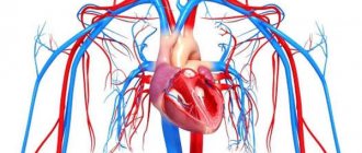
Anatomy of the heart
The human heart is a muscular organ responsible for transporting blood in the body. With contractile movements, the heart pumps blood to the lungs, where it is enriched with oxygen, and delivers oxygen-rich blood to all organs and tissues.
Anatomically, the heart is divided into 4 cavities
Anatomically, the heart is divided into four cavities: two atria and two ventricles. Functionally, the heart can be divided into two parts.
One part collects venous blood from tissues and sends it to the lungs, the other returns oxygen-rich blood to all cells of the body.
The heart is located in a connective tissue sac called the pericardium. The pericardium softens heart contractions and protects heart tissue.
With some injuries or diseases, the pericardium becomes filled with fluid and mechanically blocks the contractile activity of the heart, which can lead to death.