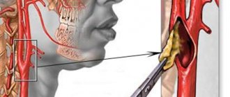
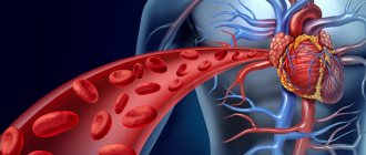
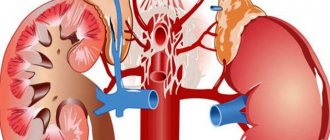
Arteries are vessels in the circulatory system through which blood enriched with oxygen and nutrients flows to all organs. The carotid artery (located in the neck near the thyroid gland) transports blood to the organs of the brain and tissues of the neck.
Disruption of these vessels (narrowing, blockage or inflammation) can lead to blurred vision, loss of speech or complete paralysis. When blood flow is stopped for a long time, death occurs, since about 70% of a person’s blood flows through the carotid artery.
Why are the arteries called carotid?
The carotid artery is a large vessel through which the bulk of blood is delivered to all tissues and organs of the head, including the brain.
If you compress an artery, the receptors (nerve endings) perceive this action as an increase in pressure, and as a result, the number of heart contractions decreases, followed by a decrease in pressure and breathing slows down (the body works similarly during sleep).
The volume of blood flow to the brain also decreases. Together, this leads to a state of drowsiness, and with prolonged compression, loss of consciousness and death are possible. Due to the development of a sleepy state as a result of the effect on the artery, it was called carotid.
How many carotid arteries does a person have?
On the neck, on the right and left sides, there are 2 common carotid arteries.
They are distinguished by their large diameter, regular cylindrical shape and elasticity. Each artery is divided into external (blood flows through them to the tissues of the head and neck) and internal (nourishes the brain and eyes). As a result, a person has 3 pairs of carotid arteries (common, external and internal).
Where are they located?
The carotid artery in humans is located on the sides of the neck near the thyroid gland. The left carotid artery comes from the aorta (the main artery that originates in the left ventricle), and the right one from the brachial artery (which is a branch of the aorta). In this regard, the left carotid artery is longer than the right by about 20-25 cm.
Further, near the Adam's apple (the protruding part of the thyroid cartilage), the right and left carotid arteries branch into external and internal. Above, each artery divides into numerous capillaries that supply all the tissues of the head and neck.
What are they responsible for?
The main purpose of the carotid artery is to provide the tissues of the head and neck with oxygen and nutrients.
At the same time, the functions of the internal and external carotid arteries are different, since they supply blood to different organs:
| The external carotid artery branches to the front of the head and supplies: | The internal carotid artery ascends to the base of the skull and supplies blood to: |
| Mimic and chewing muscles of the face. | Brain tissue. |
| Facial skin and epidermis under the hair. | |
| Oral cavity. Including the tongue, salivary glands and tooth roots. | Muscles of the forehead and temporal part. |
| Nasal cavity and middle ear tissues. | |
| Thyroid gland, pharynx and larynx. | Ocular tissues. |
The external and internal arteries are additionally connected to each other by numerous arteries, this ensures an uninterrupted flow of blood to the tissues if suddenly a slowdown in blood flow occurs in any part of the artery.
The vessels are also equipped with nerve endings, as a result of which the carotid arteries perform the following additional functions:
- normalization of blood pressure;
- regulation of the number of heart contractions;
- react to a lack of oxygen in the blood.
If you know how to act correctly on the carotid artery, you can lower blood pressure without taking medications. It is important that this procedure is only allowed to be performed by a specialist, since stronger or longer manipulations can lead to death.
What is the carotid artery?
In fact, we have two carotid arteries. One of them is located on the right side of the neck, the other on the left. The one on the left is slightly longer, it begins in the aortic arch, and the one on the right begins at the brachiocephalic trunk.
The general function of the carotid artery is to transport blood from the heart muscle to the brain and other peripheral organs that are located in the head region. It is thanks to it that our brain is stably supplied with oxygen. Compression of the carotid artery (for example, by a tight collar or tie) can cause noticeable discomfort.
The external part of the carotid artery passes above the larynx towards the front of the head. In the area of the Adam's apple, it divides into two branches, one of which supplies blood to the brain, and the second to the face and eyes. The terminal branches form a network of capillaries, thanks to which in certain life situations our eyeballs can turn red and the skin of our face can become flushed.
The interior of the carotid artery directly delivers oxygen-rich blood to the brain cells. It enters the skull in the temple area.
Under the influence of stress, hot weather and other external factors, blood flow in the internal artery may increase. In this case, we experience a surge of strength and emotional uplift. But if the intensity of blood flow exceeds the norm for a long time, the process of decline will begin and the person will fall into a state of weakness.
It is easy to feel the pulse in the area of the carotid artery. To do this, you need to find a point located in the hollow under the cheekbone, on the right or left side of the Adam's apple. If a person has highly developed muscles, this may take longer because the carotid artery may be blocked by muscles. Usually this method is resorted to if they cannot detect a pulse in the wrist.
How to find it on your neck yourself?
To locate the carotid artery in the neck, it is necessary to visually determine the location of the Adam's apple. Further to the right or left of it, in the area next to the depression under the lower jaw, you need to place your index and middle fingers (these fingers are more sensitive). With proper manipulation, pulsating impulses of blood through the vessel will be felt.
Where is the carotid artery located in the human neck?
The pressure should be gentle, without squeezing the arteries.
It is recommended to independently identify the carotid artery on the right side using the index and middle finger of the right hand. It is important that the pulse on the carotid artery can be felt better than on the wrist, so it is better to measure the number of heart beats per minute on the carotid artery (especially in unconscious people).
How to lose consciousness
Of course, such an experiment is not suitable for all cases. Agree, in order to attract the attention of strangers, this method is irrelevant. You won’t be squatting hard next to a young man, and then blowing into your finger with all your might. There is another fairly effective method: first you need to squat down, then stand up sharply, hold your breath and at the same time try to tense all the muscles of the body as much as possible. Most likely, loss of consciousness will follow immediately after this.
There is another option to lose consciousness artificially. It’s worth warning everyone right away, this is a rather dangerous method. You just need to squeeze your carotid artery. First, you need to take several deep breaths and exhales, and then, holding your breath for a few seconds, press on the carotid artery. This method must be used very carefully.
Carotid artery diameter
The carotid artery in humans is located not only in the neck; its capillaries are distributed throughout all tissues of the head. The normal diameter of the common vessel in adults (from the aorta to the branch site) is 5.98 mm. With an increase in blood pressure, the diameter of the vessel can increase in size up to 6.15 mm. The diameter of the external and internal carotid arteries is smaller and is about 5 mm.
The diameter of the capillaries can reach up to 0.01 mm. With age, connective tissue grows inside the vessels, as a result, the patency of the arteries worsens, but the outer diameter does not increase or decrease in size.
BCA segments
The carotid artery (internal part) has a conditional division into segments. The division begins from the bifurcation (the place where the main carotid artery divides into the external and internal) and to the point where the internal carotid artery branches into small vessels and capillaries. Next, we briefly consider where these areas are located and what functions they perform in humans.
Segments of the internal carotid artery
The branching of the main carotid artery into external and internal occurs approximately near the third cervical vertebra.
From this section the internal artery is divided into the following segments:
| Segment name | Start of segment | End of segment | What organs and tissues does it nourish? |
| Cervical | Bifurcation | External opening of the temporal bone canal | The site has no branches, so it does not feed tissue. But in this segment, the carotid artery borders on the laryngeal, hypoglossal and vagus nerves. |
| Rocky | Temporal bone region | Ragged hole (located at the base of the skull) | Located in the carotid canal. Nourishes the tympanic cavity (inner ear). |
| Ragged hole | The segment is located in the torn hole | It is the shortest segment and has no branches, so it does not provide tissue nutrition. | |
| Cavernous | Ragged hole | Proximal ring of dura mater | Surrounded by the cavernous sinus. The section has a C-shaped bend, and is called the siphon of the internal carotid artery (the more detailed purpose of the segment is discussed below). Nourishes brain membrane cells, pituitary gland and nerve fibers. |
| Wedge-shaped | Cavernous sinus | Distal ring. | The segment is short in length and normally has no branches. In extremely rare cases, the presence of a branch of the ophthalmic artery (that is, it nourishes the tissues of the eye) was noted. |
| Ophthalmic | Distal ring | Origin of the posterior communicating artery | The segment nourishes the pituitary gland and the eyeball. |
| Communicative | Branch site of the communicating artery | Branches into small vessels | Is the final segment. Participates in the nutrition of the brain and has connections with the vertebral arteries. |
The internal carotid artery, branched inside the skull, nourishes not only brain tissue, but also the optic nerve, pituitary gland and hypothalamus, thereby ensuring their normal functioning.
Anatomical location and topography
The place where the carotid artery is located in the neck is the anterolateral surface of the neck, directly under or around the sternocleidomastoid muscle. It is noteworthy that the left common carotid (carotid) artery branches immediately from the aortic arch, while the right one comes from another large vessel - the brachiocephalic trunk, emerging from the aorta.
Location of the common carotid artery
The area of the carotid arteries is one of the main reflexogenic zones. At the site of bifurcation there is the carotid sinus, a tangle of nerve fibers with a large number of receptors. When pressure is applied to it, the heart rate slows down, and with a sharp blow, cardiac arrest may occur.
Note. Sometimes, to stop tachyarrhythmias, cardiologists apply pressure to the approximate location of the carotid sinus. This makes the rhythm become less frequent.
Carotid sinus and topography of nerves relative to the carotid arteries
Bifurcation of the carotid artery, i.e. its anatomical division into external and internal can be topographically located:
- at the level of the upper edge of the laryngeal thyroid cartilage (“classic” version);
- at the level of the upper edge of the hyoid bone, just below and in front of the angle of the lower jaw;
- at the level of the rounded corner of the lower jaw.
We previously wrote about blockage of the coronary artery and recommended bookmarking this article.
Important. This is not a complete list of possible places for bifurcation of a. carotis communis. The location of the bifurcation can be very unusual - for example, under the mandibular bone. Or there may be no bifurcation at all, when the internal and external carotid arteries immediately depart from the aorta.
Diagram of the carotid artery. “Classical” version of bifurcation
The internal carotid artery supplies the brain, the external carotid artery supplies the remaining structures of the head and anterior surface of the neck (periorbital region, masticatory muscles, pharynx, temporal region).
Options for the branches of the arteries supplying the organs of the neck from the external carotid artery
The branches of the external carotid artery are represented by:
- the maxillary artery (from 9 to 16 arteries depart from it, including the palatine descending, infraorbital, alveolar arteries, middle meningeal, etc.);
- superficial temporal artery (supplies blood to the skin and muscles of the temporal region);
- pharyngeal ascending artery (from the name it is clear which organ it supplies with blood).
Also explore the topic of vertebral artery syndrome in addition to the current article.
External carotid artery - diagram
We also recommend studying this topic:
The structure of the right and left subclavian arteries
Trifurcation of the left internal carotid artery is a normal variation that can occur in two types: anterior and posterior. In the anterior type, the internal carotid artery gives rise to the anterior and posterior cerebral arteries, as well as the basilar artery. In the posterior type, the anterior, middle and posterior cerebral arteries emerge from the internal carotid artery.
Important. People with this type of vascular development have a high risk of developing an aneurysm, because blood flow is unevenly distributed throughout the arteries. It is known for sure that about 50% of the blood is “poured” into the anterior cerebral artery from the internal carotid.
Branching of the internal carotid artery - anterior and lateral
Siphon of the internal carotid artery
The carotid artery in humans, which is located in the cavernous area (siphon of the internal carotid artery) and is responsible for feeding the brain and nerve fibers, is most often subject to the development of pathologies. These include: pronounced tortuosity, narrowing or, conversely, expansion of the diameter of the vessel.
These deviations manifest themselves as headaches, deterioration of memory or speech, as well as weakness of the limbs and sometimes loss of consciousness. Also in this area, vessel ruptures most often occur due to excessive stretching or thinning of the vessel, which is dangerous due to the development of hemorrhage with a fatal outcome.
Symptoms and treatment of carotid artery blockage
Carotid artery blockage, also called carotid artery stenosis, is a reduction in the inner surface of the carotid artery due to the formation of atherosclerotic plaque. In other words, a blocked carotid artery is the result of a blocked blood vessel in the neck due to the formation of plaque on its walls.
What is the carotid artery?
The carotid artery is the largest blood vessel system in the body and belongs to the systemic circulation. The common carotid artery consists of 2 large branches called the internal and external arteries. Its location is recognized as the most vulnerable. Trauma to a vessel gives the body a signal that the pressure is rising, and the system begins to significantly lower the pressure, the heart begins to slow down, and the person may lose consciousness. If the artery is significantly injured, death occurs within minutes.
Even a child should know where the carotid artery is located in a person and how to avoid damage to it. It is located on both sides of the neck and is a paired vessel that together enriches the brain with oxygen and blood. The right side of the artery originates from the brachiocephalic trunk, and the left side originates from the aortic arch. Both vessels have an identical structure and direct blood flow from the heart vertically upward to the brain stem, passing through the neck area.
The external carotid artery performs the following main functions:
- Enriches the salivary glands with blood.
- Supplies blood and oxygen to the thyroid gland.
- Provides blood flow to the muscles of the face and tongue.
- Delivers blood cells to the occipital region and parotid region.
- Distributes nutrients between the upper jaw and temporal region.
The main artery of the human body consists of 2 parts:
- The external artery is the anterior part of the paired vessel, it is located closer to the facial part and originates just above the larynx. The external vessel has 3 branches of the external carotid artery: the superficial temporal artery (one of the terminal branches coming to the parotid gland and branching in its thickness), the maxillary (directed to the deep facial region) and the ascending pharyngeal artery (located between the pharynx and the muscles of the styloid process ). The outer part of the artery supplies blood to the eyes, face and tissues.
- The internal carotid artery is the posterior part of the vessel and is responsible for the timely supply of blood and oxygen to the brain cells. It rises up the neck and enters the skull in the area of the temporal lobe. In the cranial cavity, it is divided into small branches that supply blood flow to the pituitary gland and the dura mater. The carotid artery has many branches and is a large vascular artery, which is responsible for the supply of oxygen and blood bodies to all human organs, including the most important one - the brain.
Arteries and veins play a fairly important role in the human body. They help transport blood, which contains a large amount of oxygen. The carotid arteries ensure the full functioning of all organs located on the head.
Arteries are vessels that, when compressed, cause oxygen starvation. The anatomy of the artery is quite complex. There are internal and external aortas. They are also characterized by the presence of the vagus and hypoglossal nerves. Experts tell us how many carotid arteries a person has.
This problem is also sometimes called "vertebrobasilar syndrome." To explain briefly, it occurs according to this scheme:
- In a person, due to the influence of any negative factors, one of the arteries supplying the brain with blood is pinched, and sometimes two are affected at once;
- As a result, the normal supply of nutrients and oxygen becomes impossible;
- A variety of symptoms begin to appear that, at first glance, have nothing to do with the spine - for example, darkening of the eyes, dizziness;
- In the future, if left untreated, an ischemic stroke is possible, as well as the appearance of other diseases, since absolutely all organs are inextricably linked with the brain and its condition.
Often, problems with blood supply to the brain do not begin for exclusively “vertebral” reasons. Risk factors can include both the familiar atherosclerosis and, for example, congenital hypoplasia of the vertebral artery.
The comprehensive course includes:
Treatment options
- Black and white ultrasound image of blood vessels;
- Dopplerography based on measurements of blood velocity.
- Angiography is performed after injection of a contrast agent into the artery. It is needed to increase the accuracy of identifying a problem area with moderate damage to the walls of the carotid arteries. The diagnostic complex additionally includes MRI and CT to assess the physical condition of the choroid and its structure.
Methods of surgical treatment of carotid artery pathologies
- Excision of arteriovenous shunt. In the surgical treatment of thrombosis of the external carotid artery, this technology is ineffective because it is fraught with serious complications.
- The method of stenting the carotid arteries is to restore vascular patency by deploying a stent (a thin metal mesh). The most common, well-proven technique.
- Removing the thrombosed or tortuous area and replacing it with plastic material. The operation is associated with the risk of bleeding and a high probability of relapse in the future (re-formation of a blood clot). For these reasons, the technique is not widely used.
- Creation of a new path for blood flow through an artificial shunt between the internal carotid and subclavian arteries.
Operations on the carotid artery are performed in specialized surgical departments. The choice of method is determined by the attending physician, taking into account the condition, age, degree of damage to the carotid artery, and damage to the patient’s brain.
- Electroencephalography;
- Rheoencephalography;
- Doppler ultrasound examination of the vessels of the head and neck;
- MR angiography (magnetic resonance angiography), including with the introduction of a contrast agent;
- CT (computed tomography).
What happens if you press on the carotid artery?
With gentle pressure on the carotid artery, you can achieve a decrease in pressure and slow down the pulse. If the pressure is stronger and blocks the flow of blood for a short period of time (no more than 30 seconds), then loss of consciousness occurs.
After about 5 minutes, blood flow in the carotid artery is restored and the person returns to normal. With longer exposure to the vessel, disruption of brain tissue activity may develop (due to lack of oxygen). If both carotid arteries are compressed, cell death occurs and death occurs as a result.
It is important that, due to the likelihood of squeezing the carotid artery, it is not recommended to tie ties tightly and wear clothes with tight collars.
Consequences of compression of the carotid artery
With prolonged pressure on one carotid artery, the pressure decreases, the pulse and breathing slow down, as a result the person feels:
- darkening or ripples in the eyes;
- dizziness with loss of balance;
- low energy and faintness;
- temporary loss of consciousness.
When pressing on both carotid arteries at once, the nutrition of brain tissue is disrupted, which can lead to partial or complete disruption of their activity, resulting in paralysis or coma. If the compression process lasts more than 1 minute, then there is a high probability of death of brain cells, followed by death.
Symptoms
One of the first manifestations of carotid artery stenosis is often dizziness and imbalance.
Manifestations of stenosis are nonspecific and its symptoms are the same as for cerebral ischemia. When the lumen of the arteries is blocked by less than 50%, the stenosis is practically asymptomatic and almost does not affect the patient’s quality of life. Manifestations of cerebral ischemia increase gradually and the first signs of cerebrovascular accident are the following symptoms:
- dizziness;
- deterioration in sleep quality;
- balance disorders;
- headache;
- irritability;
- lethargy;
- difficulties in perceiving and reproducing information.
The progression of narrowing of the carotid arteries causes the occurrence of transient ischemic attacks, accompanied by the following manifestations:
- numbness of the face and limbs;
- visual disturbances from the affected artery: darkening of the eyes, blurring of the contours of the object in question, flickering of dots or spots;
- slurred speech and difficulty understanding spoken speech;
- difficulty swallowing;
- dizziness accompanied by nausea and vomiting;
- episodes of feeling suddenly weak;
- fainting.
The duration of such attacks can vary - from several minutes to an hour. All their manifestations disappear within 24 hours. The occurrence of attacks is always a reason for mandatory seeking medical help, since even at this stage of the disease the risk of developing an ischemic stroke increases significantly. In some patients, against the background of transient ischemic attacks, microstrokes may occur, the manifestations of which disappear within a month.
If left untreated, the stenosis progresses, and the disease is accompanied by signs of chronic cerebral ischemia. Typically, patients do not attach much importance to the symptoms that appear and attribute their occurrence to fatigue or age. Due to insufficient blood supply, the patient’s relatives may notice the following changes in his behavior:
- weakening of memory;
- decreased tolerance to stress;
- deterioration in concentration;
- character changes;
- Difficulty performing normal activities.
When the carotid artery is critically blocked, blood flow completely stops, which leads to the development of ischemic stroke. This vascular catastrophe may be accompanied by severe headaches or occur suddenly. Signs of a stroke include the following:
- speech and swallowing disorders;
- paresis and paralysis;
- sensitivity disorders;
- fainting.
In severe cases, loss of consciousness ends in cerebral coma, which is accompanied by disturbances in the activity of the heart, blood vessels and respiratory organs.
Obliterating diseases of the carotid arteries
Obliterating diseases of the carotid arteries are pathologies accompanied by impaired blood flow through the vessels as a result of their narrowing, blockage or inflammation. These pathologies can be expressed by lack of vision in one eye, impaired sensitivity of the limbs, and causeless headache. Treatment of the disease is carried out after a complete examination of the patient.
Stenosis
Arterial stenosis is a narrowing of the lumen of blood vessels with a subsequent deterioration in the patency of blood flow.
The pathology is most often caused by the formation of cholesterol plaques on the walls of the arteries, which are formed due to the following reasons:
- improper diet. Consumption of fatty and sweet foods in large quantities, as well as the lack of fiber in the menu (present in fresh vegetables and fruits);
- predisposition to the disease at the genetic level or the presence of cases of heart attack or stroke;
- being overweight. With obesity, the body's metabolism is disrupted, resulting in an increased likelihood of the formation of cholesterol plaques;
- frequent drinking and smoking. Bad habits clog the body with toxins and also accompany disruption of metabolic processes in the body;
- diabetes. The disease is accompanied by a violation of blood composition with subsequent deterioration of the blood vessels;
- regular increase in blood pressure;
- the presence of a benign or malignant tumor near the artery, which compresses the vessel from the outside as it grows;
- deviations in vascular development. The arteries are narrowed from birth;
- autoimmune pathologies;
- increased blood viscosity, pathology does not cause narrowing of the arteries, but leads to a slowdown in blood flow;
- age after 70 years;
- menopause in women. The period is accompanied by metabolic disorders;
- dysfunction of the thyroid gland;
- frequent and prolonged stress (accompanied by a malfunction of the whole organism);
- sedentary lifestyle.
If these factors are present, it is recommended to undergo an annual examination, since the symptoms of the pathology do not appear immediately, but when plaques cover approximately 50% of the lumen in the vessels and treatment of the disease takes a longer period and complications may develop.
Contact a specialist is required if the following signs are present:
- periodic lack of vision in the right or left eye. If left untreated, complete blindness may develop;
- decreased sensitivity and weakening of muscles, on the one hand. If the patency in the left carotid artery is impaired, then the symptoms of paralysis will appear on the right side of the body and vice versa;
- impaired speech pronunciation and deterioration of the swallowing process;
- rapid fatigue even with little physical activity;
- impaired attention and memory;
- deterioration in the process of falling asleep and quality of sleep;
- headaches accompanied by tinnitus.
If there is no treatment, the patency of the vessel may become minimal and will be accompanied by the following symptoms:
- the person becomes irritable, suspicious, and loses interest in life;
- weakening or memory loss;
- uneven, unsteady gait.
Without drug treatment, stenosis can result in the development of dementia, while the patient completely loses all acquired skills, even self-care.
Blockage
Blockage of the carotid artery is expressed by the lack of blood flow through the vessel. This deviation may be caused by a large accumulation of cholesterol plaques or a broken blood clot.
The reasons for the development of blockage are:
- eating fatty foods and lack of dietary fiber in the diet, as well as lack of physical activity;
- too narrowed diameter of the arteries due to their improper formation in utero;
- obesity, frequent drinking or smoking. These factors accelerate the narrowing of the lumen in the vessels;
- increased platelet count and increased blood viscosity. As a result, there is a high probability of blood clots;
- spasm of a vessel or its compression as a result of tumor growth near the artery;
- chronic pathologies with inflammatory processes that lead to disruption of the body due to weakened immunity;
- taking medications to thicken the blood;
- age after 75 years.
To prevent the development of blockage, it is recommended to promptly eliminate the cause and undergo annual examinations by specialists.
Signs of pathology appear sharply and are expressed as:
- sudden loss of consciousness;
- lack of vision;
- sharp and severe headache;
- numbness of tissues on the face;
- speech is pronounced unintelligibly;
- paralysis of limbs or half of the body.
Spontaneous urination may also occur. In this case, emergency medical care is required, as there is a possibility of death.
Inflammation
Impaired blood flow in the carotid artery can be caused by tissue inflammation. As a result of the pathology, swelling develops inside the vessels, which is accompanied by a narrowing of the lumen. The pathology most often develops as a complication from infectious diseases (typhoid, influenza, scarlet fever) or as a result of surgical intervention in the neck area (if disinfection rules are violated).
Inflammation of the carotid artery may be accompanied by the following symptoms:
- body temperature is in the range of 37.3-40 degrees;
- constant weakness;
- weight loss due to lack of appetite;
- 24-hour headache that does not improve after taking painkillers;
- deterioration in the quality of vision (blurred images, doubling of objects);
- uncertain and shaky gait;
- development of facial asymmetry;
- speech disorder;
- When you press on the area of the carotid artery, severe pain occurs.
Periodic fainting is possible. If left untreated, inflammation can lead to complete closure of the lumen in the vessel, followed by death.
Diseases
The external carotid filament does not directly supply blood to the brain. The non-stop opening of anastomoses, even with insufficiency of the circle of Willis, is explained by the good blood supply to this branch. Pathologies are characteristic mainly of the internal canal, although otolaryngologists, plastic and neurosurgeons in practice encounter problems with the functioning of the external canal. These include:
- congenital facial and cervical hemangiomas;
- malformation;
- arteriovenous fistula.
Chronic diseases, such as atherosclerosis, syphilis, muscular fibrous dysplasia, cause serious changes in the internal trunk. Possible causes of diseases of the carotid bloodstream are:
- inflammation;
- presence of plaque;
- artery blockage;
- formation of cracks in the canal wall (dissection);
- proliferation or delamination of the vessel lining.
The result of negative processes is a narrowing of the carotid artery. The brain begins to receive less nutrients and oxygen, then the clinical development of cell hypoxia, ischemic stroke, and thrombosis occurs. Against this background, the following diseases of SA are distinguished:
- pathological arterial branching;
- trifurcation, which means division into three shoots;
- aneurysm;
- thrombus in the carotid artery.
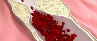
Atherosclerosis
The normal appearance of the arterial wall is smooth and elastic. The formation of plaques helps to reduce the lumen of the trunk. The increase in deposits leads to a pronounced narrowing of the vessel. Carrying out diagnostics, doctors diagnose the patient with atherosclerosis of the carotid arteries. This condition belongs to a number of serious diseases that provoke stroke and atrophy of brain tissue, and therefore requires immediate treatment. The presence of plaques in the carotid filament can be determined by the following symptoms:
- a sharp increase in cholesterol levels;
- frequent headaches;
- fainting;
- vision problems;
- rapid pulse;
- severe noise in the ears;
- numbness of the limbs;
- convulsions, confusion;
- speech disorder.
Carotid artery syndrome
A disease characterized by spasm of the vascular walls is recognized by medicine as carotid artery syndrome. Its occurrence is associated with the accumulation of a cholesterol layer along the edges of the channel, division of the membrane into several layers, and stenosis. Less commonly, the origin of the disease is caused by genetic predisposition, hereditary factors, and injuries.
Dissection of the inner surface of the artery becomes the root cause of ischemic stroke in people of different age groups. Patients over fifty years of age are at risk, but recent research by scientists shows that the percentage of strokes in young people is growing. Prevention of the development of SA syndrome involves giving up bad habits and maintaining an active lifestyle.
Aneurysm
An expansion of the arterial zone with local thinning of the covering is called an aneurysm. The condition is preceded by inflammatory reactions, muscle atrophy, and sometimes the disease is congenital. It is formed in the intracranial zones of the internal carotid branch and looks like a sac. The worst consequence of such a formation is rupture, leading to death.
An aneurysm should not be confused with carotid chemodectoma, which is a benign tumor. According to statistics, 5% of cases result in cancer. The developmental path originates in the bifurcation area, continuing its movement under the jaw. During its life, the nuisance does not manifest itself in any way, so it is diagnosed by pathologists.
Diagnostic methods
The carotid artery in humans is located not only in the neck, but is also located in the tissues of the head. Therefore, if a violation of the condition of the vessel is suspected, it must be examined along the entire area from the aorta to the branching into capillaries.
To identify pathology, it is necessary to go through the following diagnostic stages:
1. Collection of data from the patient’s words and visual inspection:
- what symptoms are present and when they appeared;
- the presence of a hereditary predisposition to these diseases;
- pressure measurement;
- test for coordination of movement and thinking;
- the presence of chronic pathologies;
- what medications are taken;
- presence of bad habits.
2. Taking tests. Data from blood and urine tests can reveal the presence of infection in the body and check the chemical composition of the blood (including platelets and cholesterol).
3. Hardware examination:
- duplex scanning using ultrasound. The procedure allows you to assess the presence of plaques, the magnitude of vasoconstriction and the quality of blood flow;
- MRI or CT with contrast. Allows you to more accurately determine the diameter of the artery lumen;
- Electroencephalography. The activity of brain cells is measured;
- Rheoencephalography. The device checks the condition of all blood vessels in the brain.
To make a complete diagnosis, it is necessary to undergo at least 2 hardware examinations.
Diagnostics
After examining the patient's complaints, the doctor examines the patient. With carotid artery stenosis, the following symptoms are found:
- asymmetrical pulsation in the carotid and temporal arteries;
- vascular murmur in the artery bifurcation zone;
- decreased pressure in the central retinal artery on the affected side (when examined by an ophthalmologist).
To examine the patient and assess the degree of damage to the carotid arteries, the following studies are performed:
- general and biochemical blood test;
- general urine analysis;
- ECG;
- Vascular ultrasound with Doppler ultrasound (USDG);
- angiography, MR or CT angiography;
- CT and MRI of the brain (if an ischemic stroke is suspected).
The gold standard for diagnosing carotid artery stenosis is angiography. This study allows you to obtain accurate data about the narrowing zone, its extent and degree. The results of angiography are especially important for drawing up a surgical treatment plan.
Treatment of carotid artery diseases
Treatment is prescribed only by a specialist after a complete examination.
If stenosis or blockage is confirmed, the following treatment methods are prescribed:
| Types of treatment | Brief description of the method | Restrictions on treatment | Efficiency |
| Diet | Foods high in fat and cholesterol are completely excluded from the menu. It is recommended to eat fresh vegetables/fruits, cereals and lean meats/fish. | If there are diseases of the digestive tract, the menu should be compiled by a specialist. Fasting is prohibited. | The diet prevents the formation of new cholesterol plaques. To eliminate old ones, drug treatment is required. Therefore, it is recommended to combine the diet with medication. |
| Medicines |
| Drugs can be prescribed in combination. Therefore, it is important to consider their compatibility. The dosage and course are prescribed strictly by the attending physician. Increasing the dosage, course, or replacing medications yourself is strictly prohibited. | An improvement in the condition is noted after completing the course and is confirmed by repeated examination of the patency of the carotid arteries. |
| Surgical cleansing of blood vessels | If cholesterol plaques cannot be eliminated with medication, they are removed surgically. | The operation is prescribed only if there are no contraindications. | If you follow a diet and take prophylactic medications, the risk of cholesterol plaques recurring is minimal. |
| Restoring patency | This procedure is used if the narrowing of the vessel is caused by other reasons (pathologies during intrauterine development or frequent spasms of the artery). To do this, a metal frame is installed in the damaged area. And if the segment is severely damaged, then it is replaced with a prosthesis. |
If the patency of the arteries is impaired as a result of the development of an inflammatory process in them, then the specialist may prescribe the above medications together with Prednisolone or Dexamethasone.
Surgical methods of treatment
If atherosclerotic plaques block an artery by 70 percent or more, or if the patient has already suffered a mini-stroke, surgical treatment options are considered. For blockages between 50% and 69%, doctors may recommend surgery based on the patient's age and health.
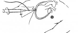
Carotid angioplasty with stenting
A newer procedure for treating blocked carotid arteries than endarterectomy is carotid angioplasty with stenting. A minimally invasive procedure, it involves inserting a catheter into the carotid artery through a blood vessel in the groin. Once the catheter is in place, a small balloon is inflated in the artery to open the artery, and a stent is then placed at the site of the blockage. A stent is a tiny mesh of wire that keeps the artery open. To prevent plaque particles from moving to other parts of the body during the procedure, the surgeon uses an embolic filter to catch them.
Endarterectomy
This is a standard surgical procedure used to treat a blocked carotid artery, in which fatty plaques in the carotid artery are removed through an incision in the neck. After gaining access to the artery by cutting tissue, the surgeon clamps the artery and opens it longitudinally. He then physically removes the plaque by scraping, and finally widens the artery with a diamond-shaped flap and stitches it together.
So, in order to prevent a fatal stroke or paralysis, it is necessary to monitor the appearance of symptoms of a blocked carotid artery and promptly provide appropriate treatment. To avoid carotid artery blockage, it is important to take care of your health and stay in good physical shape. Measures such as avoiding alcohol and tobacco, eating a low-fat and low-cholesterol diet, and exercising regularly can greatly help prevent this disease.
Source: buzzle.com
How to measure the pulse on the carotid artery?
To obtain reliable heart rate readings on the carotid artery, a person must be at rest (if measured immediately after exercise, stress, and even after eating, the value will be incorrect). The patient should sit and lean on the back of the chair/chair.
Next, you need to find the carotid artery and count the pulsations for 1 minute. The indicator should be in the range of 60-80 beats. It is recommended to take measurements on the right and left sides of the neck. The values should not differ much. If the indicator goes beyond the limits (subject to measurement in compliance with the rules) within 5-7 days, then an examination by a specialist is necessary.
It is important that when measuring the pulse on the carotid artery, you should not apply too much pressure to the vessel.
How to stop bleeding?
If there is bleeding from the carotid artery, death can occur within 2-3 minutes. Therefore, it is important to stop blood loss in a timely manner. To do this, the victim must be seated.
If the artery on the right side of the neck is damaged, the patient must raise his left arm above the ready one, apply a thick gauze bandage to the site of bleeding and secure it with a bandage, wrapping it to the left arm near the shoulder. Record the time of application of the tourniquet. The data is necessary for medical staff.
The carotid artery not only delivers nutrients and oxygen to the tissues of the neck and head, but also regulates heart rate, breathing and blood pressure.
If the patency of the vessel is disrupted (depending on where the artery is located), blindness, oxygen starvation of brain tissue, and even death can occur. Therefore, for timely detection of pathology, it is important to undergo an annual examination, especially if a person has a predisposition.
Author: Kotlyachkova Svetlana
Article design: Vladimir the Great