The human circulatory system, like that of all living creatures belonging to the class Vertebrates, is a closed system of tubes and cavities through which blood circulates non-stop. The circulatory system includes the heart and various sized arteries, veins, and blood capillaries.
The concept of the human circulatory system was introduced by the English physician W. Harvey in 1616.
The role of the circulatory system in the body is such that the cardiovascular system is often called transport or distribution. The heart of an adult healthy person pumps approximately 6 liters of blood through the vascular circulatory system, the total length of which is about 100,000 km.
Circulatory system
If you look at the pattern of blood distribution throughout the body, its cyclical path is striking. If we do not take into account the placental blood flow, then among those isolated there is a small cycle that ensures respiration and gas exchange of tissues and organs and affects the human lungs, as well as a second, large cycle that carries nutrients and enzymes.
The task of the circulatory system, which became known thanks to the scientific experiments of the scientist Harvey (in the 16th century, he discovered the circulatory circuits), generally consists of organizing the movement of blood and lymph cells through the vessels.
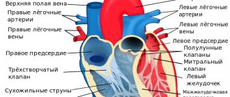
Vessels draining into the atria of the heart
Vessels flow into the right atrium, through which venous blood flows with a high content of CO2 and waste. They close the large circle of blood circulation.
There are two such veins:
- Inferior vena cava . It is the largest venous vessel, which is often called the venous trunk. “Waste” blood from the legs, tissues and organs located in the pelvis and abdomen is collected here.
- Superior vena cava . It is a short trunk formed by two brachiocephalic venous vessels. It opens with an orifice into the right atrial chamber at the level of the third right rib. Venous blood from the arms, head, tissues and organs of the chest flows here.
The thickness of the upper and lower hollow venous vessels flowing into the heart is different, since they experience different loads. In addition, there are gender differences in the topographic and anatomical characteristics of the thickness of their walls.
For your information. Histological maturity of the tissue of the walls of the inferior and superior vena cava occurs at the age of 10, and starting from the age of 70, there is a gradual reduction in the number and atrophy of circularly located smooth muscle cells.
By the way, answering the question: “What vessels flow into the right atrium?” – we should remember the lymphatic system. Its ducts connect into trunks, which open into veins located near the clavicular bones.
Thus, venous blood flowing in the area between the mouths of the lymphatic trunks and the superior vena cava has the highest degree of bactericidal activity.
Veins draining into the atria of the heart
Paired vessels flow into the left atrium, closing the pulmonary circulation:
- The two left (superior and inferior) pulmonary veins drain oxygenated blood into the left atrium. Their mouths are located vertically, one above the other, and are located in the left posterolateral region of the wall of the atrial chamber. The upper vessel collects blood from the upper lobe of the left lung, and the lower left pulmonary vein, respectively, from the lower lobe of the left lung.
- The two right (superior and inferior) pulmonary veins drain blood into the left atrial chamber of the heart from the right lung. The length of these vessels is greater than that of the left pulmonary veins, since before they enter the posterior wall of the left atrium near the interatrial septum, they still pass through the thickness of the posterior wall of the right atrium (pictured below).
The thickness of the walls of the vessels that flow into the left atrium is almost the same in men and women. However, there are also differences. The thickness of the wall between the orifices and the diameter of the superior venous vessel are greater in women, and the diameters of both inferior pulmonary veins are greater in men.
Location of the pulmonary veins
We also note that during intrauterine development the pulmonary circulation does not work. Oxygen-enriched blood comes from the mother through the umbilical vein and is drained through the umbilical arteries of the placenta.
The pulmonary circulation, starting with the pulmonary column and ending with the pulmonary veins, begins to function immediately after birth, as soon as the baby takes his first breath.
Pulmonary circulation
From above, venous blood from the right atrial chamber enters the right cardiac ventricle. Veins are medium-sized vessels. Blood passes in portions and is pushed out of the cavity of the cardiac ventricle through a valve that opens in the direction of the pulmonary trunk.
From it, blood exits into the pulmonary artery, and, as it moves away from the main muscle of the human body, the veins flow into the arteries of the pulmonary tissue, turning and breaking up into a multiple network of capillaries. Their role and primary function is to carry out gas exchange processes in which alveolocytes take up carbon dioxide.
As oxygen is distributed through the veins, the blood flow begins to exhibit arterial characteristics. So, through the venules, the blood flows to the pulmonary veins, which open into the left atrium.
Structure of the human cardiovascular system
The anatomical structure of the human cardiovascular system has many features. For example, the appearance and functionality of a person's circulatory system may differ between individuals, even if they are closely related. Thus, the size and location of the heart in the mediastinum is individual for men and women, adults and children, as are the sizes of veins and arteries.
Similar anatomy is observed in the topography of the organs of the cardiovascular system: the heart is localized in the chest, the largest vessels depart from it, which then branch into smaller ones. Lymphatic vessels are located almost parallel to them.
Until a certain point, anatomists considered the circulatory and lymphatic systems to be a single whole. They were finally separated only towards the end of the 19th century.
Over time, the structure of the human circulatory system can change under the influence of external factors. The most common are age-related changes in the cardiovascular system, which occur gradually. Acquired pathologies are considered less common, although they have more severe health consequences. All this gives grounds to call the cardiovascular system not a completely stable system of the body.
Heart
Among all the organs of the circulatory system, the heart occupies a central position. It is the “pump” that ensures the continuity of blood flow in the vessels. The heart is a hollow organ consisting of muscles that contract rhythmically under the influence of impulses sent by the medulla oblongata. Inside, it is divided by a system of partitions and valves into four parts: the left and right ventricles, the left and right atrium.
The heart wall consists of three layers:
- The endocardium is the inner layer consisting of several types of cells. The surface of muscle fibers, tendon threads and valves is covered with endothelial cells, and underneath there is a basement membrane and loose fibrous subendothelium. Beneath these layers is a thin layer of mixed muscle and elastic fibers, connected through a thin layer of connective cells to the myocardium.
- Myocardium is the middle layer of the heart, consisting of striated muscles. The cells of this type of tissue are connected into spirally arranged threads surrounding all the chambers of the heart. The bulk of myocardial muscle cells are of the contractile muscle type. Less than 1/3 of the muscle mass of the heart is represented by conducting and secretory cardiomyocytes. Between all types of cardiomyocytes there are connective tissue spaces penetrated by a network of capillaries.
- The epicardium is the outer layer of the heart, consisting of a loose layer of connective cells and a denser layer of mesothelial cells. Connective tissue contains nerve fibers and blood vessels. The surface of the heart is covered with a layer of fatty tissue.
All layers of the heart are supported by a fibrous skeleton, formed by several rings of dense connective tissue and bundles of collagen, cartilaginous plates and elastic fibers.
Heart sounds
As the heart contracts and relaxes, it makes sounds. In cardiology (the science that studies the structure, functions and diseases of the heart) they are called tones. Two heart sounds are identified:
- Systolic - occurs when the cusps of the bicuspid and tricuspid valves vibrate and the tendons of the heart stretch. Its main features are high duration and low level of sound vibrations.
- Diastolic - occurs at the moment of complete closure of the valves of the aorta and arteries of the pulmonary trunk. Its features are short duration and high level of sound vibrations.
Normally, heart sounds are harmonious and rhythmic. The average heart rate in a healthy person at rest is between 60 and 70 beats per minute.
Vessels
The human circulatory system consists of hollow tubes of different sizes, which are divided into two types: main and those involved in metabolic processes. The main circulatory system is large vessels that perform exclusively a transport function and are divided into two types:
- arteries that carry blood from the heart to the organs and tissues of the body;
- veins that carry blood from organs and tissues to the heart.
The arterial network consists of the main artery of the circulatory system - the aorta, as well as many smaller branches that gradually transform into arterioles. The wall of vessels of this type is thick and elastic, with a pronounced muscle layer, due to which they resist blood pressure and forcefully push it to distant areas.
The venous circulatory system consists of large, medium and small veins. Large-diameter vessels are located near the heart, and when moving away from it they branch into smaller ones. The veins gradually become thinner and become venules.
The circulatory system, consisting of arteries and veins, is closed by a microcirculatory bed consisting of arterioles, capillaries, and venules, as well as arteriovenular anastomoses. This part of the channel carries out exchange functions. Here the release of oxygen from blood cells and the diffusion of carbon dioxide and waste products from tissues occur.
Systemic circulation
Let's trace the big blood cycle. The systemic circulation begins from the left cardiac ventricle, which receives an arterial flow enriched in O2 and depleted in CO2, which is supplied from the pulmonary circulation. Where does blood go from the left ventricle of the heart?
After the left-sided ventricle, the subsequent aortic valve pushes arterial blood into the aorta. It distributes O2 in high concentration throughout all arteries. Moving away from the heart, the diameter of the artery tube changes - it decreases.
All CO2 is collected from the capillary vessels, and the flows of the large circle enter the vena cava. From them, the blood again enters the right atrium, then into the right ventricle and pulmonary trunk.
Thus, the systemic circulation ends in the right atrium. And to the question - where does the blood go from the right ventricle of the heart, the answer is to the pulmonary artery.
Blood circulation in humans
Arteries
- These are vessels that carry blood from the heart.
They have a thick muscle layer. Veins
are vessels that carry blood to the heart. They have a thin muscle layer and valves.
Capillaries
- These are single-layer vessels in which the exchange of substances between blood and tissues occurs.
Arterial blood
- This is blood saturated with oxygen.
Venous blood
is saturated with carbon dioxide. In the pulmonary circulation, venous blood flows through the arteries, and arterial blood flows through the veins.
The human heart has four chambers
, consists of two atria and two ventricles (in the left half of the heart there is arterial blood, in the right - venous).
The leaflet valves are located between the ventricles and atria
, and between the arteries and ventricles -
semilunar.
The valves prevent blood from flowing backwards (from the ventricle to the atrium, from the aorta to the ventricle).
The thickest wall is at the left ventricle, because it pushes blood through the systemic circulation. When the left ventricle contracts, a pulse wave is created, as well as maximum blood pressure.
Blood pressure:
in the arteries the largest, in the capillaries the average, in the veins the smallest.
Blood speed:
the highest in the arteries, the smallest in the capillaries, the average in the veins.
Big circle
blood circulation: from the left ventricle, arterial blood flows through the arteries to all organs of the body. In the capillaries of the large circle, gas exchange occurs: oxygen passes from the blood into the tissues, and carbon dioxide passes from the tissues into the blood. The blood becomes venous, flows through the vena cava into the right atrium, and from there into the right ventricle.
Small circle:
From the right ventricle, venous blood flows through the pulmonary arteries to the lungs. Gas exchange occurs in the capillaries of the lungs: carbon dioxide passes from the blood into the air, and oxygen from the air into the blood, the blood becomes arterial and flows through the pulmonary veins into the left atrium, and from there into the left ventricle.
You can also read
DETAILED SUMMARY: Circulatory systems of vertebrates, Circulatory system of mammals
ASSIGNMENTS OF PART 2 OF THE USE ON THIS TOPIC
Part 1 tasks
Choose one, the most correct option. Why can't blood get from the aorta to the left ventricle of the heart?
1) the ventricle contracts with great force and creates high pressure 2) the semilunar valves fill with blood and close tightly 3) the leaflet valves are pressed against the walls of the aorta 4) the leaflet valves are closed and the semilunar valves are open
Answer
2
Choose one, the most correct option. Blood enters the pulmonary circulation from the right ventricle through
1) pulmonary veins 2) pulmonary arteries 3) carotid arteries 4) aorta
Answer
2
Choose one, the most correct option. Arterial blood flows through the human body
1) renal veins 2) pulmonary veins 3) vena cava 4) pulmonary arteries
Answer
2
Choose one, the most correct option. In mammals, blood is enriched with oxygen in
1) arteries of the pulmonary circulation 2) capillaries of the systemic circle 3) arteries of the systemic circle 4) capillaries of the systemic circle
Answer
4
Choose one, the most correct option. The vena cava in the human body drains into
1) left atrium 2) right ventricle 3) left ventricle 4) right atrium
Answer
4
Choose one, the most correct option. Valves prevent blood from flowing back from the pulmonary artery and aorta into the ventricles.
1) tricuspid 2) venous 3) bicuspid 4) semilunar
Answer
4
ARTERIES - VEINS 1. Establish a correspondence between the signs and blood vessels: 1) vein 2) artery. Write numbers 1 and 2 in the order corresponding to the letters.
A) has a thin muscle layer B) has valves C) carries blood from the heart D) carries blood to the heart E) has elastic elastic walls E) withstands high blood pressure
Answer
112122
2. Establish a correspondence between the structural features and functions and types of vessels: 1) artery, 2) vein. Write numbers 1 and 2 in the order corresponding to the letters.
A) has valves B) the wall contains fewer muscle fibers C) carries blood from the heart D) carries venous blood in the pulmonary circulation D) communicates with the right atrium E) carries out blood flow due to contraction of skeletal muscles
Answer
221122
ARTERIES - VEINS - CAPILLARIES Establish a correspondence between the features of blood vessels and their types: 1) artery, 2) vein, 3) capillary. Write numbers 1-3 in the order corresponding to the letters.
A) the wall consists of one layer of cells B) endothelial cells fit tightly to each other, forming smooth walls C) the walls have valves D) the walls are thin, elastic, contain muscles E) has the smallest diameter
Answer
31223
VEINS Select three options. Veins are blood vessels through which blood flows
1) from the heart 2) to the heart 3) under greater pressure than in the arteries 4) under less pressure than in the arteries 5) faster than in the capillaries 6) slower than in the capillaries
Answer
245
VEINS IN EXC. FROM ARTERIES 1. Choose three correct answers out of six and write down the numbers under which they are indicated. Veins, as opposed to arteries
1) have valves in their walls 2) can collapse 3) have walls made of a single layer of cells 4) carry blood from organs to the heart 5) withstand high blood pressure 6) always carry blood that is not saturated with oxygen
Answer
124
2. Choose three correct answers out of six and write down the numbers under which they are indicated. Veins, unlike arteries, are characterized by
1) leaflet valves 2) transport of blood to the heart 3) semilunar valves 4) high blood pressure 5) thin muscle layer 6) fast blood flow
Answer
235
VENOUS BLOOD Choose three correct answers out of six and write down the numbers under which they are indicated. Elements of the human circulatory system containing venous blood are
1) pulmonary artery 2) aorta 3) vena cava 4) right atrium and right ventricle 5) left atrium and left ventricle 6) pulmonary veins
Answer
134
ARTERIAL - VENOUS 1. Establish a correspondence between the type of human blood vessels and the type of blood they contain: 1) arterial, 2) venous
A) pulmonary arteries B) veins of the pulmonary circulation C) aorta and arteries of the systemic circulation D) superior and inferior vena cava
Answer
2112
2. Establish a correspondence between a vessel of the human circulatory system and the type of blood that flows through it: 1) arterial, 2) venous. Write numbers 1 and 2 in the order corresponding to the letters.
A) femoral vein B) brachial artery C) pulmonary vein D) subclavian artery E) pulmonary artery E) aorta
Answer
211121
3. Establish a correspondence between the sections of the human circulatory system and the type of blood passing through them: 1) arterial, 2) venous. Write numbers 1 and 2 in the order corresponding to the letters.
A) left ventricle B) right ventricle C) right atrium D) pulmonary vein E) pulmonary artery E) aorta
Answer
122121
ARTERIAL IN EXC. FROM VENOUS Select three options. In mammals and humans, venous blood, unlike arterial,
1) is poor in oxygen 2) flows in a small circle through the veins 3) fills the right half of the heart 4) is saturated with carbon dioxide 5) enters the left atrium 6) provides body cells with nutrients
Answer
134
PRESSURE SEQUENCE 1. Establish the sequence of human blood vessels in order of decreasing blood pressure in them. Write down the corresponding sequence of numbers.
1) inferior vena cava 2) aorta 3) pulmonary capillaries 4) pulmonary artery
Answer
2431
2. Establish the order in which the blood vessels should be arranged in order of decreasing blood pressure in them
1) Veins 2) Aorta 3) Arteries 4) Capillaries
Answer
2341
3. Establish the sequence of arrangement of blood vessels in order of increasing blood pressure in them. Write down the corresponding sequence of numbers.
1) inferior vena cava 2) aorta 3) pulmonary artery 4) alveolar capillaries 5) arterioles
Answer
14532
SPEED SEQUENCE Arrange the blood vessels in order of decreasing speed of blood movement in them
1) superior vena cava 2) aorta 3) brachial artery 4) capillaries
Answer
2314
BIG Choose three correct answers out of six and write down the numbers under which they are indicated. Large circle of blood circulation in the human body
1) begins in the left ventricle 2) originates in the right ventricle 3) is saturated with oxygen in the alveoli of the lungs 4) supplies organs and tissues with oxygen and nutrients 5) ends in the right atrium 6) brings blood to the left half of the heart
Answer
145
Choose three correct answers out of six and write down the numbers under which they are indicated. Which parts of the circulatory system belong to the systemic circulation?
1) pulmonary artery 2) superior vena cava 3) right atrium 4) left atrium 5) left ventricle 6) right ventricle
Answer
235
LARGE SEQUENCE 1. Establish the sequence of blood movement through the vessels of the systemic circulation. Write down the corresponding sequence of numbers.
1) hepatic portal vein 2) aorta 3) gastric artery 4) left ventricle 5) right atrium 6) inferior vena cava
Answer
423165
2. Determine the correct sequence of blood circulation in the systemic circulation, starting with the left ventricle. Write down the corresponding sequence of numbers.
1) Aorta 2) Superior and inferior vena cava 3) Right atrium 4) Left ventricle 5) Right ventricle 6) Tissue fluid
Answer
416235
3. Establish the correct sequence of blood passage through the systemic circulation. Write down the corresponding sequence of numbers in the table.
1) right atrium 2) left ventricle 3) arteries of the head, limbs and torso 4) aorta 5) inferior and superior vena cava 6) capillaries
Answer
243651
4. Establish the sequence of blood movement in the human body, starting with the left ventricle. Write down the corresponding sequence of numbers.
1) left ventricle 2) vena cava 3) aorta 4) pulmonary veins 5) right atrium
Answer
13254
5. Establish the sequence of passage of a portion of blood in a person, starting with the left ventricle of the heart. Write down the corresponding sequence of numbers.
1) right atrium 2) aorta 3) left ventricle 4) lungs 5) left atrium 6) right ventricle
Answer
321645
6f. Establish the sequence of blood movement through the systemic circulation in humans, starting from the ventricle. Write down the corresponding sequence of numbers.
1) left ventricle 2) capillaries 3) right atrium 4) arteries 5) veins 6) aorta
Answer
164253
GREAT CIRCLE ARTERIES Select three options. Blood flows through the arteries of the systemic circulation in humans
1) from the heart 2) to the heart 3) saturated with carbon dioxide 4) saturated with oxygen 5) faster than in other blood vessels 6) slower than in other blood vessels
Answer
145
SMALL SEQUENCE 1. Establish the sequence of blood movement in a person through the pulmonary circulation. Write down the corresponding sequence of numbers.
1) pulmonary artery 2) right ventricle 3) capillaries 4) left atrium 5) veins
Answer
21354
2. Establish the sequence of circulatory processes, starting from the moment when blood moves from the lungs to the heart. Write down the corresponding sequence of numbers.
1) blood from the right ventricle enters the pulmonary artery 2) blood moves through the pulmonary vein 3) blood moves through the pulmonary artery 4) oxygen enters from the alveoli into the capillaries 5) blood enters the left atrium 6) blood enters the right atrium
Answer
256134
3. Establish the sequence of movement of arterial blood in a person, starting from the moment it is saturated with oxygen in the capillaries of the pulmonary circle. Write down the corresponding sequence of numbers.
1) left ventricle 2) left atrium 3) veins of the pulmonary circle 4) capillaries of the pulmonary circle 5) arteries of the systemic circle
Answer
43215
4. Establish the sequence of movement of arterial blood in the human body, starting with the capillaries of the lungs. Write down the corresponding sequence of numbers.
1) left atrium 2) left ventricle 3) aorta 4) pulmonary veins 5) pulmonary capillaries
Answer
54123
5. Establish the correct sequence of passage of a portion of blood from the right ventricle to the right atrium. Write down the corresponding sequence of numbers.
1) pulmonary vein 2) left ventricle 3) pulmonary artery 4) right ventricle 5) right atrium 6) aorta
Answer
431265
SMALL CIRCLE ARTERY Select three options. Blood flows through the arteries of the pulmonary circulation in humans
1) from the heart 2) to the heart 3) saturated with carbon dioxide 4) saturated with oxygen 5) faster than in the pulmonary capillaries 6) slower than in the pulmonary capillaries
Answer
135
LARGE - SMALL VESSELS 1. Establish a correspondence between the sections of the circulatory system and the circle of blood circulation to which they belong: 1) Systemic circulation, 2) Lesser circulation. Write numbers 1 and 2 in the correct order.
A) Right ventricle B) Carotid artery C) Pulmonary artery D) Superior vena cava E) Left atrium E) Left ventricle
Answer
212121
2. Establish a correspondence between the vessels and human circulatory circles: 1) pulmonary circulation, 2) systemic circulation. Write numbers 1 and 2 in the correct order.
A) aorta B) pulmonary veins C) carotid arteries D) capillaries in the lungs E) pulmonary arteries E) hepatic artery
Answer
212112
3. Establish a correspondence between the structures of the circulatory system and the human circulation circles: 1) small, 2) large. Write numbers 1 and 2 in the order corresponding to the letters.
A) aortic arch B) hepatic portal vein C) left atrium D) right ventricle E) carotid artery E) alveolar capillaries
Answer
221121
LARGE - SMALL SIGNS Establish a correspondence between the processes and the circles of blood circulation for which they are characteristic: 1) small, 2) large. Write numbers 1 and 2 in the order corresponding to the letters.
A) Arterial blood flows through the veins. B) The circle ends in the left atrium. B) Arterial blood flows through the arteries. D) The circle begins in the left ventricle. D) Gas exchange occurs in the capillaries of the alveoli. E) Venous blood is formed from arterial blood.
Answer
112212
SEQUENCE WITH ORGANS Establish the sequence of movement of the hormone thyroxine through the human circulatory system, starting from the moment of its formation until reaching the target organ. Write down the corresponding sequence of numbers.
1) left ventricle 2) superior vena cava 3) right atrium 4) medulla oblongata 5) pulmonary trunk 6) thyroid capillary
Answer
623514
HEART SEQUENCE Establish the sequence of events that occur in the cardiac cycle after blood enters the heart. Write down the corresponding sequence of numbers.
1) contraction of the ventricles 2) general relaxation of the ventricles and atria 3) blood flow into the aorta and artery 4) blood flow into the ventricles 5) contraction of the atria
Answer
54132
LEFT VENTRICLE 1. Select three options. A person has blood from the left ventricle of the heart
1) when it contracts, it enters the aorta 2) when it contracts, it enters the left atrium 3) supplies the cells of the body with oxygen 4) enters the pulmonary artery 5) under high pressure enters the systemic circulation 6) under low pressure enters the pulmonary circulation
Answer
135
2. Choose three correct answers out of six and write down the numbers under which they are indicated. From the left ventricle of the heart
1) blood enters the systemic circulation 2) venous blood comes out 3) arterial blood comes out 4) blood flows through the veins 5) blood flows through the arteries 6) blood enters the pulmonary circulation
Answer
135
RIGHT VENTRICLE Choose three correct answers out of six and write down the numbers under which they are indicated. Blood leaks from the right ventricle
1) arterial 2) venous 3) through arteries 4) through veins 5) towards the lungs 6) towards the cells of the body
Answer
235
LEFT - RIGHT Establish a correspondence between the characteristics and chambers of the human heart: 1) left ventricle, 2) right ventricle. Write numbers 1 and 2 in the order corresponding to the letters.
A) The pulmonary arteries depart from it. B) It enters the systemic circulation. B) Contains venous blood. D) It has thicker muscle walls. D) A bicuspid valve opens into it. E) Contains oxygen-rich blood.
Answer
212111
Analyze the table “Structure of the Heart”. For each cell indicated by a letter, select the corresponding term from the list provided.
1) Contracting, provides blood flow through the systemic circulation 2) Left atrium 3) Separated from the left ventricle by a bicuspid valve 4) Right atrium 5) Separated from the right atrium by a tricuspid valve 6) Contracting, directs blood to the left ventricle 7) Pericardial sac
Answer
451
Choose three correctly labeled captions for the picture that depicts the internal structure of the heart. Write down the numbers under which they are indicated.
1) superior vena cava 2) aorta 3) pulmonary vein 4) left atrium 5) right atrium 6) inferior vena cava
Answer
126
Choose three correctly labeled captions for the picture that depicts the structure of the human heart. Write down the numbers under which they are indicated.
1) superior vena cava 2) leaflet valves 3) right ventricle 4) semilunar valves 5) left ventricle 6) pulmonary artery
Answer
246
Establish a correspondence between the structural and functional features and the chambers of the heart indicated in the figure. Write numbers 1 and 2 in the order corresponding to the letters.
A) is the end of the systemic circulation B) is the beginning of the systemic circulation C) is filled with venous blood D) is filled with arterial blood E) has a thin muscle wall
Answer
12121
Establish a correspondence between the chambers of the heart, indicated in the figure by numbers 1 and 2, and their structural features and functions. Write numbers 1 and 2 in the order corresponding to the letters.
A) is the end of the pulmonary circulation B) is the end of the systemic circulation C) is filled with venous blood D) is filled with arterial blood E) is connected to the pulmonary vein
Answer
12211
Establish a correspondence between the chambers of the heart, indicated in the figure by numbers 1 and 2, and their structural features and functions. Write numbers 1 and 2 in the order corresponding to the letters.
A) is the end of the pulmonary circulation B) is the beginning of the pulmonary circulation C) is filled with venous blood D) is filled with arterial blood E) has a thinner muscle wall
Answer
12211
Choose three correct answers out of six and write down the numbers under which they are indicated. Human pulse
1) is not associated with the speed of blood flow 2) depends on the elasticity of the walls of blood vessels 3) is palpated on large arteries close to the surface of the body 4) accelerates blood flow 5) is caused by the rhythmic oscillation of the veins 6) is not associated with the contraction of the heart
Answer
123
Establish the sequence of carbon dioxide transport from the moment it enters the blood. Write down the corresponding sequence of numbers.
1) left ventricle 2) capillaries of internal organs 3) vena cava 4) capillaries of the alveoli
Answer
2341
Establish a correspondence between human blood vessels and the direction of blood movement in them: 1) from the heart, 2) to the heart
A) veins of the pulmonary circulation B) veins of the systemic circulation C) arteries of the pulmonary circulation D) arteries of the systemic circulation
Answer
2211
© D.V. Pozdnyakov, 2009-2020
Human circulatory organs
These include the heart and blood vessels (veins, arteries and capillaries). Let's consider the most important organ in the human body.
The heart is a self-governing, self-regulating, self-correcting muscle. The size of the heart depends on the development of skeletal muscles - the higher their development, the larger the heart. The structure of the heart has 4 chambers - 2 ventricles and 2 atria, and is placed in the pericardium. The ventricles are separated from each other and between the atria by special heart valves.
Responsible for replenishing and saturating the heart with oxygen are the coronary arteries, or as they are called “coronary vessels”.
The main function of the heart is to act as a pump in the body. Failures are due to several reasons:
- Insufficient/excessive volumes of incoming blood.
- Injuries to the heart muscle.
- External compression.
The second most important vessels in the circulatory system are blood vessels.
Vessels of the circulatory system
All vessels included in the circulatory system are divided into groups:
- Arterial vessels;
- Arterioles;
- Capillaries;
- Venous vessels.
Arteries
Arteries include those vessels that transport blood from the heart to the internal organs. There is a common misconception among the population that the blood in the arteries always contains a high concentration of oxygen. However, this is not the case; for example, venous blood circulates in the pulmonary artery.
Arteries have a characteristic structure.
Their vascular wall consists of three main layers:
- Endothelium;
- Muscle cells located underneath;
- A membrane consisting of connective tissue (adventitia).
The diameter of the arteries varies widely - from 0.4-0.5 cm to 2.5-3 cm. The entire volume of blood contained in vessels of this type is usually 950-1000 ml.
As they move away from the heart, the arteries divide into smaller vessels, the last of which are the arterioles.
Capillaries
Capillaries are the smallest component of the vascular bed. The diameter of these vessels is 5 microns. They penetrate all tissues of the body, ensuring gas exchange. It is in the capillaries that oxygen leaves the bloodstream and carbon dioxide migrates into the blood. This is where the exchange of nutrients takes place.
Vienna
Passing through organs, capillaries merge into larger vessels, first forming venules and then veins. These vessels carry blood from the organs towards the heart. The structure of their walls differs from the structure of arteries; they are thinner, but much more elastic.
A feature of the structure of veins is the presence of valves - connective tissue formations that block the vessel after the passage of blood and prevent its reverse flow. The venous system contains much more blood than the arterial system - approximately 3.2 liters.
Linear and volumetric blood flow velocity
When considering blood velocity parameters, the concepts of linear and volumetric velocities are used. There is a mathematical relationship between these concepts.
Where does blood move at the fastest speed? The linear velocity of blood flow is in direct proportion to the volumetric velocity, which varies depending on the type of vessels.
The highest blood flow speed is in the aorta.
Where does blood move at the slowest speed? The lowest speed is in the vena cava.
Time for complete blood circulation
For an adult whose heart beats about 80 times per minute, the blood makes the entire journey in 23 seconds, distributing 4.5-5 seconds on the small circle and 18-18.5 seconds on the big one.
The data is confirmed experimentally. The essence of all research methods lies in the principle of labeling. A traceable substance that is not found in the human body is injected into a vein and its location is dynamically determined.
This is how long it takes for the substance to appear in the vein of the same name, located on the other side. This is the time for complete blood circulation.
CORONARY ARTERIES AND VEINS
The heart is a very independent organ of the human body, consisting of special cells - cardiomyocytes. To provide the heart with everything it needs, it has its own blood supply system. The most basic knowledge about the human structure clearly indicates the presence of two blood circulation circles in the body. There is no anatomist who would disagree with you on this. Clinicians, cardiologists, and everyone who views the heart as an object of professional study from different angles, clarify that there are three circles of blood circulation in the human body: large, small and coronary (coronary, also called the cardiac circle). Like any other circulatory system, the coronary circle consists of vessels (the structure of the vessels themselves has already been discussed: veins, arteries and capillaries).
CORONARY ARTERIES
The arteries that make up the coronary circle originate at the base of the aorta, in the sinuses of Valsalva, which are kind of “pockets” of the semilunar valves. There are two arteries (Fig. 1) – right (1) and left (4). The right coronary artery lies between the two cavities of the heart - the right ventricle and the right atrium in the coronary sulcus, and gives them its branches. This coronary artery passes to the posterior surface, then bends sharply and goes down to the apex, thus supplying blood to the posterior walls of both ventricles. Speaking about the left coronary artery (4), it is worth noting that after leaving the aorta, it diverges into 2 thick branches.
One branch (2) immediately goes to the apex of the heart along the anterior interventricular groove, providing blood to the anterior walls of the ventricles. The location of the second branch (3) runs along the coronary groove between the left atrium and ventricle and, providing them with blood, bends around the heart on the left side. At the apex of the organ, an anastomosis is observed - the junction of one vessel with another; it is here that the right coronary artery merges with the descending branch of the left coronary artery lying in front. Fig.1. Arteries of the heart (sternocostal surface)
CORONARY VEINS
When the heart wall receives oxygen and nutrients through the brought blood, gets rid of carbon dioxide and unnecessary compounds, the blood collects in the veins of the coronary circle (Fig. 2). There are more venous vessels in the coronary circle than arteries. Those of them that are larger flow into a thick, elongated and rounded reservoir - the coronary sinus (6), lying in the coronary sulcus behind.
Fig.2. Veins of the heart (diaphragmatic surface)
Among the coronary veins, it is worth highlighting five main ones:
1. The great vein (2) originates at the apex, then rises along the anterior interventricular groove along the descending branch of the left coronary artery, turns along the coronary groove to the left and back, where it flows into the coronary sinus;
2. The middle vein (4) also originates at the apex, but runs along the posterior interventricular groove to the coronary sinus;
3. The small vein (5) lies in the coronary sulcus on the posterior right, flowing either directly into the sinus or into the middle vein;
4. Oblique vein (1) – itself small, it collects blood from the posterior parts of the left atrium;
5. The posterior vein of the left ventricle (3) drains from the posterior wall of this section.
Speaking about the coronary sinus , we note that it opens with a special opening (7) into the right atrium. There, smaller venous trunks, named after A. Tebezia and R. Viessen, independently end with tiny holes.
BLOOD CIRCULATION, LYMPH OUTFLOW AND INNERVATION
An essential feature of the hemocirculation (blood circulation) of the heart is that during systole there is an almost absolute cessation of blood movement through the vessels.
This occurs due to compression of the vessels on all sides by an impressive array of myocardium. During diastole, the myocardium relaxes, due to which blood flow resumes. Lymphatic drainage from the deep myocardial-endocardial and superficial epicardial networks of lymphocapillaries is carried out through the left lymphatic vessel into one of the lower tracheobronchial nodes, and through the right vessel into one of the anterior mediastinal lymph nodes. Cardiac nerves begin from the cervical and thoracic nodes of the sympathetic chains, parasympathetic innervation - from the branches of the vagus nerve; Extra-organ (superficial and deep) and intra-organ (6 subepicardial, intramyocardial and subendocardial) cardiac plexuses are built from these elements.
SCHEMATIC IMAGE OF THE CORONARY CIRCLE
So, the coronary circulation can be represented in the form of a small diagram that will look like this: