Effective diagnosis of cardiovascular diseases is of particular importance in modern society due to their increasing scale and danger. Thanks to modern special methods for diagnosing these insidious diseases, successfully used by foreign and domestic medicine, doctors save and prolong the lives of tens of millions of patients.
Instrumental diagnostic methods
Accurate modern diagnosis of heart and vascular diseases has been developed quite flexibly by medical science and medical practice. It is an in-depth examination of a patient with symptoms of these diseases using proven instrumental methods. Such methods have repeatedly proven their objectivity and the need for their use in studying the mechanical and electrical activity of the heart and its functions, the vascular system and other important human organs.
A positive recent trend is the addition of traditional research methods (ECG and other types of cardiography) with new ones. Among them, it is worth noting orthogonal electrocardiography, rheography, etc., which help to significantly expand the usual range of examination of the patient’s body, giving doctors maximum important information about his condition, risks and prospects. Thus, the diagnosis was significantly simplified, which simplifies the treatment process. This required additional knowledge from doctors and other medical workers not only of a professional nature, but also in purely applied areas - measurement technology, physiology, mathematics, physics, electronics and others.
Angiocardiography
When choosing treatment, diagnosing cardiovascular diseases in this way is indicated if it is necessary to study not only the heart, but also the large vascular elements that feed it. A probe is used, inserted through arteries and veins in the periphery. If you need to study an organ in the right half, puncture of the femoral or brachial vein on the right is indicated; if on the left, on the opposite side of the body. This method of assessing the patient’s condition gives an accurate idea of the content of various gases in the blood and helps to clarify pressure indicators not only in the blood vessels, but also in the cardiac cavity. The doctor will have accurate data on volumes per minute, emissions. They record phono- and electrocardiograms and determine in which direction the blood is discharged.
As part of the diagnosis, treatment and prevention of cardiovascular diseases, various methods of studying the condition using catheters are used very often, since they allow the introduction into the circulatory system of substances that are reflected in contrast when examined by X-rays. Angiocardiography will be no exception, in which a small amount of special compounds are infused into the patient’s body through a catheter, helping to take a series of accurate radiographs.
Electrocardiogram
Without obtaining an ECG, it is impossible to make an accurate medical diagnosis of the heart condition, so it can be considered a key diagnostic method in cariology. Few people have not experienced this method on themselves. Its essence lies in the fact that several special sensors are attached to the patient’s body in the chest area and on his limbs. With their help, the cardiograph mechanism captures and records the electrical activity of the heart. The result is graphically displayed on a special tape in the form of curved lines. An experienced cardiologist reads these notes without any problems, immediately determining what exactly in the work of the heart deviated from the norm, that is, already at this stage he can make a preliminary diagnosis.
Using an ECG, myocardial ischemia, the presence of necrosis (infarction), its depth, extent and other parameters are very accurately determined. In addition, it is easy to determine the presence of electrolyte disorders and arrhythmias of all types. Pericarditis, cardiac tamponade, and other defects are also predicted. Sometimes an ECG is done with stress: sensors are attached to the patient and transmit data while he runs, walks or exercises on a bicycle ergometer.
Electrocardiogram
Electrophysiological study
An effective diagnostic tool is also electrophysiological examination (EPS) of the heart, which began to be practiced in the 60s of the last century. Since then, its forms and methods have been significantly improved and are used everywhere. The procedure is carried out using special catheter electrodes and recording sensors, studying the condition of the inner cardiac surface. The results obtained in this way allow the doctor to diagnose the disease with fairly high accuracy, effectively treat it therapeutically or surgically, and also predict various arrhythmias and types of cardiac conduction disorders. In addition, non-invasive EPI of the TEES type, which uses esophageal electrodes, has been effectively used for more than 30 years.
ECHO-KG
An echocardiogram is a modern method of functional diagnostics of cardiovascular diseases, in which the doctor receives basic useful data through the use of ultrasound equipment. The event helps to imagine how much the heart’s ability to be an active pump is preserved. Scanning with a special device provides data on the structure of the fibers that form the main muscle of the body. If the heart valves are transformed or cannot function normally, ECHO-CG will clearly reflect this fact. Based on the results of the study, the doctor will know what the contractility of the organ as a whole is, how large the heart cavities are, and how thick the walls are in the structure of the organ.
ECHO-CG is a modern and reliable, safe method that gives an accurate idea of the presence of an aortic aneurysm, thrombus, or tumor process. Using this approach, hypertension, ischemia, and heart defects can be diagnosed.
Echocardiogram
To make the diagnosis of heart disease accurate, a special ultrasound examination in the electrocardiogram format is also used. To do this, a special sensor is moved on the patient's chest. On the monitor of the device, the doctor observes clear images of the blood vessels and heart, from which he can determine their condition, features, as well as the quality of the work of the heart muscle. Examination by ultrasound (echocardiography) is maximally polished taking into account the latest achievements of foreign and domestic medical science and clinical observations of patients with cardiac pathologies. Therefore, ultrasound diagnostics is now practiced in many cardiology clinics and specialized centers. Moreover, the ultrasound procedure (heart diagnosis) is completely painless for the patient and takes approximately 20 minutes.
Ultrasound of the heart
Monitoring
Some time ago, scientist Holter proposed a research technique that soon proved to be one of the most effective and gives an accurate idea of the patient’s condition. This is an instrumental diagnosis of cardiovascular diseases, requiring long-term monitoring of the activity of the heart muscle. The event lasts at least 24 hours. Sometimes the patient is prescribed a three-day continuous check. This method allows you to analyze the human condition as deeply as possible. If coronary disease occurs, all its episodes are recorded. In case of arrhythmia, observation allows us to clarify the nuances of the course.
Holter technology testing involves placing electrodes on the chest of a person in need. They are attached to a special small device, which the patient wears on his belt throughout the study period. The unit is secured with a small belt. Sometimes they are attached to the shoulder. This method of diagnosing diseases of the cardiovascular system gives an accurate picture of all changes during the time period being studied. Whatever the processes occurring in the heart muscle, the unit will record everything that happens. The patient is required to keep a diary of actions throughout the entire procedure. It records what is happening. The doctor’s task is to compare the results given by the equipment and the patient’s diary. Based on them, cardiac activity and ability to resist stress are assessed.
24-hour Holter monitoring
As practice shows, a patient can have a heart attack at any time of the day, including when it is not possible to visually monitor his condition (for example, at night). To record such dangerous cardiac interruptions, so-called 24-hour Holter monitoring is practiced. It is a permanent recording of an ECG throughout the day. During the application of the method, a portable cardiograph is attached to the patient's body. It allows you to record all the necessary data. At the same time, the patient records each of his actions throughout the examination, keeping notes in a diary. Subsequently, the doctor analyzes the ECG readings and projects them onto the patient’s records. Such a comparison allows us to accurately determine exactly when, why and how pathological deformities occurred in the patient. 24-hour Holter analysis of the cardiovascular system is also widely and effectively used in practice. It is considered mandatory for all patients with cardiac arrhythmias and a tendency to faint.
Not only with instruments: laboratory tests
The relevance of laboratory diagnostics of cardiovascular diseases cannot be underestimated. For research activities, the patient's blood from a vein is required. In order for the result to be accurate, you should give up alcohol one day before. Children under one year of age should not eat in the 40 minutes preceding blood sampling. Children under five years of age should fast for three hours. Older people need a twelve-hour fast. You can drink water without gas and additives. They stop taking medications within 24 hours, and stop physical exercise within half an hour. It is necessary to exclude emotional experience. You are not allowed to smoke half an hour before the event.
Laboratory diagnostics for diseases of the cardiovascular system involves a comprehensive study aimed at identifying several key indicators of organ performance. Based on the results of clinical screening, the doctor will know how many red blood cells, leukocytes, and platelets are in the circulatory system and what their characteristics are. The data helps determine stroke, heart attack, ischemia, inflammation, anemia. Identifying the fat profile of the blood gives an idea of metabolic abnormalities in the body. Based on the results of the analysis, the doctor will assess how high the risk of atherosclerosis is.
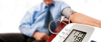
24-hour blood pressure monitoring
It is often necessary to diagnose a patient using the ABPM technique - 24-hour blood pressure monitoring. The essence of this technique is its automatic measurement throughout the day. For this purpose, a special device is used that is attached to the body of an adult patient or child. The results obtained in this way make it possible to assess the dynamics of pressure over a 24-hour period in those who have arterial hypertension or autonomic dysfunction.
Coronary angiography
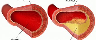
This method is often characterized in diagnostics as the “gold standard”, since it allows one to find out with high accuracy what the coronary arteries are, whether they have atherosclerotic narrowings, their length and other features. Taking into account all these important factors together, the doctor has the opportunity to clarify the condition of the patient’s heart and blood vessels, and then adjust the treatment process and optimize its tactics. Coronary angiography is not prescribed to all patients, but mainly only to those in whom the question of the need for surgical treatment of coronary heart disease, that is, bypass surgery or stenting, is being decided.
Coronary angiography of the heart vessels
The method is used both planned and urgently (for example, if a myocardial infarction is suspected). Due to the fact that it is a serious type of minimally invasive surgical intervention, its widespread use is not practiced. The essence of the method is that the patient is given a puncture in the femoral vessel or in the radial artery. Through this puncture, the doctor inserts a catheter into the bloodstream. Based on the resulting images, you can clearly analyze the real state in which the vessels are located.
Laboratory research methods for diseases of the heart and blood vessels. Their diagnostic value.
To diagnose and clarify the etiology of various diseases of the cardiovascular system, laboratory tests are of great importance.
Blood test.
Diagnosis of prolonged septic endocarditis is greatly facilitated by identifying leukopenia, anemia (the amount of hemoglobin decreases to 50-80 g/l, red blood cells - to 2-106-3-106) and hematuria; the latter may be in the nature of micro- and sometimes macrohematuria. In addition, if prolonged septic endocarditis is suspected, it is customary to perform blood cultures on nutrient media, as well as use the Bittorf-Tushinsky test, in which a large number of endothelial cells - histiocytes - are found in blood smears taken from the patient’s earlobe.
It is well known that myocardial infarctions from the first day of the disease are accompanied by neutrophilic leukocytosis (1-104—-1.2-104 or more) with a shift to the left, followed by a high ESR on the 3-4th day. Biochemical blood tests during myocardial infarction reveal high levels of aldolase and transaminase.
Atherosclerosis is characterized by hypercholesterolemia, and to clarify a syphilitic infection it is necessary to examine the blood for the Wasserman reaction.
Pericarditis, as a rule, occurs with leukocytosis and elevated ESR numbers.
In the presence of rheumatism (recurrent rheumatic carditis, endocarditis, vasculitis), ESR, along with other clinical signs, makes it possible to monitor the exacerbation, development and course of the rheumatic process; a slowdown in ESR usually indicates an improvement in the process.
In addition to ESR, the diagnosis of rheumatism is clarified by a formol test, as well as symptoms indicating increased vascular permeability.
Urine examination.
A healthy body excretes about 75-80% of the water introduced through food and drink through urine. With heart failure, some of the water does not reach the kidneys and is retained in the tissues, and the daily amount of urine decreases. In addition, with a functionally insufficient heart, nocturia is observed, i.e., urine excretion in significant quantities at night.
FKG. Principle of the method. Phonocardiogram analysis.
Phonocardiography is a method of recording sound phenomena arising in the heart during its activity. It is an essential addition to cardiac auscultation, as it allows you to register sounds that are not perceived by the human ear.
In phonocardiography, sound vibrations arising in the heart are recorded in the form of a curve - a phonocardiogram (PCG) using a phonocardiograph apparatus. It consists of a microphone, an amplifier, a system of frequency filters and a recording device.
The microphone perceives sound vibrations and turns them into electrical signals. The latter are amplified and transmitted to a system of frequency filters, which make it possible to separately record sound vibrations of a certain frequency: low-, mid- and high-frequency. Next, vibrations of a certain frequency are transmitted to a recording device, where they are recorded in the form of a curve on paper.
FCG is recorded in conditions of complete silence, in the patient’s supine position, while holding the breath in the exhalation phase. The microphone is alternately placed at those points on the chest where the heart valves are usually heard during auscultation, and additionally at those points in the chest where sound phenomena are most clearly expressed. Analysis of FCG and diagnostic conclusion on it are carried out only taking into account auscultatory data. For correct interpretation of the PCG, an ECG is simultaneously recorded simultaneously with it.
Normal PCG consists of oscillations reflecting the I and II heart sounds, between which there is a straight line corresponding to the systolic and diastolic pause (Fig. 61). During the diastolic pause, fluctuations caused by the third and fourth heart sounds are sometimes recorded.
Tone I is represented by several oscillations that occur after the Q wave of a synchronously recorded ECG. Its oscillation frequency is 70-150 Hz. Initial oscillations of the first tonal amplitude are associated with atrial systole. The main, central, part of the first tone is represented by two to three high-amplitude oscillations, which are determined at the level of the S wave and correspond to the vibrations of closed atrioventricular valves. Following the main part of the first tone, additional vibrations of lower amplitude are recorded, caused by vibration of the myocardium and the vascular component. The intensity of sound and, in particular, tone is determined by the amplitude of vibrations.
The amplitude of the first tone is highest at the apex of the heart, where it is 1 V2-2 times higher than the amplitude of the second tone; Based on the heart, the amplitude of the first tone can be very small. When assessing the first sound at the apex of the heart, attention is paid to how much its central part lags behind the Q wave of a synchronously recorded ECG. Normally, this interval Q - I tone does not exceed 0.04-0.06 s. It corresponds to the time between the onset of ventricular excitation and the closure of the mitral valve. When pressure in the left atrium increases (for example, with mitral stenosis), the mitral valve closes later, and the Q-I interval increases.
Tone II is represented by a group of oscillations appearing at the end of the G wave of a synchronous ECG. Its oscillation frequency is in the range of 70-150 Hz. The first higher oscillations correspond to the closure of the aortic valve, and the following ones, of lower amplitude, are due to the closure of the pulmonary valve. The amplitude of the second tone is highest at the base of the heart, where it exceeds the amplitude of the first tone.
FCG is of great help in diagnosing many diseases of the cardiovascular system and, first of all, heart defects. It allows you to clarify and supplement auscultation data. This is especially important in case of tachycardia, arrhythmias, when with the help of auscultation alone it is difficult to decide in which phase of the cardiac cycle certain sound phenomena occurred.
Dopplerography
Dopplerography has become an effective method for studying cardiac patients. This term defines a type of ultrasound examination of the body, through which it is possible to identify facts of blood flow disturbances in the vessels. The name of the method comes from a well-known physical effect. It consists of the peculiarities of ultrasonic waves, when reflected from moving objects, changing their frequency in proportion to the speed of movement of these objects. In this case, red blood cells serve as a reflective shield for sound waves. Depending on how they move and the speed of blood flow, a corresponding image appears on the monitor. It characterizes the condition of the blood vessels. In addition, using Doppler ultrasound, it is also possible to diagnose the condition of the blood vessels of such important human organs as the head, neck, spine, limbs, etc.
The procedure is carried out using an ultrasound machine equipped with a special sensor. Such a sensor sends waves through a person's skin into the tissue of his body. The data collected and summarized by the device eloquently characterizes the movement of blood. By combining them with information from a regular ultrasound, the machine creates an image that gives doctors reason to say whether there are blood flow problems or not. If there is a blockage, diagnosis allows you to accurately determine its size and other nuances. Let us note that the procedure itself is absolutely painless for the patient, does not pose any danger to him and, as a rule, takes no more than half an hour.
Aortography
Aortography has reliably proven itself as another effective modern diagnostic method. Here, the aorta is examined x-ray, obtaining a comprehensive image of the aorta after it is filled with a special contrast agent. The main role here is played by the radiation of radioactive waves: it is thanks to them that images are obtained that clearly show the state of the vessels filled with such a contrast agent. Cardiac aortography allows diagnosis when the patient’s heart has tumor diseases and other deviations from normal parameters.
It should be noted that in practice, in addition to those listed, other proven and well-proven methods for diagnosing cardiovascular diseases are successfully used. We are talking about radiography, which makes it possible to find dark spots in the patient’s lungs - evidence that the pulmonary circulation is marked by stagnation, magnetic resonance imaging, computed tomography and others. Therefore, in the event of any abnormalities in the functioning of the heart and blood vessels, we strongly advise you to immediately contact a doctor so that, with their assistance, you can undergo a full examination: such prevention of your health will never hurt!
Initial examination and collection of complaints
The initial stage of examining a patient with cardiovascular disease consists of collecting complaints, anamnesis and a physical examination by a doctor. These techniques are carried out in the emergency room by a cardiologist. First, he asks the patient about the presence and nature of complaints, their dynamics. How long do they persist in the patient, at what time of day they worsen and what are they provoked by.
An important step in diagnosis is collecting a family history. Based on it, the doctor assesses the hereditary nature of the pathology.
An objective (physical) examination is carried out in three stages - palpation, percussion (tapping) and auscultation. First, the cardiologist visually examines the patient for the presence of pathological pulsations, swelling, and changes in skin color. The specialist pays attention to height, weight, harmony of general development and condition, and features of respiratory movements. The main groups of lymph nodes, liver and spleen are examined by palpation. Liver enlargement may be a marker of blood stagnation in the systemic circulation due to valvular insufficiency or ventricular pathology.
After this, the doctor begins percussion - tapping the borders of the heart. Their displacement may indicate an increase in a certain part of the heart muscle. Such hypertrophy may result from overloading part of the heart. The third and most important stage of an objective examination is auscultation.
Using a stethoscope, the cardiologist listens to noises, tones, rhythm patterns, features of blood flow and valve function. The appearance of additional tones or noise may be a sign of valve defect, stenosis or insufficiency, or atherosclerosis of one of the vessels (for example, the aorta).
To comprehensively check the functioning of the heart and blood vessels, the doctor checks the pulse at several levels - the carotid arteries in the neck, the radial arteries in the wrist, the femoral arteries in the groin. If diabetes is present, the pulsations of the arteries of the feet are also checked to evaluate diabetic angiopathy (vascular degeneration). Pulse measurement must be symmetrical - on both sides.