Coding of atrial fibrillation in the ICD
Atrial fibrillation is a disorder of normal heart rhythm, which is characterized by rapid, erratic excitation and contraction of the myocardium.
I 49.0 – according to ICD 10, the code for atrial fibrillation, which belongs to class IX “Diseases of the circulatory system”. Normally, in a healthy person, with each contraction of the heart, the atria should first contract, and then the ventricles. Only in this way is it possible to adequately ensure hemodynamics. If this rhythm is disturbed, arrhythmic and asynchronous contraction of the atria occurs, and the functioning of the ventricles is disrupted. Such fibrillations lead to exhaustion of the heart muscle, which can no longer work effectively. Restrictive and then dilated cardiomyopathy may develop.
Heart rhythm disturbances in ICD 10 are coded as follows:
- I 49.0 – “Ventricular fibrillation and flutter”;
- I 49.1 – “Premature contraction of the ventricles”;
- I 49.2 – “Premature depolarization emanating from the junction”;
- I 49.3 – “Premature atrial depolarization”;
- I 49.4 – “Other, unspecified premature reductions”;
- I 49.5 – “Sick sinus syndrome”;
- I 49.7 – “Other specified heart rhythm disturbances”;
- I 49.8 – “Heart rhythm disturbances, unspecified.”
In accordance with the established diagnosis, the necessary code is indicated on the title page of the medical history. This encryption is the official and uniform standard for all medical institutions; it is used in the future to obtain statistical data on the prevalence of mortality and morbidity from specific nosological units, which has prognostic and practical significance.
Disease prevention
One of the most important dangers with this disease is the likelihood of blood clots forming in the vessels, which increases the likelihood that you will have a stroke. But regardless of the type of disease, treatment should be carried out in a hospital.
Traditional methods
It is very important after discovering this disease to switch exclusively to a healthy lifestyle . This includes proper nutrition, elimination of all bad habits and other clinical recommendations. Also, in some situations, a diet is prescribed to reduce the amount of animal fats.
Physical activity in this case can have both positive and negative properties. Excessive exercise in this case is prohibited, but moderate exercise in the morning is approved.
Bad habits, as already stated above, must be eliminated as much as possible.
In order to eliminate all possible attacks, you need to improve your emotional background and completely protect yourself from stress . In most cases, for this purpose they often resort to the use of certain medications.
You need to do everything to regain your normal body weight. Unconventional methods
Non-traditional methods include the use of traditional medicine, in particular herbal remedies. The recipes in such cases are very simple and accessible to everyone. You can take 5 grams of hawthorn and pour boiling water over it. Let this infusion simmer for about 15 minutes. After the tincture has cooled, you need to strain it and remove all excess from it. Use the medication half an hour before meals.
You can also use lovage, combining it with hawthorn and calendula. All this is filled with boiling water. The tincture should be used 6-7 times a day, one spoon.
Reasons for the development of rhythm pathology
Atrial fibrillation can occur for various reasons, but the most common are:
- congenital and acquired heart defects;
- infectious myocarditis (bacterial, viral, fungal heart disease);
- IBS atrial fibrillation (usually as a serious complication of acute myocardial infarction);
- hyperproduction of thyroid hormones - thyroxine and triiodothyronine, which have an inotropic effect;
- drinking large amounts of alcohol;
- as a consequence of surgical interventions or invasive research methods (for example, fibrogastroduodenoscopy);
- arrhythmias after strokes;
- when exposed to acute or chronic stress;
- in the presence of dysmetabolic syndrome - obesity, arterial hypertension, diabetes mellitus, dyslipidemia.
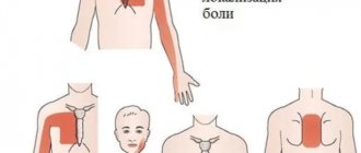
Attacks of arrhythmia are usually accompanied by a feeling of interruptions in the heart and an arrhythmic pulse. Although often a person may not feel anything, in such cases the diagnosis of pathology will be based on ECG data.
OTHER HEART DISEASES (I30-I52)
Included: acute pericardial effusion
Excludes: rheumatic pericarditis (acute) (I01.0)
Excluded:
- some current complications of acute myocardial infarction (I23.-)
- postcardiotonic syndrome (I97.0)
- heart injury (S26.-)
- diseases specified as rheumatic (I09.2)
Excluded:
- acute rheumatic endocarditis (I01.1)
- endocarditis NOS (I38)
Excluded: mitral (valvular):
- disease (I05.9)
- deficiency (I05.8)
- stenosis (I05.0)
for an unknown reason, but mentioning it
- aortic valve disease (I08.0)
- mitral stenosis or obstruction (I05.0)
lesions specified as congenital (Q23.2-Q23.9)
lesions specified as rheumatic (I05.-)
Excluded:
- hypertrophic subaortic stenosis (I42.1)
- for unknown cause, but with mention of mitral valve disease (I08.0)
- lesions specified as congenital (Q23.0, Q23.1, Q23.4-Q23.9)
- lesions specified as rheumatic (I06.-)
Excluded:
- without specifying the reason (I07.-)
- lesions specified as congenital (Q22.4, Q22.8, Q22.9)
- specified as rheumatic (I07.-)
Excluded:
- lesions specified as congenital (Q22.1, Q22.2, Q22.3)
- disorders specified as rheumatic (I09.8)
Endocarditis (chronic) NOS
Excluded:
- endocardial fibroelastosis (I42.4)
- cases specified as rheumatic (I09.1)
- congenital aortic valve insufficiency NOS (Q24.8)
- congenital aortic valve stenosis NOS (Q24.8)
Included: endocardial damage due to:
- candida infection (B37.6+)
- gonococcal infection (A54.8+)
- Libman-Sachs disease (M32.1+)
- meningococcal infection (A39.5+)
- rheumatoid arthritis (M05.3+)
- syphilis (A52.0+)
- tuberculosis (A18.8+)
- typhoid fever (A01.0+)
Excluded:
- cardiomyopathy, complicating: pregnancy (O99.4)
- postpartum period (O90.3)
Excluded:
- cardiogenic shock (R57.0)
- complicating: abortion, ectopic or molar pregnancy (O00-O07, O08.8)
- obstetric surgical interventions and procedures (O75.4)
Excluded:
- complicating: abortion, ectopic or molar pregnancy (O00-O07, O08.8)
- obstetric surgical interventions and procedures (O75.4)
- NOS (R00.0)
Read also: What medications can cause arrhythmia
Excluded:
- bradycardia: NOS (R00.1)
- sinoatrial (R00.1)
- sinus (R00.1)
- vagal (R00.1)
- abortion, ectopic or molar pregnancy (O00-O07, O08.8)
Excluded:
- conditions complicating: abortion, ectopic or molar pregnancy (O00-O07, O08.8)
- obstetric surgical interventions and procedures (O75.4)
- kidney disease (I13.-)
Excluded:
- any conditions listed in sections I51.4-I51.9 due to hypertension (I11.-)
- any conditions listed in sections I51.4-I51.9 due to hypertension with renal disease (I13.-)
Excludes: cardiovascular disorders NOS in diseases classified elsewhere (I98*)
In Russia, the International Classification of Diseases
10th revision (
ICD-10
) was adopted as a single normative document for recording morbidity, reasons for the population’s visits to medical institutions of all departments, and causes of death.
ICD-10
introduced into healthcare practice throughout the Russian Federation in 1999 by order of the Russian Ministry of Health dated May 27, 1997. No. 170
The release of the new revision (ICD-11) is planned by WHO in 2022.
Consequences of arrhythmia
Atrial fibrillation in ICD 10 is quite common and has a poor prognosis, subject to inadequate monitoring and treatment. The disease can be complicated by the formation of blood clots and the development of chronic heart failure.
Arrhythmia is especially dangerous in coronary heart disease, arterial hypertension and diabetes mellitus - in these cases, thromboembolism can lead to cardiac arrest, heart attack or stroke.
Heart failure can develop quite quickly and manifest itself as hypertrophy of the myocardial walls, which will aggravate existing ischemia. Arrhythmia in ICD 10 is a common complication of acute myocardial infarction, which can be a direct cause of death. The above facts indicate the seriousness of the disease and show the need for constant and correct therapy. All kinds of antiarrhythmic drugs, potassium-containing drugs, and antihypertensive drugs are used for treatment. Great importance is given to taking anticoagulants and antiplatelet agents. Warfarin and acetylsalicylic acid are used for these purposes - they prevent the development of blood clots and change the rheology of the blood. It is very important to establish the primary cause of the development of atrial fibrillation and block its action in order to prevent all sorts of complications.
Save the link, or share useful information on social media. networks
What to do during an attack?
The patient and his relatives should know what to do if a paroxysm occurs. The following procedures help to completely remove or reduce the intensity of the painful condition:
- abdominal compression;
- holding your breath;
- pressing on the eyeballs.
At the same time, it is necessary to call an ambulance. The doctor injects the patient intravenously with Korglikon, Strophanthin, and the drugs Ritmilen, Aymalin or Novocainamide. Sometimes an attack is relieved by intravenous administration of potassium chloride.
Read also: Yoga for arrhythmia
In terms of prognosis, arrhythmias are extremely ambiguous; it is recommended to limit the intake of stimulants (caffeine), avoid smoking and alcohol, and independently select antiarrhythmic and other drugs
The prognosis for treatment of paroxysmal atrial fibrillation depends on the disease that caused the disturbance in the rhythm of atrial contraction.
Lack of therapy and failure to provide timely assistance to a patient during an attack of paroxysmal atrial fibrillation can result in the development of dangerous conditions that lead to death.
Content
What is flicker and flutter
Reasons for development
Classification
Clinical picture
Complications
Diagnostics
Treatment
To optimize international disease statistics, the World Health Organization created the International Classification of Diseases (ICD). Doctors use the tenth revision of the edition. In the category of cardiovascular pathology, atrial fibrillation is listed under the name “atrial fibrillation and flutter” (ICD 10 code - I 48).
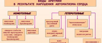
Arrhythmia ICD code: I 44 - I 49 - disturbance of the speed of heart contractions, their regularity as a result of functional or organic damage to specialized conducting myocardiocytes. Under normal conditions, electrical impulses are transmitted from the sinus node to the atrioventricular node and to the fibers of the heart muscle through bundles of conducting muscle fibers.
Damage can affect any of these structures and is manifested by characteristic changes in the ECG line and clinical picture. Most often, sinus arrhythmia develops with regular heart contractions (ICD 10 code - I 49.8).
What is flicker and flutter
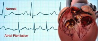
Atrial fibrillation is a disordered contraction of the atria with varying levels of blood filling during diastole. Most of the conductive waves, due to their large number, do not propagate to the ventricular myocardium.
A circular conduction wave causes atrial flutter with a contraction rate ranging from 0 to 350 per minute. This condition occurs 30 times less frequently than flickering. Waves during flutter can reach the conduction system of the ventricles, causing them to contract in the correct or incorrect rhythm.
Depending on the speed of the heart rate, atrial fibrillation can be bradysystolic (with a slowdown of the rhythm below 60 beats), normosystolic (from 60 to 90 beats per minute) and tachysystolic (over 90 beats).
Reasons for development
Heart rhythm disturbances in the form of atrial fibrillation develop as a result of morphological changes in the myocardial conduction system, with endogenous and exogenous intoxication, and some other diseases. A rare option is idiopathic (causeless) atrial fibrillation, when a visible background for its development has not been established.
Diagnostics
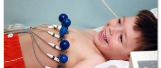
The primary stage of diagnosing arrhythmia can be carried out by a therapist or cardiologist using an electrocardiogram
Atrial fibrillation is a sign of serious illness. If a person has atrial fibrillation, they may need emergency care. However, to carry out the necessary therapy, the correct diagnosis must be established.
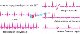
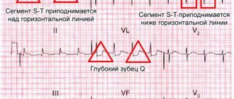
The most important method for diagnosing paroxysmal atrial fibrillation is electrocardiography. The ECG shows the main signs indicating the disease.
Holter monitoring, exercise tests, listening to heart sounds with a phonendoscope, ultrasound and ECHO CG are used as auxiliary diagnostic methods.
Read also: Love arrhythmia read online
Only a competent specialist can prescribe the correct treatment. For paroxysmal atrial fibrillation, it may be necessary to use different techniques. They are selected individually for each patient.
The choice of treatment method directly depends on the duration of paroxysms and the frequency of their occurrence.
If atrial fibrillation bothers a person for no more than 2 days, then doctors take measures to restore sinus rhythm. At a later stage, treatment is required to help prevent the development of life-threatening complications.
In difficult situations, the patient is prescribed therapy, the main goal of which is to restore the correct rhythm of atrial contractions. Additionally, you need to take medications that can thin the blood.
Drug treatment
Class III antiarrhythmic drug, has antiarrhythmic and antianginal effects
Paroxysmal heart rhythm disturbances, which affect the entire cardiovascular system, can be combated using medications. To reduce heart rate and restore disturbed rhythm. the drug Cordarone is used. It has a minimal number of side effects, so it is suitable for treatment for most patients.
When diagnosed with atrial fibrillation, Novocainamide is often prescribed. The drug is slowly introduced into the human body. During the procedure, it is forbidden to rush, as the injection can sharply reduce blood pressure, thereby aggravating the situation. In some cases, Digoxin is prescribed, which is able to control ventricular contraction.
If the prescribed drug showed a good result for the first time, then when using it for a new attack you should not expect the same effect. Each time the effect of the medication will weaken.
Electropulse therapy
Electropulse therapy is used to treat atrial fibrillation, the procedure is carried out in the clinic in one day, the patient should not eat anything for 6 hours before the session.
To eliminate attacks of arrhythmia, a method of electrical pulse treatment was developed. It is prescribed if the medication course does not give the expected result. Electrical discharge is indicated for patients who have developed complications due to another paroxysm.
Electropulse treatment is performed according to the standard scheme:
- Initially, the patient is put into a state of medicated sleep and anesthesia (the procedure is characterized by high pain).
- 2 electrodes are installed on his chest area.
- Next, you need to set the required mode, which corresponds to the category of atrial contractions;
- All that remains is to set the current indicator and carry out the discharge.
After the discharge, the heart begins its work again. From now on, its functions are performed a little differently. The electric current “recharges” the conduction system, which is why it is forced to begin sending rhythmic impulses of excitation to the sinus node.
Practice shows that this treatment option in most cases guarantees a positive result.
Surgical intervention
If attacks of the disease occur too often, the patient will require surgical intervention. It is used to relieve symptoms of pathology and eliminate its cause. Thanks to this method, attacks of arrhythmia are stopped, since the surgeon destroys the source of pathological excitation in the heart.
Relieving paroxysm and preventing new attacks is the main goal of the operation.
Surgery (catheter ablation) is performed using a catheter that is inserted through an artery. If necessary, the operation is repeated after a certain period of time.
general information
Short description
Currently, cardiomyopathies are classified mainly by pathophysiology or, if possible, by etiological and pathogenetic factors.
Cardiomyopathies are defined as diseases of the myocardium associated with its dysfunction. They are divided into hypertrophic, dilated and restrictive cardiomyopathies and arrhythmogenic right ventricular cardiomyopathy (WHO, 1995).
Protocol code : PT-026 “Cardiomyopathy” (Part II. Heart rhythm disturbances*) Profile: therapeutic Stage: PHC
II. Heart rhythm disturbances (Atrial fibrillation. Ventricular cardiac arrhythmias. Sudden cardiac death)
(Guidelines of the American College of Cardiology, American Heart Association, European Society of Cardiology - 2001)
– Professional medical reference books. Standards of treatment
– Communication with patients: questions, feedback, making an appointment
Download the application for ANDROID
– Professional medical reference books
– Communication with patients: questions, feedback, making an appointment
Download the application for ANDROID
Classification
Classification of cardiomyopathies (World Heart Federation, 1995)
– associated with recognized cardiovascular disease.
Clinic: asymptomatic or shortness of breath, chest pain (coronary syndrome), syncope or presyncope and palpitations. Arrhythmias and VS are typical.
3. Restrictive cardiomyopathy. Definition: characterized by impaired filling and reduced diastolic volume of one or both ventricles with normal or nearly normal systolic function and wall thickness, massive interstitial fibrosis may be present: – idiopathic;
Ischemic cardiomyopathy is a dilated cardiomyopathy with impaired contractile properties that is not explained by extensive coronary artery disease or ischemic injury.
Valvular cardiomyopathy presents with impaired ventricular function out of proportion to changes in load.
Hypertensive is often present with left ventricular hypertrophy and is accompanied by manifestations of dilated or restrictive cardiomyopathy and heart failure.
II. Heart rhythm disturbances
Atrial fibrillation
PVCs of classes 3-5 are called high grade PVCs and have an unfavorable prognosis.
1. Unsustained ventricular tachycardia (VT) – three or more ventricular complexes following each other, lasting less than 30 seconds. with a ventricular contraction frequency of more than 100 beats/min. (cycle duration less than 600 ms).
2. Sustained VT – VT lasting more than 30 seconds. or requiring resuscitation measures.
5. VT reentry according to the type of bundle branch block . VT from the His-Purkinje system, most often in the form of left bundle branch block, complicates cardiomyopathies.
6. Bidirectional fusiform VT (torsades de pointes) - polymorphic VT, having the form of a slow polymorphic ventricular flutter without discernible QRS complexes or T waves. Ventricular activity is characterized by a constantly changing amplitude, as if rotating around an isoelectric line. Associated with long QT syndrome.
8. Ventricular fibrillation is rapid and completely disorganized ventricular activity without discernible QRS complexes or T waves on the ECG.
CHAPTER 18. DISORDERS OF RHYTHM AND CONDUCTION OF THE HEART
SUPRAVENTRICULAR ARRHYTHMIAS
SUPRAVENTRICULAR EXTRASYSTOLE
SYNONYMS
DEFINITION
Supraventricular extrasystole is a premature excitation and contraction of the heart relative to the main rhythm (usually sinus), caused by an electrical impulse that occurs above the level of the branching of the His bundle (i.e. in the atria, AV node, trunk of the His bundle). Repeated supraventricular extrasystoles are called supraventricular extrasystoles.
ICD-10 CODE
EPIDEMIOLOGY
The frequency of detection of supraventricular extrasystole in healthy people during the day ranges from 43 to 91-100% and increases slightly with age; frequent supraventricular extrasystole (more than 30 per hour) occurs only in 2-5% of healthy people.
PREVENTION
Prevention is mainly secondary and consists of eliminating extra-cardiac causes and treating heart diseases that lead to supraventricular extrasystole.
SCREENING
Active detection of supraventricular extrasystole is carried out in patients with potentially high significance or in the presence of typical complaints using ECG and Holter ECG monitoring throughout the day.
CLASSIFICATION
There is no prognostic classification of supraventricular extrasystole. Supraventricular extrasystole can be classified:
• by frequency of occurrence: frequent (more than 30 per hour, i.e. more than 720 per day) and rare (less than 30 per hour);
• according to the regularity of occurrence: bigeminy (every 2nd impulse is premature), trigeminy (every 3rd), quadrigeminy (every 4th); in general, these forms of supraventricular extrasystole are called allorhythmia;
• by the number of extrasystoles occurring in a row: paired supraventricular extrasystoles or couplets (two supraventricular extrasystoles in a row), triplets (three supraventricular extrasystoles in a row), while the latter are regarded as episodes of unstable supraventricular tachycardia;
Registration is required to continue.