Characteristics of pulse value
To understand what the pulse value is, let us remind you of its two other characteristics:
- Filling;
- Voltage.
The filling of the pulse depends on the amount of blood that is ejected from the heart into the aorta during its contraction. This indicator (cardiac output) is determined by the contractility of the heart muscle and heart rate. With poor contractility or tachycardia, the pulse will have reduced filling; with normal values of these values, the pulse will have good filling. In addition, this property may change with a decrease in the volume of blood circulating in the vessels. For example, with severe acute blood loss, an “empty” pulse may be detected.
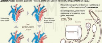
Tension is the force of blood pressure on the vascular wall. With high blood pressure the pulse is tense, with low blood pressure it is soft or of low tension.
What does the heart rate depend on:
- on blood pressure level;
- on the strength of contractions of the heart muscle;
- on heart rate;
- on the volume of blood in the vascular bed.
This figure increases with hypertension, especially if high blood pressure is combined with a low pulse. With arrhythmias, the magnitude of pulse waves decreases, as the volume of blood that is ejected by the heart in one defective contraction decreases.
Rhythmic heartbeat and pulse - what is it?
For example, with atrial fibrillation (atrial fibrillation), a phenomenon called pulse deficiency may be observed: its indicator, determined on the radial artery in the wrist, will be less than the heart rate. With a very fast heartbeat, which occurs with the tachysystolic form of atrial fibrillation, during some myocardial contractions, practically no blood is ejected into the aorta, and the pulse value is very small. Such a pulse wave will not be detected in a peripheral vessel.
How to determine the pulse value? You can learn to do this only experimentally, by comparing the quality of pulse waves in people with different heart diseases.
Normally, some force must be applied to clamp the radial artery while palpating the pulse. If the pulse value is large, then this is almost impossible to do. When the pulse waves are small, it is very easy to compress the artery and stop the blood flow.
Contraindications
Due to a sharp increase in blood pressure and stress on the heart, muscles, joints and spine, a VEM examination is difficult to carry out or dangerous to health. All contraindications are divided into absolute and relative.
The absolute ones include:
- suspicion of acute myocardial infarction;
- acute myocardial infarction on ECG;
- severe cardiac or respiratory failure (in the final stages);
- cerebrovascular accident;
- unstable angina (progressive blockage of the coronary arteries);
- vascular aneurysm (sac-like expansion that threatens to rupture);
- sharp narrowing of the aortic valve;
- acute thrombophlebitis (inflammation of the veins with the formation of blood clots);
- any acute infectious disease.
Relative ones include:
- heart rhythm disturbances (100-160 heart beats per minute, life-threatening arrhythmias);
- severe arterial hypertension (pressure above 160/100 mm Hg);
- frequent fainting;
- mental illness;
- diseases of the nervous system;
- pathology of joints and muscles.
Important: If there are contraindications to bicycle ergometry, there is another device for diagnosing coronary artery disease - the Holter. It allows you to record ECG throughout the day in the patient’s usual mode of physical activity.
Pulse value (frequency)
A normal pulse can take on different values depending on the level of physical activity, blood pressure, state of wakefulness or sleep, age, and concomitant diseases.
What should be the normal heart rate for a healthy person at rest: according to WHO standards, this figure ranges from 60 to 100 beats per minute. In some sources, 90 per minute is taken as the upper limit of the norm. This was what was believed in previous years in the medicine of the Soviet Union.
In adult women and men, the normal values are the same. Children have a natural increase in resting heart rate. The younger the child, the higher his heart rate.
Pulse in children by age: tables of heart rate norms and reasons for heart rate deviations from it
Maximum and submaximal value
The maximum pulse value is considered by cardiologists to be the limit after which even a healthy person may experience insufficient blood supply to the heart muscle due to too short a period of myocardial relaxation (short diastole).
The arteries supplying the myocardium fill with blood precisely at this time, when the heart muscle is relaxed and does not compress them. When the pulse value exceeds the maximum, blood simply does not have time to pass into the vessels of the heart.
The maximum value is defined as 220 – age in years.
The submaximal frequency (“magnitude” – such a replacement of terms will not be entirely correct) of the pulse is 75–80% of the maximum.
The submaximal rate is also called the “should” rate (SHR).
This is the highest heart rate at which a healthy person never experiences insufficient blood supply to the myocardium.
On the contrary, in patients with coronary artery disease, an increase in pulse to a submaximal frequency is often accompanied by signs of ischemia of the heart muscle.
Therefore, it is the submaximal, or proper, pulse value that is the upper limit of the norm to which the heart is “accelerated” during ECG tests with physical activity (bicycle ergometry or treadmill test) to diagnose angina.
In this case, an additional correction of the required pulse value is carried out towards an increase, since the test is performed under constant ECG monitoring.
Sizing chart for children
Table with maximum heart rate values for children by age:
Age, years Maximum safe heart rate
| 0 – 5 | 215 – 220 beats. in a minute |
| 6 – 10 | 210 – 214 beats. in a minute |
| 11 – 15 | 205 – 209 beats. in a minute |
| 16 – 18 | 202 – 204 beats. in per minute |
If a child plays sports, a coach or sports doctor regularly checks the heart rate to ensure that it does not exceed the maximum value.
If a child does not train specifically, there is no particular need to evaluate his pulse during exercise. It is enough to ensure that after active games or running he does not have the following signs:
- prolonged shortness of breath;
- attack of dry cough;
- blue discoloration of the nasolabial triangle;
- weakness or fainting.
If you suspect poor exercise tolerance, the child should be shown to a pediatric cardiologist, have an ECG done, and, if necessary, conduct 24-hour ECG monitoring and echocardiography (ultrasound of the heart).
Size tables for adults
Maximum and submaximal heart rate values in healthy adults in the table by age:
Age, yearsMaximum valueSubmaximum value
| 30 – 35 | 185 | 148 |
| 36 – 40 | 180 | 144 |
| 41 – 45 | 175 | 140 |
| 46 – 50 | 170 | 136 |
| 51 – 55 | 165 | 132 |
| 56 – 60 | 160 | 128 |
| 61 – 65 | 155 | 124 |
| 66 and older | 150 | 120 |
The submaximal value is the upper limit of normal during physical activity.
Table of values after a heart attack
Submaximal (proper) pulse value in patients who have had myocardial infarction by year:
Age, yearsMenWomen
| 35 – 40 | 141 | 136 |
| 41 – 45 | 136 | 132 |
| 46 – 50 | 132 | 128 |
| 51 – 55 | 128 | 123 |
| 56 – 60 | 123 | 118 |
| 65 and older | 118 | 115 |
Myocardial infarction: symptoms, forms and standards of treatment after surgery
When working at submaximal intensity, both healthy and sick adults are not recommended to exceed the required heart rate for normal blood supply to the heart.
Myocardial hypertrophy
With chronic overload (pressure, volume), the heart muscle in certain areas begins to thicken, and the chambers of the heart begin to stretch. On the ECG, such changes are usually described as hypertrophy.
- Left ventricular hypertrophy (LVH) is typical for arterial hypertension, cardiomyopathy, and a number of heart defects. But even normally, athletes, obese patients and people engaged in heavy physical labor may experience signs of LVH.
- Right ventricular hypertrophy is an undoubted sign of increased pressure in the pulmonary blood flow system. Chronic cor pulmonale, obstructive pulmonary diseases, cardiac defects (pulmonary stenosis, tetralogy of Fallot, ventricular septal defect) lead to RVH.
- Left atrial hypertrophy (LAH) – with mitral and aortic stenosis or insufficiency, hypertension, cardiomyopathy, after myocarditis.
- Right atrial hypertrophy (RAH) – with cor pulmonale, tricuspid valve defects, chest deformities, pulmonary pathologies and PE.
- Indirect signs of ventricular hypertrophy are deviation of the electrical axis of the heart (EOC) to the right or left. The left type of EOS is its deviation to the left, that is, LVH, the right type is RVH.
- Systolic overload is also evidence of hypertrophy of the heart. Less commonly, this is evidence of ischemia (in the presence of angina pain).
A pathological Q wave is characterized by an increase in its amplitude by more than 1/4 of the amplitude of the R wave in the same lead and an increase in its duration by more than 0.03 seconds. The presence of a pathological Q wave may indicate myocardial ischemia.
The P wave characterizes the passage of the sinus impulse through the conduction system of the atria, an increase in height of more than 0.4 mV, pointedness - hypertrophy of the right atrium, observed more often in diseases of the lungs (P-pulmonale), expansion of more than 0.04 seconds, double hump - hypertrophy of the left atrium is observed more often for mitral defects (P-mitrale).
The QRS complex characterizes the passage of a sinus impulse through the ventricular conduction system
Left ventricular hypertrophy (LVH) is accompanied by an increase in the amplitude of the R wave over 2.5-3.0 mV in leads II and aVF, an increase in the duration of the QRS complex to 0.05-0.06 seconds, and a decrease in the ST segment below the isoline may also be observed, The EOS may be shifted to the left. what if RI{amp}gt;RII or RI{amp}gt; RaVF is hypertrophy, and an increase in RI, RII, RIII is dilatation
Right ventricular hypertrophy (RVH): displacement of the EOS to the right, the presence of an S wave in leads I, II, III and aVF, signs of right atrium hypertrophy, an increase in the amplitude of the Q wave by more than 0.5 mV in leads II, III and aVF.
Low QRS complexes: obese animals, fluid accumulation in the pericardium, pleura, ascites, hypothyroidism, hyperkalemia, pneumothorax, some respiratory diseases, external and internal fluid loss - hypovolemia, normal variant (in cats).
Alternating high and low QRS complexes are observed when fluid accumulates in the pericardium. This should not be confused with a gradual increase in amplitude during inspiration.
Notches on the R waves indicate bundle branch block if not an artifact.
How is the test performed?
The test is carried out in laboratory conditions. Everyone is given time to bring the cardiovascular system into stable tension. It takes everyone a different amount of time. Participants are constantly monitored and their heart rate is measured, 2 minutes after the start and at the 3rd minute. If the difference is more than 5 beats, then another approach is performed and so on until a stable result is achieved - less than 5 beats.
It is especially important that the pulse is within the range of 115-150 beats; these are optimal indicators for the ratio of heart contractions and oxygen consumed by the muscles.
Purpose of the study
A test has been developed that shows the endurance and physical fitness of an athlete. Under constant loads, the coach understands what the limit point is for the athlete. This study is carried out to avoid occupational injuries. The fact is that after excessive exercise, muscles stop fully absorbing oxygen and destruction of cells, tissues, and vessel walls occurs. If an athlete does 3 approaches and his heart rate drops, it means that his physical fitness is at a high level and his body is not under stress.
Preparation
Bicycle ergometry is carried out in the functional diagnostics room. It is equipped in such a way that it has everything necessary to provide assistance if the patient suddenly becomes ill during the study. In addition to medications, there is a defibrillator in the office, which is used to provide emergency assistance in case of sudden cardiac arrhythmia or cardiac arrest.
To carry out VEM you need:
- exercise bike;
- electrocardiograph for recording ECG;
- manometer for measuring blood pressure;
- phonendoscope for listening to heart sounds.
The VEM study is scheduled in the first half of the day. The patient must come to the test hungry. On the day of the study, you should not smoke, be nervous, and do not donate blood for tests.
For VEM, the patient is prepared as follows:
- 2-3 days before the test, stop taking beta blockers (drugs that slow the heart rate and lower blood pressure);
- the day before the test, stop taking nitroglycerin, which dilates the coronary vessels, and drugs that lower blood pressure (including diuretics);
- 12 hours before the test does not perform physical activity.
You cannot stop taking medications on your own, because this can be dangerous to health, and in addition, the doctor could prescribe a VEM to determine the effectiveness of the drugs. Therefore, if the patient has not been warned about changing medications, you should consult a doctor.
How is it determined?
To determine the submaximal pulse, equipment is used that shows the number of contractions of the heart muscle, but this study does not give results with 100% accuracy. There is a complex mathematical formula that calculates a person’s submaximal endurance, but there is also a simplified version of the calculation. To do this, you should take a fixed maximum heart rate of 220 and subtract the athlete’s age; the data obtained is the maximum permissible heart rate. Other indicators are also taken into account.
What is a joint venture?
Submaximal heart rate is a test that all professional track and field athletes undergo to objectively assess their maximum capabilities. The procedure is carried out in laboratory conditions and indicates how much oxygen the body is ready to process in order to synthesize ATP in muscle tissue. The results are calculated based on the maximum heart rate - 85% for a healthy person and 70-75% for a person with heart problems.
Sources
- https://PulsNorma.ru/izuchenie-pulsa/velichina-pulsa.html
- https://s-storm.ru/submaksimalnaja-chss-dostignuta-chto-jeto-znachit/
- https://gupmok.ru/submaksimalnaja-chss-dostignuta-chto-jeto-znachit/
[collapse]
Indications for performing submaximal stress testing
Submaximal stress test is indicated for:
- atypical chest pain;
- nonspecific changes in the ECG taken at rest, in the absence of pain or its atypical nature;
- lipid metabolism disorders;
- mass epidemiological studies of the population and preventive examinations of healthy people;
- selection and evaluation of the effectiveness of therapeutic and rehabilitation measures;
- determination of tolerance to physical activity.