Causes of development and treatment of sural vein thrombosis
Thrombophlebitis is a common problem in modern society.
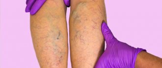
The pathological condition is characterized by the appearance of a coagulated blood clot that blocks the arterial lumen. As a result, blood circulation is impaired. Thrombosis of the sural veins is the main consequence of this disorder. According to most patient complaints, the appearance of blood clots occurs in the sural veins. At rest, the muscle tissue of the calves relaxes, and the sural veins fill with blood. On the contrary, contraction of muscle tissue in the calf area promotes blood flow. Such blood circulation in the venous vessels of the legs is considered natural and normal. Thrombosis of the sural veins of the lower extremities is more common in old age. The appearance of thrombosis at a younger age is facilitated by severe injuries, surgical operations, pregnancy, smoking, excess weight, and prolonged bed rest.
Forecasts and prevention of pathology
If you do not treat the pathology, the blood clot will break off and enter the pulmonary artery, and this will be quickly fatal. When the veins are completely clogged, a heart attack and stroke may occur, as well as other diseases that can make a person disabled.
When the disease arose due to temporary injuries, then after treatment and rehabilitation the pathology will never make itself felt. But, if the main cause has not disappeared even after treatment, then the disease may remind itself again. Non-occlusive deep vein thrombosis of the lower extremities is also dangerous, but its treatment is much easier.
Of course, it is better to prevent a disease than to fight it. But such a serious pathology can claim life, so you need to carefully consider preventive measures. In order to avoid the risks of illness, the time period when it is in one position should be reduced so that stagnation does not form. During this position, it is important to constantly warm up. This will prevent the occurrence of stagnation, which provokes the occurrence of blood clots.
In addition, you should stop wearing clothes that are tight and uncomfortable. This rule also applies to shoes. Walking in the fresh air before bed can also be an excellent preventive measure. You need to go on a special diet to normalize blood clotting and metabolic processes. This will not only improve the condition of blood vessels, but also reduce weight. It is important to establish a drinking regimen that helps thin the blood. Foods that retain fluid in the body should be eliminated, as this can lead to edema.
By adhering to these simple rules, you can reduce the risk of such a disease.
The causes and methods of treating thrombosis are described in the video in this article.
Clinical picture of the disease
At the initial stage of development, thrombophlebitis of the sural veins is asymptomatic. For a long time, the pathology of the sural veins does not manifest itself externally, although the blood circulation process in the walls of the vessels already has disruptions.
The progression of the disease gradually causes the death of tissue particles that are no longer fully supplied with the necessary nutrients. Danger arises from cases where a formed blood clot is rejected from the walls of a vessel and travels through the blood to other organs. It is for this reason that doctors strongly recommend undergoing medical examinations on time.
Main symptoms of thrombosis
There are a number of characteristic signs indicating damage to this type of pathology. At first, the clinical picture is blurred, and it is very difficult to discern the true causes of certain symptoms. In addition, the signs directly depend on the spread of the blood clot, the degree of damage, and location.
At the initial stage, the symptoms can be confused with banal fatigue, because the manifestations are minor and do not bring particularly noticeable discomfort.
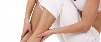
After a certain time, pain in the calf muscles is observed, at the next stage you may notice increased swelling of the leg, or both limbs. This may continue for several days. Other manifestations of the disorder include:
- aching and bursting pain, especially when bending the leg;
- pain when walking, sometimes walking becomes impossible;
- constant swelling of the legs;
- enlarged veins;
- change in skin color. It may turn blue;
- burning and itching of the limb;
- heat;
- change in vein density.
Also, at a certain stage, the feet often swell, to which the patient pays special attention. Even at the beginning of the disease, if you touch the affected skin, you can feel pain, and quite severe.
A pronounced clinical picture is observed when the clot fills the space more and more. Over time, the severity of symptoms increases.
Accompanying chest pain can be a fairly serious manifestation. This may be a sign of a pulmonary embolism.
Forms of thrombophlebitis of the lower extremities
Thrombosis can take on various forms, each with its own characteristics and duration.
- Acute thrombophlebitis. It is characterized by a sudden appearance and a duration of thirty days.
- Subacute. It is recognizable by its long-term clinical symptoms, which last up to six months.
- Chronic thrombophlebitis is expressed by the appearance of painful sensations in the veins of the legs, which can be caused by various factors and causes.
- Migratory. This form is characterized by the appearance of periodic signs of thrombophlebitis of the lower extremities.
Based on the location of vessels and veins susceptible to thrombophlebitis, the following are distinguished:
According to the level of closure of the venous lumen, thrombosis is divided into:
The formation of blood clots in occlusive thrombophlebitis of the deep veins of the legs is an absolute blockage of the venous lumen. In this condition, there is a risk of absolute cessation of blood circulation. Professional medical procedures are urgently required to avoid the progression of further spread of the disease.
The form of non-occlusive thrombosis is caused by the formation of floating thrombi. In such cases, the clots are concentrated at the base of the vessel, as they are a parietal type of thrombus. With this form, blood flow is not impaired, because the free venous area is washed with blood and has no significant obstacles.
Since most often thrombosis of the sural veins of the lower extremities is asymptomatic, it is rarely possible to establish a diagnosis at the beginning of the disease. Only after a long period of time does thrombophlebitis show its signs. This period is characterized by a change in the color of the skin of the legs in places where blood clots form in the vessels.
Symptoms of the disease
Symptoms of the disease include the following:
- painful sensations in the lower extremities, which are aching and bursting in nature (the pain begins to intensify during flexion of the limbs);
- painful sensations while walking, which can make the process much more difficult, sometimes even impossible;
- pronounced constant dense swelling;
- enlarged saphenous veins;
- blue color of the skin;
- burning sensation in the lower limb;
- high temperatures.
In most cases, the patient begins to complain of swelling of one limb, but sometimes swelling of two is possible. Swelling can make itself felt in just a couple of days. The degrees and levels of swelling may vary. For example, after a night's sleep they may decrease in size.
Symptoms and diagnosis of occlusive thrombosis of the veins of the lower extremities
This type of disease causes swelling of the leg due to blockage of the venous lumen by a blood clot.
This phenomenon can occur in various areas for a single reason - due to the appearance of blood clots in the veins of the legs, thighs, and ankles. There are cases of complete swelling of the limb.
Medicine identifies the following symptoms of the disease:
- pain;
- restriction in movements;
- feeling of “cast iron” legs, heaviness.
A specific sign of thrombosis is the unexpectedness of its development. This type of disease contributes to the appearance of venous swelling, leading to negative disorders of the body. A few days are enough, and the patient loses the ability to stand on his own. Medicine has established the facts of the dangerous consequences of a latent form of thrombophlebitis, when a pulmonary artery is damaged by a blood clot. This can happen at any time if a blood clot breaks off and moves through the blood towards the pulmonary artery. A blockage of the vessel occurs, and there is a risk of a heart attack.
The progression of the disease is accompanied by more severe symptoms and venous stagnation. These manifestations are dangerous due to blockage of the vascular lumen and disruption of metabolism in tissues. In this case, there is a risk of gangrene.
Timely diagnosis and treatment of deep vein thrombosis ensure complete normalization of the functions of unimpeded blood flow. Cases of damage to the patient’s vessel walls indicate the presence of fibrotic disorders. They are characterized by changes in the functions of the valves and incompetence of the veins of the lower leg.
Thrombophlebitis of the sural veins can subsequently turn the veins into tubes. This complication occurs due to the weakening of the function of the veins to prevent the reverse flow of blood. At the same time, the pressure in the veins increases, and venous insufficiency becomes a chronic form of the disease. An examination of a person with signs of thrombosis of the lower extremities is carried out on the basis of the general clinical and symptomatic picture of the disease. At an appointment with a specialist, the affected area is examined and palpated.
- radiography;
- proximal pelvic venography;
- magnetic resonance therapy;
- ultrasound scanning. It is carried out to determine the level of lumen in the vessel. This examination method allows you to determine the location of blood clots.
According to indications, in some cases differential diagnosis is prescribed.
Diagnosis of the disease
When making a diagnosis, the doctor is guided by the following data:
- Visual inspection.
- Patient complaints.
- Laboratory research.
When diagnosing thrombosis, the main methods are:
- Ultrasound duplex scanning of veins . Allows you to make a diagnosis quickly and accurately. With the help of ultrasound, the doctor receives all the necessary information about the state of the patient’s blood flow. The diagnosis is made based on the condition of the vessels being examined.
- Multislice computed tomography with contrast . Allows maximum visualization of the affected area. Using a CT scan, the doctor examines in detail the appearance of the vessel and the condition of the blood clot.
- MRI angiography . Used when inflammation of surrounding tissues is suspected. The method allows us to understand which parts of the body are involved in the pathological process.
A comprehensive blood test is also performed:
- general blood analysis;
- coagulogram (determines the state of blood flow, including blood clotting).
To make a diagnosis of thrombophlebitis, as a rule, an external examination of the patient is sufficient. To clarify the diagnosis and determine the extent of damage, duplex scanning is used.
Treatment of thrombosis
Treatment of deep vein thrombosis of the legs involves exclusively traditional therapy in a medical institution by prescribing the necessary medications. In cases of complications of the disease, as well as its neglect, surgical treatment methods are used.
There is a treatment regimen developed over years of practical medical experience. Doctors note that the method of intravenous administration of heparin has a positive effect on treatment. The daily dose of the drug has its own characteristics depending on the age, gender and body weight of the patient. The course of treatment with heparin is determined within 10 days. After the first half of the course of treatment, the patient is prescribed additional indirect anticoagulants.
Simultaneously with treatment, the patient is prescribed strict bed rest. The treatment is quite long. As you recover, the doctor prescribes physical therapy classes to restore the functions of blood flow through the vessels of the veins. Exercises are performed exclusively under the supervision of a physical therapy specialist.
Surgery to remove blood clots is effective in preventing recurrence. Typically, surgery is performed within 7 days from the moment the blood clot appears. In rare cases, surgical bypass surgery is used. This type of operation in the treatment of thrombosis is considered difficult.
Recently, a lot of information has appeared about traditional medicine methods in the treatment of thrombophlebitis. Don't rely on random healing recipes. A sick person not only wastes time experiencing on his body the “wonderful” properties of homemade poultices made from infusions of herbs, mushrooms, berries, etc., but can also cause even greater harm to his health. And the complications and consequences of the disease can be quite serious. It is worth keeping in mind that treatment of thrombosis in the sural veins is only possible with timely diagnosis and proper therapy in a medical facility.
Principles of treatment
With such a vascular disease, the patient must be hospitalized immediately. Therapy can be conservative and surgical.
On a note!
It will be possible to save the patient’s life if he strictly follows all the doctor’s instructions, especially regarding bed rest.
The duration of bed rest is determined by the doctor based on the patient’s condition. The minimum period is 7 days. It is important to position your leg correctly relative to your body. It should be fixed at an angle of 50-60 degrees.
A conservative method of treating occlusive thrombosis involves the use of the following medications:
- Anticoagulants (Warfarin, Heparin),
- Non-steroidal anti-inflammatory drugs (Diclofenac),
- Thrombolytics (Purolase, Streptokinase),
- Phlebotonics (Phlebodia, Detralex).
Surgical intervention on the veins is used if the patient’s condition is serious and conservative treatment has proven ineffective. The following operations are performed on vessels:
- Phlebectomy, in which the affected vein is completely or partially removed,
- Thrombectomy, the blood clot itself is removed, and the vessel is cleaned and preserved,
- Endovascular thrombectomy is a minimally invasive procedure in which a balloon is inserted into the vessel and the clot is pulled into it.
Only the attending physician can decide which method of conservative or surgical treatment will be effective for occlusive thrombosis. The first type of operation is the most traumatic, as patients take a long time to recover and there is a greater likelihood of complications.
Basic information about the disease
Blood clots form in the veins with blood vessels, through which thrombosing barriers are gradually built. They do not allow blood to circulate normally, and in the future they may end up floating freely in the bloodstream, as a result of which they can be carried to the pulmonary artery. But this is the worst option. More often, the problem affects directly the site of the lesion, which causes a malnutrition in nearby tissues. The affected area dies.
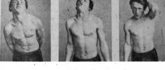
The development of an acute form of the disease does not symbolize anything good, even if the legs are physiologically healthy and have not previously had signs of varicose veins. This indicates that there is a possibility of detecting a malignant tumor. In medicine we are talking about Fischer's symptom. There is no edema, and no enlargement of the lower limb is noted. But there are a number of signs characterized by acute thrombophlebitis. It is diagnosed based on the following symptoms:
- varicose veins;
- pain with a cord-like cord, which is visualized in relation to other tissues;
- redness with swelling of the skin above the induration;
- the presence of reddened bumps, compactions;
- constant preservation of nodes horizontally;
- a position forced to take for pain relief of a limb;
- a slight increase in temperature at a subfebrile level
Diagnosis and treatment of the disease
Most often, the formation of occlusive thrombosis occurs in the vessels of the leg. If treatment is not started in time, they spread to other vessels.
In order to determine the presence of a blood clot, the doctor can send the patient for examinations that will show an accurate picture of the pathological process in the body. The main task of diagnostic measures is to determine the location and size of blood clots. It is also necessary to determine the extent of their distribution.
If you identify the problem in a timely manner, you can avoid consequences and save not only your health, but also your life. When there are certain symptoms, you need to consult a doctor. To diagnose thrombosis, the following is used:
- Studies on D-dimer.
- General blood test (determines the presence of an inflammatory process).
- Duplex scanning (allows you to determine their exact condition, determine the exact blood flow and its parameters).
- Coagulogram, which can determine the ability of blood to clot.
- Finding a blood clot. By injecting a special substance into a vein, you can view the affected areas.
- Ultrasound examination of the lower extremities.
- X-ray examination of the lungs.
- Electrocardiograms.
Read also: Burdock for varicose veins reviews
Based on the results of these studies, an accurate diagnosis can be determined and treatment prescribed. Therapy is carried out using conservative methods. For the first week of treatment, you must adhere to bed rest.
The patient must be prescribed the use of special medications that relieve inflammation. For example, using Melbek and Olfen is effective. Also, you need to use anticoagulants, which can regulate the ability of blood to clot. These could be Clexane and Heparin.
Also during treatment it is common to use:
- rheological medications (Trental);
- glucocorticoids;
- prostaglandin drugs.
In parallel with taking the necessary medications, you need to wear special compression garments that can improve blood flow. Thrombolysis is also possible. This procedure involves the introduction of a special substance into a vein using a catheter, which helps dissolve blood clots. If the disease has become severe, it is necessary to undergo surgery to remove blood clots. For the following therapeutic measures, physical therapy must be prescribed.
If the therapy was carried out in a timely manner, within six months the patient can regain the original abilities of the veins.
About the varieties of the disease
The degree of blockage of the lumen remains uneven, therefore the names of the species are determined:
With occlusive thrombosis, localized in the area of the deep veins of the leg, complete blockage of the venous lumen is characterized, and this entails a stop in the blood flow. Initially, this is visualized by the tightening of the crural vessel inward. Lack of action leads to a worsening of the situation until it spreads to the rest of the veins.
In the non-occlusive form, the formation of floating thrombi is likely, wall-mounted and fixed near the base of the vein. The disease proceeds without impaired blood flow, there are no obstacles to flushing blood through the vessel.
The development of the disease is identified by the following signs:
- pain;
- stiffness;
- feeling of heaviness in the legs.
In practice, it happens that a person is unable to get out of bed due to illness. As it progresses, severe symptoms and venous congestion are noted. Due to the blockage in the vascular lumen with impaired tissue metabolism, there is a possibility that gangrene will begin.
Causes of the disease
Occlusive thrombosis is always dangerous not only for health, but also for human life. If therapy is ignored, the formation breaks off and moves through the vessels, which can cause thromboembolism and lead to the death of the patient.
In addition, complete blockage of the lumen can lead to a heart attack, stroke and subsequent disability of the patient.
Any type of thrombosis is considered an extremely dangerous disease that can lead to disastrous consequences. Regardless of whether the deep or superficial vein is affected, pathology occurs due to similar reasons. This requires a certain influence of factors.
All of them together have a certain negative effect on blood vessels. They are divided into certain groups, depending on their occurrence.
There are objective reasons, called Virchow's triad. These include:
- slow blood flow. Such a process becomes a provocateur of stagnation in the blood;
- high blood density. This property of blood is typical for patients with oncology, liver pathologies, and metabolic disorders in the body;
- change in the structure of the venous wall. Pathology appears due to the influence of many factors, including surgery and injury.
Another very important reason is radiation during the treatment of tumors. This process provokes the appearance of such a pathology. The use of certain medications that make the blood thicker also affects.
There are other reasons that can influence the development of the disease. They relate to external factors.
Experts attribute the following to secondary causes of occlusive thrombosis:
- Age. Under the influence of age-related changes, the structure of blood vessels changes.
- Postponed birth.
- Obesity.
- Suffered leg fractures.
- Performed operations on the joints of the legs.
- Smoking.
- Use of certain medications.
- Presence of oncological tumors.
- Alcohol abuse.
- Lack of physical activity in life.
- Congenital tendency to thrombus formation.
- Obliterating atherosclerosis.
These reasons affect almost every person. Because of this, the disease affects more and more young people.
People who have sedentary jobs are also susceptible to the disease.
Signs of deep vein phlebothrombosis
Primary thrombosis in the deep veins of the legs often appears in the area of the sural venous sinuses. In these places, which are a venous cavity located in the thickness of the calf muscles, where it flows into a deep vein. With muscle contractions, the sural sinuses are emptied. The patient has to be immobilized; a blood clot often forms in these places. Venous lesions are perceived in the same way as arterial lesions in the area of the vessels of the lower extremities. In both cases, it is important to perform differential diagnosis using special examination capabilities.
Phlebothrombosis manifests itself in a form called “pseudoembolic” or white painful phlegmasia. The symptoms of the disease are similar to thrombosis in the arteries of the legs. It is expressed by sharp pain at the site of the lesion, with a feeling of coldness and numbness of the limb. Swelling, before our eyes the leg begins to swell, it becomes difficult to move it in moderation. The arterial pulse value becomes weaker in the foot area.
There are blue painful phlegmasia, which has other symptoms. It can also be diagnosed in the presence of extensive thrombosis, in case of damage to a vein in the pelvis, or in the vein of the lower limb. The color is close - something between black and purple. There is even a possibility of the formation of blisters containing a serous or bloody liquid mass. It doesn’t take long to reach a state of shock or venous gangrene.
Symptoms
Signs of pathology are not always obvious. Occlusive deep vein thrombosis, occurring in an acute form, can cause the following symptoms:
- Swelling in the area of the blockage;
- Feeling of heaviness;
- Signs of inflammation;
- Change in skin tone;
- Pain in the area of the saphenous vein under the knee;
- Periodic increase in temperature;
- General weakness, exhaustion.
Read also: Where a blood clot can break off
Initial symptoms often do not prompt a person to see a doctor, as he thinks that his leg just hurts after a day of work. In the meantime, the blood clot can only grow and thicken.
Progressive occlusive thrombosis causes the following symptoms:
- Night cramps;
- Enlargement and thickening of venous nodes;
- Signs of varicose veins;
- Pain in the lower leg;
- Enlarged lymph nodes.
When these signs appear, treatment must begin immediately. To select therapy, an examination is required.
Treatment of deep venous thrombosis
It is not possible to cure blood clots at home. There is immediate concern that the blood clot may penetrate higher, reaching the lungs or heart. There are a number of conservative methods, with:
- bed rest, followed by the introduction of moderate activity, for which the leg is bandaged with elastic bandaging;
- diet therapy to limit the access of food irritants and allergens;
- thrombolytic therapy to resolve a blood clot
- anticoagulant treatment as a subsequent step after thrombolytics to prevent thrombosis in blood vessels;
- hemorrhagic therapy aimed at improving blood clotting, reducing viscosity, incl. Aspirin;
- NSAIDs (nonsteroidal anti-inflammatory drugs), such as Diclofenac and Ketoprofen;
- venotonic drugs in the form of Troxevasin, Detralex or Venorutin to normalize the tone of venous vessels in the form of general and local treatment;
- symptomatic treatment with cardiotropic, painkillers and other drugs.
As an emergency, thrombectomy is performed - the blood clot is removed from the vessel if thrombosis has just occurred (up to several days).
After the acute symptoms of thrombosis have been relieved, a planned operation is performed. There are several preferred options: with the installation of a vena cava filter; This is a plication in the inferior vena cava. The second option is relevant if it is impossible to install a vena cava filter. The presence of strict indications will be the reason for surgical intervention.
Prevention
Prevention of occlusive thrombosis includes the following measures:
- Proper nutrition to maintain blood formula;
- Active lifestyle with moderate exercise;
- Rejection of bad habits;
- Taking anticoagulants as prescribed by a doctor;
- Wearing special compression stockings.
These simple recommendations will help maintain vascular health. If you suspect the development of the disease, you should visit a doctor.
It is possible to get rid of occlusive thrombosis. But treatment should be started immediately, as complications can be fatal. After diagnosis, the doctor will select a treatment method and give additional recommendations.
Treatment of sural vein thrombosis
If thrombosis of the sural veins has an uncomplicated course, then treatment is possible exclusively with conservative methods. But at the same time, the patient must observe strict bed rest for two weeks, but no less.
The following drugs are used for therapy:
Direct anticoagulants. Low molecular weight Heparin remains the drug of choice. It helps reduce blood viscosity, as well as increased release of antithrombin in the body. As a result, the clot dissolves. Heparin is administered intravenously. The doctor determines the dosage on an individual basis.
Indirect anticoagulants. These are drugs such as Warfarin and Coumadin. They prevent thrombin from being produced. It should be taken into account that taking Warfarin is associated with the risk of bleeding, so the patient should be under strict medical supervision.
Enzyme drugs that have thrombolytic properties. These include Streptokinase and Urokinase. They allow the blood clot to dissolve and prevent blood clotting. Medicines are administered only in the form of injections.
Drugs that are designed to improve the quality of blood, enhance its microcirculation, and reduce blood viscosity. These are medicines such as: Reosorbilact, Reopoliglucin, etc.
NSAIDs are drugs that are aimed at reducing inflammation, thinning the blood and reducing pain. For this purpose, medications such as Voltaren, Indomethacin, Aspirin, etc. can be used.
Traditional treatment
Leg vein disease is not a new problem. For centuries, healers and healers have tried to find a cure for this type of illness. Many of them turned out to be quite effective. But in an effort to recover, there is no need to use only folk remedies.
Therapy must be comprehensive, it must include all available treatment methods. Including diet, physiotherapy, alcohol and smoking cessation. Only in this case can treatment with folk remedies be effective.
Before starting treatment with herbs, infusions and other traditional medicine, you should consult a doctor, because the patient may have an allergic reaction to the elements of the remedy.
For varicose veins, it is recommended to use horse chestnut extract to strengthen the walls of blood vessels. This product is so effective and popular that it is sold in regular pharmacies.
Another treatment for varicose veins is rubbing the feet with a tincture of lilac leaves. The mixture is prepared simply: a 0.5 liter container is filled with the flowers of the plant and filled with alcohol. After the product has been infused in the dark for 10 days, it can be safely used in the morning and evening. The course of such treatment is 30 days.
Thrombophlebitis is treated with a mixture of honey and apples. In this case, the apples must be grated, mixed with honey (for a glass of apples - 1 spoon of honey) and allowed to brew for at least 5 hours in a dark place. It is believed that this remedy helps blood clots in the veins to dissolve on their own within 2 weeks.
Endovasal coagulation of the lower extremities: complications, side effects, results, video
What is endovasal coagulation of the veins of the lower extremities?
Our understanding of varicose veins and how to treat patients with varicose veins has changed dramatically over the past decade. This was facilitated by duplex ultrasound scanning, which is used to view blood flow in the veins.
Normally, blood circulates from the heart to the legs through the arteries and in the opposite direction through the veins. Using Doppler ultrasound (USDG), it is possible to visualize the vein and determine the direction of blood flow.
This procedure is performed before treating varicose veins to determine the direction of blood flow that is causing the deformed veins.
In the legs (as in other parts of the body), veins contain one-way valves that allow blood to return from the legs to the heart, thanks to gravity. If the valves stop functioning properly, blood pools in the veins of the legs, causing the veins to become enlarged.
After a physical examination and duplex ultrasound, an individual plan is developed to treat varicose veins and redirect blood flow to nearby healthy veins. This can be done using one of the following procedures:
- Laser obliteration (coagulation, EVLT, EVLO) – removal of deformed veins using laser energy/heat;
- Radiofrequency obliteration – removal of deformed veins using radio wave energy/heat;
- Sclerotherapy is a chemical removal/destruction technique used to close small varicose veins.
The above procedures can be combined under one term - endovasal (endovenous) coagulation (obliteration, ablation). Coagulation of varicose veins often helps people with chronic venous insufficiency. This procedure is also used to eliminate a cosmetic defect.
How to prepare for coagulation of varicose veins?
The specialist doctor who will perform the procedure should familiarize you with the following points during the consultation:
- You should be aware of all possible complications regarding treatment and costs.
- You should tell your doctor about all the drugs you take, including herbal supplements. It is also necessary to talk about allergies, if any, about reactions to anesthesia during any procedure performed.
- Immediately before the procedure, you may need to stop taking blood thinning medications: aspirin, nonsteroidal anti-inflammatory drugs (NSAIDs), etc.
- You should wear loose and comfortable clothing. During the procedure, you will need to remove all jewelry in the area that will be treated.
- Don't forget to wear compression stockings immediately after the procedure. The required degree of compression of the stockings should be discussed in consultation with the treating physician before ablation.
- Immediately after the procedure, you will need to walk for about 30-40 minutes. You should also move daily for the next 14 days.
- You will need to wear compression stockings for 14 days after the procedure - for the first 4 days you will need to wear stockings day and night, the remaining days only during the day.
- You may feel some discomfort or pain in the area of the treated vein after surgery.
- The procedure is performed on an outpatient basis using local anesthesia.
What happens to varicose veins during laser or radiofrequency ablation?
- While standing, your affected veins will be imaged under ultrasound guidance.
- The legs should be clean, shaved, sterilized, and then covered with a surgical drape.
- If laser photocoagulation will be used, you will be provided with protective glasses.
- Anesthesia will be injected into the area where the affected veins are located.
- Under ultrasound guidance, a laser or catheter is inserted into the affected vein through a small incision in the skin.
- After laser or radiofrequency energy is applied, the catheter is removed from the skin. The affected veins must be sealed and blood flow from these veins is redirected to nearby healthy veins. To stop the bleeding, the doctor applies pressure to the legs and a small cut in the skin is sealed. Compression stockings are also put on immediately.
- During sclerotherapy: a chemical solution is injected with a very thin needle into the affected vein.
Side effects after coagulation of varicose veins
Features of the classification of the disease and localization of lesions
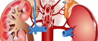
Usually the disease affects the inferior vena cava system. The formation of a blood clot often occurs in deep vessels of the leg, namely in the sural, anterior and posterior great tibial vein. Often, the formation of a blood clot occurs in other parts of the bloodstream of the lower extremities. Typically, damage to the vessels of the leg tends to spread. Gradually, the popliteal and main veins of the thigh are involved in the process.
In rare cases, pathological changes in the circulatory system of the upper extremities are noted. A diagnosis such as thrombosis of the lateral vein of the forearm is made infrequently. It is usually associated with improper technique for administering subcutaneous injections. Therefore, when it comes to the formation of blood clots, in most cases they mean damage to the right or left lower limb.
In accordance with the morphological characteristics and characteristics of the location of the thrombus in the vascular bed, non-occlusive thrombosis (also called parietal or floating) and occlusive thrombosis are distinguished. It is the first type of disease that is considered the main cause of the development of the most dangerous complication - pulmonary embolism (or PE for short). The fact is that a floating thrombus has only one point of fixation; therefore, it can easily break off and begin moving through the circulatory system, so such a disease requires mandatory treatment in a hospital setting. An occlusive thrombus with a moving upper part is also dangerous. The location of such clots has a characteristic feature; they are localized in the area of vascular dilatation, for example, at the junction of the deep veins of the leg into the popliteal vein or the transition of the superficial femoral vein into the common vein, etc.
Read also: Varicose veins in children
Vein thrombosis
Have you been trying to cure VARICOSIS for many years?
Head of the Institute: “You will be amazed at how easy it is to cure varicose veins by taking the product every day for 147 rubles...
Read more "
Thrombosis is a pathology that develops due to excessive blood clotting and is accompanied by a violation of the passage of fluid through the vessel. Due to the occurrence of this disease, secondary changes in organs and soft tissues often develop.
Many people are only familiar with the concept of “lower extremity venous thrombosis,” but doctors distinguish more types of this pathology. The common mechanism of their development allows us to combine all diseases into one large group.
Causes
Recent medical research has been able to identify three main causes of thrombosis. These include:
- disruption of the integral structure of the vascular wall (damage can be caused by any factor: from mechanical trauma to infection);
- decrease in the speed at which blood passes through a vessel in a certain place (develops due to compression or venous insufficiency);
- increasing the innate ability of blood to clot (the pathology can be either congenital or acquired under the influence of certain chemicals, including medications).
In addition to the mandatory conditions, doctors identify a number of risk factors that contribute to the development of pathology. These include:
- elderly age;
- reduced physical activity for any reason: a sedentary lifestyle or forced long-term bed rest;
- various abdominal surgeries in the patient’s history (including cesarean section);
- the presence of various bad habits contributes to thrombosis;
- some medications, for example, those based on hormones, can provoke an increase in blood clotting;
- Long-term infections can lead to damage to the walls of blood vessels.
The formation of blood clots is always dangerous with a high risk of developing heart thrombosis, so it is necessary to take into account the risk factors for developing the disease and, if possible, eliminate them.
Symptoms
The symptoms of thrombotic lesions can be very diverse. The patient's complaints will depend on the stage of the process and the location of the vessel in which the lumen is blocked. Symptoms of venous thrombosis of the lower extremities cannot be compared, for example, with how portal vein thrombosis develops, which must be taken into account when making a diagnosis.
Symptoms of deep vein thrombosis in the lower extremities are usually the following:
- severe pastiness and gradually increasing swelling of the limb;
- numbness and paresthesia;
- heaviness in the legs when moving, gradually developing pain syndrome.
Gradually, as the signs of deep vein thrombosis worsen, trophic changes appear in the tissues, which in some cases lead to amputation of the limb.
Separately, acute venous thrombosis is distinguished, which is regarded as an emergency condition and requires immediate medical attention. Symptoms of the pathology will be:
- acute, sharp pain in the limb, intensifying in response to pressure;
- reduction of all types of sensitivity and the occurrence of paresthesia;
- absence of pulsation below the thrombosed area;
- severe coldness of the skin of the extremity.
Due to the occurrence of acute thrombosis, contractures may develop, which will indicate irreversible changes.
Deep vein thrombosis of the leg or any other part of the lower limb is not the only type of thrombosis. There are additional types, each of which has its own characteristic features.
Varieties
Blood clots that block the lumen of a vessel can form not only in the veins. It is also important to remember that blood clots can form in any part of the human body, including the vessels of the upper extremities, the brain, and cardiac thrombosis.
In medical practice, there are 4 main types that are most common and should be given special attention.
Intestinal form
The intestinal form of thrombosis is of arterial origin. This means that the artery is blocked, and in this case the superior mesenteric artery suffers. With thrombophlebitis, endocarditis, and atherosclerosis, the risk of developing the disease increases significantly.
Symptoms may include:
- severe, sharp pain in any area of the abdomen (localization may vary depending on the area in which the blockage has formed);
- attacks of nausea and vomiting, the appearance of diarrhea (bloody inclusions may be found in the stool);
- a jump in body temperature and increased heart rate (tachycardia).
The progression of intestinal thrombosis resembles obstruction in its symptoms, so only a specialist can make an accurate diagnosis.
Phlebothrombosis
Phlebothrombosis is another name for deep thrombosis. In most cases it develops in the veins of the lower extremities, its symptoms have already been mentioned. Doctors separately identify a pathology in which blood clots are found in the pelvis and have the ability to fluctuate with the blood flow (float). Such blood clots can become emboli when detached from the vessel wall and lead to cardiac thrombosis.
Damage to the upper extremities is much less common in practice. Symptoms of the initial stage include hyperemia of the skin, slight swelling and pain at the site of the lesion.
Ileofemoral thrombosis
Many patients mistakenly confuse ileofemoral thrombosis with blockage of the veins of the upper extremities. This is not entirely true. This pathology is a consequence of occlusion of the femoral and iliac veins. In most cases, the disease is acute; the patient complains of blue discoloration of the skin on the affected side, swelling of the limb, and a jump in body temperature. Complete closure of the lumen of the vessel can lead to tissue necrosis, but if the lumen is minimally preserved, the symptoms may regress.
To avoid cardiac thrombosis and other complications, it is recommended that the patient be treated in a hospital setting.
Blockage of the cavernous sinus
Blockage of the cavernous sinus is one of the most dangerous types of thrombosis, as a blood clot blocks a cerebral vein. In most cases, the cause of the disease is an infectious pathology that began in the area of the nose or eyes and spread to the brain area. Manifestations of the disease are quite specific. The patient reports severe headaches, seizures, and protruding eyes. People with this pathology also often experience fluctuations in body temperature and various nervous system disorders. Without timely treatment, coma is possible.
Which doctor treats thrombosis?
The choice of the attending physician depends on what type of thrombosis has developed in the patient. If the disease has affected the arteries, then you should contact an angiologist, who also specializes in diseases of the lymphatic system.
100% works! Varicose veins are removed by a cream made at home from three components - this is...
This is the only way VARICOSIS will disappear in 1 week! Do it every night at night...
Treatment of deep vein thrombosis of the lower extremities will be carried out by a phlebologist whose specialization is various veins.
Both phlebologist and angiologist are narrow areas of medicine, and it can be difficult to find specialists in this field. If there are no doctors, the patient can be treated by a therapist or, if surgery is necessary, by a surgeon.
Diagnostics
Acute deep vein thrombosis of the lower extremities is usually diagnosed based on a clear clinical picture. The patient needs help immediately, and therefore there is often no time for detailed studies. If the situation is not life-threatening, then the following may be used:
- venography (x-ray contrast examination, in which the contrast agent goes up the vein, has a high degree of accuracy);
- ultrasound duplex scanning, which is used most often to make this diagnosis;
- thromboelastography, with which you can understand how the blood clotting process occurs and whether there are any violations in it;
- radionuclide scanning.
X-rays help in diagnosing intestinal vein thrombosis. In some cases, when the clinical picture and test data do not provide a sufficient understanding of the situation, diagnostic laparoscopy is performed. With its help, you can clarify the diagnosis, as well as choose the optimal treatment method.
Treatment
How to treat thrombosis? The choice of technique largely depends on what stage the disease is at and what symptoms bother the patient. Today, doctors in most cases prefer conservative therapy, which is safer and is accompanied by a lower risk of complications. However, if the need arises, surgical intervention is also possible.
Conservative treatment
Treatment of deep vein thrombosis begins with the selection of medications. The goal of drug therapy is to:
- reducing the activity of increasing thrombus size;
- fixation of the clot to the vascular wall to prevent its detachment;
- eliminating spasm, swelling and inflammation;
- normalization of blood circulation processes.
First of all, direct-acting anticoagulants are used. While taking medications from this group, the patient must constantly undergo tests so as not to miss the moment when blood clotting becomes dangerously low, which will threaten the development of bleeding.
The main drug from the group of direct anticoagulants is Heparin. On its basis, medications are prepared for oral administration, as well as ointments and creams. Read more about Heparin ointment→
In addition to Heparin, Hirudin, which is an extract of leech saliva, is often used. It has also proven to be a good direct anticoagulant.
In addition to direct anticoagulants, the following are used:
- indirect anticoagulants, which have a more pronounced effect and are easier to use;
- agents that improve the rheological properties of blood;
- thrombolytics, which promote rapid resorption of the blood clot and restoration of normal blood flow;
- non-steroidal anti-inflammatory drugs intended to eliminate the main unpleasant symptoms;
- For severe pain, painkillers can be used.
Non-drug treatment
Non-drug therapy plays a major role in the treatment of thrombosis. During the acute period, the patient is recommended to rest in bed for 5-10 days. During bed rest, the limbs are raised at a slight angle to facilitate blood outflow processes.
After the end of the acute period, dosed physical activity is recommended to prevent relapse of the disease. The exercises are performed under the supervision of a doctor; the dose of exercise therapy is also prescribed by a specialist. The patient is necessarily recommended to change a sedentary lifestyle to a more active one.
Compression therapy is widely used. With its help, it is possible to eliminate swelling, improve blood flow through the veins, and prevent damage to superficial vessels. For compression therapy, special bandages are used, which are recommended to be worn during the acute period of the disease. In case of chronic thrombosis, bandages are replaced with underwear with compression properties.
Doctors recommend compression therapy only during the daytime. At night, the body should rest from all pressure.
Surgery
In some cases, non-drug and conservative therapy are completely useless. Then the surgeons propose to perform a surgical intervention, which should completely relieve the patient of blood clots.
There are three main types of surgery:
- intervention of the Troyanov-Trendellenburg type, in which the great saphenous vein is sutured, thus blocking the progression of the blood clot through the vessel;
- vena cava filtration, in which a special filter is installed in the vein, capable of retaining blood clots (the disadvantage of the intervention is that after a few years there is a need to replace the filter, as it can become clogged);
- thrombectomy, which is the most modern type of intervention, involves removing a blood clot from the cavity of the affected vessel (in most cases, the vessel itself is not injured during the operation).
Traditional methods
There are also folk remedies to combat thrombosis. All of them are aimed mainly at eliminating the inflammatory response.
The following recipes are recommended:
- drinking verbena decoction for three months;
- drinking alcohol tincture of fresh or dried white acacia flowers for 3-4 weeks;
- ingestion of decoctions of hop cones;
- using onion juice mixed with honey, applied in a course of 2-4 weeks.
It is important to remember that folk remedies are absolutely not suitable for use without drug and non-drug therapy. They can be used only as auxiliary methods, but not as the main therapy aimed at preventing thrombosis of the heart or cerebral vessels.
Prevention
Prevention of thrombosis will be useful not only for people at risk, but also for those who are too early to think about this disease.
The following set of measures is recommended:
- drinking large amounts of liquid;
- physical activity (morning exercises and simple exercises throughout the day);
- refusal to travel by car, if possible a large amount of walking;
- foot massage and contrast shower every evening;
- proper nutrition;
- wearing compression garments.
Prevention can also include timely treatment of varicose veins, cardiovascular diseases, pathologies of the endocrine system and the musculoskeletal system.
Diet
Diet for deep vein thrombosis of the lower extremities is an important element of treatment. Patients are strictly prohibited from consuming animal fats, sweets, including chocolate, alcoholic beverages, foods rich in spices or overly fried.
Nutrition for deep vein thrombosis of the lower extremities should be based on the consumption of raw vegetables and fruits, which are rich in antioxidants. You can also eat bran and foods rich in copper and flavonoids. This will help strengthen the vascular walls and prevent relapse of the disease.
Thrombosis is a disease dangerous to human health and life. It is a mistake to believe that blood clots can block blood flow only in the lower veins, and only older people suffer from the pathology. You can encounter thrombosis at any age and, moreover, not only veins, but also arteries in any organ can be affected. When the first signs of pathology appear, you should consult a doctor.
Causes of lower vein disease
Phlebitis, varicose veins and thrombosis of the sural veins of the lower extremities is a problem that is being addressed by the best minds of humanity working in this direction. These diseases of varying severity affect 25-30% of the world's population.
Accordingly, first of all, scientists are trying to establish the causes of these pathologies.
The main reasons identified to date are:
- Injury to the walls of deep vein vessels. This happens to people engaged in activities with a high risk of injury to the legs. For example, with professional football players. It is not uncommon for players with many years of experience to retire after losing a leg due to gangrene caused by a vascular injury.
- Another reason is a blood clotting disorder, namely an increase in this indicator. In this case, there is a high risk of the formation of blood clots - blood clots so large that they can block the lumen of the largest vessel in the leg.
- Low blood flow speed can cause thrombosis or stagnation, which is a direct consequence of a sedentary lifestyle, which has become the main problem of modern man. The anatomy of the sural veins requires constant movement - walking, running, cycling. Everything in the legs is designed specifically to withstand loads. And if a person moves during the day only by elevator, escalator, car, and spends the working day in a chair, then his blood vessels in the legs weaken and are susceptible to various diseases.
- Thrombosis of the sural veins of the leg can occur against the background of hymenal disorder. Most often it is caused by incorrectly selected hormonal contraceptives. This is confirmed by the fact that the percentage of sick women is slightly higher than the number of men suffering from foot disease.
- The sural veins of the leg are destroyed under the influence of nicotine, alcohol and drugs.
- The deep veins in the body of an obese and overweight person bear an excessive load. And since this condition is often accompanied by diabetes mellitus, which destroys the walls of blood vessels, this often leads to gangrene of both limbs.
- Sural varicose veins may be a consequence of cancer.