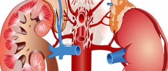
When a renal artery is blocked by a blood clot, an organ infarction develops. When the blood supply to the second kidney is preserved, it takes on the entire burden of filtering urine. In the case of a bilateral process or thrombosis of blood vessels in a single kidney, acute renal failure develops. Treatment requires complex drug therapy, dissolution of a blood clot, or surgical intervention.
Types of renal vein thrombosis and causes of development
The condition can be bilateral or unilateral (in most cases), acute or chronic.
Bilateral thrombosis is usually associated with ascending thrombosis of the inferior vena cava. This condition is extremely dangerous for the patient’s life, because leads to dysfunction of both kidneys.
Congestive heart failure often leads to renal vein thrombosis. Due to stagnation, blood clotting processes intensify, which leads to the formation of blood clots. This same group of reasons includes any factors that “thicken” the blood.
In children, renal vein thrombosis develops with severe dehydration caused by profuse diarrhea.
Renal vein thrombosis is one of the symptoms of malignant neoplasms in the kidney itself or in the retroperitoneal space - a disintegrating tumor clogs the lumen of the vessel.
In addition, thrombosis often accompanies antiphospholipid syndrome and membranous nephropathy.
Causes of the acute process
Thrombosis of the renal artery occurs when a blood clot forms in it on the surface of a destroyed atherosclerotic plaque, injury to the wall during surgery or a diagnostic procedure, or a congenital vascular anomaly (aneurysm, malformation). Most often, a blood clot forms in other parts of the circulatory system or in the cavity of the heart, and enters the kidneys with the bloodstream - thromboembolism develops. Possible causes of blockage include:
- atrial fibrillation;
- increased blood clotting activity, hereditary thrombophilia;
- antiphospholipid syndrome;
- trauma, especially a blow to the stomach;
- valve defects, bacterial endocarditis;
- malignant neoplasms;
- autoimmune diseases – periarteritis nodosa, systemic lupus erythematosus.
Very often, renal artery thrombosis is not an independent disease, but is combined with blockage of the pulmonary artery, abdominal branches of the aorta, cerebral and coronary vessels, and damage to the lower limb. Therefore, they are considered as one of the manifestations of general thromboembolic disease.
We recommend reading the article about renal artery stenosis. From it you will learn about the causes and symptoms of the pathology, the danger of stenosis, diagnosis and treatment. And here is more information about atherosclerosis of the renal arteries.
Signs of the disease
The condition begins with pain - the affected kidney swells, nephrons (vascular glomeruli that carry out filtration) begin to collapse. It can hurt on one side or on both sides (with bilateral thrombosis). If two kidneys are damaged, oliguria may develop, up to anuria (urine is not filtered). At this point, the color of the urine may change; examination reveals hematuria (traces of blood).
If thrombosis does not occur at the level of the entire kidney, but one lobe or segment, then the condition can develop over a long period of time.
Chronic renal vein thrombosis is often associated with a negligent attitude towards one’s own health - patients ignore the pain (or treat it with medications) and do not consult a nephrologist or therapist.
Pain during the chronic process becomes aching and dull. More and more protein and blood cells (proteinuria) begin to enter the urine, and edema develops. In some cases, the condition is accompanied by increased blood pressure.
If thrombosis occurs gradually (vascular atherosclerosis, thrombosis by embolus), then the condition may be asymptomatic, because the vascular system adapts - outflow occurs through collaterals (vascular bridges are formed to discharge excess blood).
How to identify the problem
This condition usually does not occur spontaneously. In the anamnesis of such patients there are factors that provoke the formation (separation) of a blood clot, episodes of vascular disorders of other localization.
As a rule, we are talking about the following diseases or conditions:
- Bedridden patients of any profile;
- Varicose veins, lymphadenopathy, thrombophlebitis/phlebothrombosis;
- Surgeries on the lower extremities, injuries (especially femur fractures);
- Malignant oncological process, regardless of its location;
- Long-term administration of loop diuretics (as a side effect).
As a result of venous thrombosis, the functioning of a section of the kidney or the entire organ is disrupted, which, without adequate treatment, can lead to the replacement of this section with connective tissue (fibrosis).
The doctor pays attention to pain in the lower back, swelling, changes in the color of the blood, venous congestion in the legs - they look for signs of outflow, obstruction of the inferior vena cava. If you complain of prolonged pain, the abdominal wall is carefully examined (there may be signs of venous collaterals, in other words, the venous pattern intensifies).
Diagnostic methods
Urography (excretory and survey) helps to determine oliguria, as well as defects in the filling of the renal collecting system (due to blood clots); in some cases, signs of formed collaterals are visible.
Cytoscopy (analysis of the cellular composition) of urine detects red blood cells in the urine. This study allows us to exclude glomerulonephritis as the cause of hematuria (the formed elements are not deformed because they are not “pressed” through the glomeruli).
A coagulogram determines blood clotting parameters. Without it, you cannot prescribe thrombolytics, anticoagulants, or, conversely, hemostatic agents (you cannot treat a blood clot without understanding the parameters of thrombus formation).
Ultrasound of the kidneys helps assess the condition of the main vessels and the kidneys themselves.
MRI and CT with contrast give a detailed picture of the blood circulation of the kidney - all vascular anomalies and hemorrhages in the parenchyma of the organ are visible. These methods are optimal for diagnosing malignant neoplasms, the symptom of which is often venous thrombosis.
Thrombosis-like conditions:
- Renal colic. In this case, we are also talking about blockage, but not of a vessel, but of the ureter (stone, blood clot, mucus plug). In this condition, protein separation in the urine is not typical. The issue is resolved using ultrasound, CT or MRI;
- Glomerulonephritis. Differs in the structure of red blood cells in the urine, the absence of venous collaterals;
- Amyloidosis of the kidneys. The difference is clearly visible on MRI with contrast, and sometimes a biopsy is performed.
Diagnostics
Correct diagnosis is of great importance for timely assistance. However, it will be more important to contact the patient when the first symptoms of pathology appear.
The most informative diagnostic method, in this case, will be an ultrasound examination with Doppler. Studying the vessels of the kidney will allow us to determine the location of obstruction of the vein/artery, the presence of a blood clot in the lumen of the vessel, and the reverse movement of blood.
To determine the condition of the organ in this situation, an MRI with a contrast agent will be used. During the study, it is possible to determine the presence of a focus of necrosis or other damage to the kidney tissue.
Laboratory tests will allow you to assess the degree of dysfunction of the excretory organs. Standard tests will be: general blood and urine analysis; determination of erythrocyte sedimentation rate, blood platelet level; creatinine level; urea level.
Drug treatment of renal vein thrombosis
Thrombolytics are prescribed to destroy a “fresh” blood clot, low molecular weight heparins (Clexane), and rivaroxaban drugs (Xarelto) to prevent subsequent thrombus formation. All these drugs are dangerous in the presence of even slight bleeding (hematuria). Children with thrombosis due to dehydration undergo appropriate water and electrolyte replacement.
If severe hematuria is observed, hemostatic agents (etamzilate, etc.) are prescribed.
Treatment of renal vein thrombosis is a risky process, because... accompanied by the phenomenon of bleeding and, at the same time, thrombosis. The doctor is forced to balance between two mutually exclusive therapeutic tactics.
Causes
Blockage of the main blood vessels of the kidney occurs as a secondary pathology (that is, a complication of the underlying disease) against the background of the presence of various diseases.
- Violation of the blood clotting process.
- Development of atherosclerotic changes in blood vessels.
- Cardiovascular failure, causing congestion.
- Malignant processes of the urinary organs.
- Presence of antibodies to phospholipids (APS syndrome).
- Various autoimmune pathologies (glomerulonephritis, thrombocytopenic purpura, lupus erythematosus, etc.).
- The presence of moving blood clots in the circulatory system (migratory thrombus).
- Metabolic disorders (protein, carbohydrate, etc.).
- Development of nephrotic syndrome.
- Injury to the kidneys, severe damage to the back area (impacts, wounds).
- Inflammatory vascular diseases.
- Inflammatory processes, pathologies and tumor-like neoplasms of the pelvic organs and retroperitoneal space.
Symptoms
With the slow development of renal artery thrombosis, symptoms may be mild or completely absent. If the disease develops rapidly, the following signs of pathology may be observed:
- nausea and vomiting;
- swelling of the limbs;
- increased body temperature;
- blood in urine;
- pain in the lumbar region;
- decrease in the amount of urine excreted;
- pale skin.
If the vessels of the kidneys are damaged, other symptoms may appear that are characteristic of other diseases, for example, venous thromboembolism.
Chronic renal vein thrombosis can manifest itself as follows:
- aching or dull pain in the lumbar region;
- arterial hypertension;
- large amounts of protein when urinating.
To diagnose a disease, the doctor interviews the patient, examines him and sends him for instrumental and hardware diagnostics. It can be:
- venography;
- CT angiography;
- blood tests;
- Analysis of urine;
- urography;
- coagulogram;
- X-ray of the renal arteries.
Symptoms
Thrombosis of large vessels of the kidneys (veins, arteries) can be unilateral (more often) or bilateral. In case of unilateral damage, the functions of the affected organ are taken over by the second kidney, and a compensation effect appears. In this regard, the symptoms are weak. If the patient has only one kidney and the blood vessels are blocked, the entire clinical picture will manifest itself clearly, with the gradual appearance of symptoms.
The danger of thrombosis of the veins of the arteries of the kidneys lies in the late detection of the condition, when the organ has been without blood supply for a long time. This is due to the lack of typical symptoms. Almost all signs of pathology are similar to symptoms of other diseases of the genitourinary system and retroperitoneal organs. Therefore, the appearance of one or more of the following signs should be a signal that you need to contact a medical facility. If thrombosis develops, delay threatens the loss of the organ.
Thrombosis of both veins and arteries of the kidney gives similar symptoms:
- The appearance of acute, pressing, aching or nagging pain in the lumbar region from the affected organ, irradiation to the genitals or leg is most often absent.
- A rapid or sudden increase in blood pressure. Particular attention should be paid to diastolic pressure values. Diastolic pressure allows us to judge the condition of blood vessels. The higher the “lower” pressure values, the worse the vascular permeability. This means the veins and arteries lose their elasticity, patency and ability to contract. Diastolic pressure is the second digit as a result of the measurement. That is, in terms of 110/70, the diastolic pressure will be the number “70”.
- The appearance of nausea, vomiting and an increase in body temperature.
These symptoms are signs of intoxication, which develops as a result of poisoning of the body with decay products. The accumulation of toxins occurs due to impaired excretory functions of the kidneys.
The appearance of blood and protein in the urine, a decrease in the amount of urine excreted - these symptoms indicate a malfunction of the affected kidney. The appearance of blood clots and streaks (“worms”) in urine is particularly dangerous and is a sign of increasing renal failure.
With thrombosis of the renal arteries, the symptoms will be severe, and the pain will resemble “renal colic.”
If the disruption of the blood supply to the kidney is not eliminated in the next few hours, the process will become irreversible. The tissues of that part of the organ that is left without nutrition undergo irreversible scarring. If the situation repeats, the kidney functions will be completely impaired. The result is the death of the organ and its subsequent removal.
Causes and symptoms of renal vein thrombosis
In children, most cases of renal vein thrombosis are thought to be caused by episodes of severe dehydration. Severe dehydration reduces blood volume and causes it to clot. Further symptoms of the disease develop very quickly.
In adults, renal vein thrombosis can be caused by damage to organs in the abdomen or back, malignant kidney tumors, the formation of scar tissue (stricture), or kidney disease that causes degenerative changes in the cells of the renal tubules (nephrotic syndrome).
Renal thrombosis is more common in patients with nephrotic syndrome, although studies have shown high variability among these patients, with rates ranging from five to sixty percent. Nephrotic syndrome is characterized by abnormally low levels of albumin (hypoalbuminemia), abnormally high levels of cholesterol in the blood (hypercholesterolemia), and fluid retention (edema). The most common form of nephrotic syndrome seen in children is characterized by swelling due to fluid retention, foamy urine, and loss of appetite.
Acute onset of renal vein thrombosis at any age causes pain in the lower back and sides of the abdomen, fever, bloody urine, decreased urine output, and sometimes renal failure.
Other symptoms include high blood pressure or a "whistling" sound on stethoscopy of the abdomen. This noise is made by the flow of blood trying to pass through a blocked vessel. The doctor may also be able to feel the enlargement of the kidney by palpating during a physical examination. Some patients have no symptoms.
When to see a doctor
Parents should contact a doctor immediately if the child has any of the following symptoms:
- high body temperature – 38.9°C or higher,
- sudden onset of lower back pain,
- sudden, severe swelling of the legs,
- labored breathing,
- blood in urine
- decreased urination,
- dry mouth,
- increased or excessive thirst,
- dark yellow urine
- irritability,
- severe weakness
- dizziness or fainting,
- sunken belly, eyes and cheeks.
Subscribe to our YouTube channel!
Treatment methods
Treatment of renal vein thrombosis is carried out in a hospital setting, and the patient must remain in bed. The doctor prescribes medications from the group of anticoagulants. They are designed to thin the blood, prevent the formation of blood clots and get rid of already formed blood clots. These include Warfarin in tablets and Heparin in the form of an injection solution.
Treatment with such drugs begins immediately after a blood clot is detected in the vessel. Therapy can last 6-12 months. Some patients with bleeding disorders take these medications for the rest of their lives.
If severe hematuria (blood in the urine) is observed, anticoagulants are contraindicated. Thrombolysis shows good effectiveness - a procedure during which special enzymes are injected into a vein to dissolve blood clots and restore kidney function.
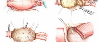
In severe cases of thrombosis of the blood vessels of the kidney, surgical treatment is performed. The following types of surgical intervention are distinguished:
- Thrombectomy. It is used if treatment with anticoagulants has not been effective. This operation is indicated for dangerous renal dysfunction and bilateral chronic thrombosis.
- Nephrectomy. It is rarely performed if a kidney infarction has developed and if there are irreversible changes in one of the organs.
- Kidney removal. It is carried out as a last resort, when the entire renal tissue is affected by an infarction and cannot be subject to other treatment. Such intervention is contraindicated in case of bilateral thrombosis.
If the disease was diagnosed in a timely manner, and if the treatment rules are followed, blood circulation is restored, then kidney function returns to normal after some time.
Why can stent thrombosis occur?
A separate group of pathology is the appearance of a blood clot at the site of the operated vessel. A stent is installed in the renal artery when it is narrowed. It is caused by thickening of the muscle layer or connective tissue membrane in fibromuscular dysplasia of the vessel. To restore blood flow, a stenting operation is used, during which a metal frame is placed into the artery after balloon expansion. It prevents a decrease in the lumen of the vessel.
Stent blockage may be caused by:
insufficient use of anticoagulants, unauthorized cessation of their use;
Installation of a stent in the renal artery
Prices
DiseaseEstimated price, $
| Prices for examination and treatment for testicular cancer | 3 730 – 39 940 |
| Prices for vaporization of prostate adenoma with a “green laser” | 16 050 |
| Prices for diagnosis and treatment of impotence | 1 320 – 50 000 |
| Prices for diagnostics of the genitourinary system in men | 5 630 |
| Testicular cancer treatment prices | 15 410 |
| Prices for the treatment of urolithiasis | 11 760 – 16 180 |
| Bladder cancer treatment prices | 21 280 – 59 930 |
| Prices for diagnosing prostatitis | 2 720 |
| Prices for diagnosing male infertility | 6 300 |
| Prices for prostate cancer treatment | 23 490 – 66 010 |
DiseaseEstimated price, $
| Prices for kidney cancer treatment | 28 720 – 32 720 |
Since the pathologies are very different, their treatment methods also differ significantly from each other. Vascular surgery in Israel carries out a variety of operations by type, level of complexity, etc. which effectively help eliminate the problem and save a person’s life.
Clinical picture and diagnosis of the disease
There are three main symptom complexes (syndromes), which to varying degrees accompany thrombosis of the renal veins and arteries:
Hypertensive syndrome
The sudden nature of the increase in blood pressure is the main symptom accompanying thrombosis of the renal vein and artery. Its severity depends on the degree of damage to the vessel and the presence of concomitant disorders - heart disease, lung disease. When diagnosing the disease, it is important to take into account the increase in blood pressure against the background of the presence of other external symptoms and changes in blood parameters and heart function.
Urinary syndrome
Changes in urine are caused primarily by increased permeability of the walls of small blood vessels due to narrowing of the lumen of a large artery or vein.
The main symptoms are as follows:
- the appearance of red blood cells in the urine;
- the appearance of protein in the urine;
- urine density is normal;
- the amount of urine excreted does not change.
The appearance of bloody urine, especially in combination with hypertension, may indicate possible thrombosis of the renal veins and arteries.
Pain syndrome
Painful sensations are not the main diagnostic sign of the disease, since their nature is similar to the manifestations of other pathologies of the abdominal organs. Pain caused by thrombosis of the renal veins and arteries occurs suddenly.
Localization may vary:
- in the lower back;
- in the kidney area;
- in a stomach;
- in the groin area;
- in the side.
Pain syndrome is often accompanied by the following symptoms:
- nausea;
- bouts of vomiting;
- constipation;
- temperature increase.
The main diagnostic measures include laboratory and instrumental studies.
Laboratory parameters of blood and urine are characterized by the following deviations:
- increased leukocyte levels;
- increased number of neutrophils;
- increased erythrocyte sedimentation rate;
- increase in urea concentration;
- increased nitrogen content in the blood;
- high creatinine concentration;
- detection of protein in urine;
- the appearance of blood in a urine test.
Among the instrumental methods, the following are highly informative:
- Renal angiography.
- Ultrasound (ultrasound examination).
- Urography.
- Renography.
- Radiography.
Symptoms and research results, which need to be analyzed only in combination, will help make the correct diagnosis and prescribe rational and effective treatment to the victim.
Diagnosis of the condition
Thrombosis and thromboembolism of the renal artery in their clinical course do not differ from inflammatory diseases. And the speed of diagnosis determines the prognosis not only for recovery, but in especially severe cases for the patient’s life. Therefore, as quickly as possible, if severe pain in the lower back occurs in the absence of painful and frequent urination, an instrumental examination should be performed.
Renal artery thrombosis
Ultrasound examination is the most informative and accessible. It should be noted that a regular ultrasound, which is most often prescribed for suspected kidney disease, does not show changes in thrombosis.
When a renal artery is blocked, a mandatory Doppler ultrasound scan of the vessels is required; it helps to detect:
- reduction or cessation of blood flow;
- turbulent blood movement;
- thrombus in the lumen of the artery;
- asymmetry of blood circulation in the two kidneys.
In addition, the diagnostic search includes:
- blood test - anemia, increased platelets, ESR, creatinine, potassium, lactate dehydrogenase;
- urinalysis - protein, red blood cells, blood, daily diuresis and urine density are reduced;
- angiography with CT – renal infarction, not used in case of impaired urine filtration;
- MRI with contrast is less dangerous than CT in renal failure; it helps to detect the focus of necrosis of renal tissue in the segment corresponding to the thrombosed artery.
CT scan.
Thrombosis of the left renal artery In case of atrial fibrillation, echocardiography, including transesophageal echocardiography, is performed. To exclude bleeding from the bladder, cystography may be prescribed.
Urgent Care
In the first three hours after thrombosis, thrombolysis can be performed with enzyme preparations - Streptokinase, Urokinase or Actilyse. They are administered into a vein or through a catheter inserted into the renal artery. If blood appears in the urine, thrombolysis is not performed. Patients are also given anti-shock and painkillers.
Subsequently, patients are transferred to Heparin or Arixtra, which is more effective in such cases.
In case of an overdose of anticoagulants, bleeding from the kidneys may develop or intensify, and the disintegration of a blood clot with subsequent blockage of smaller arterial branches is also possible. Therefore, thrombolytic therapy is carried out with constant monitoring of coagulogram and ultrasound.
Treatment
If thromboembolism of the veins and arteries of the kidney is detected, emergency care is necessary.
First of all, it is necessary to restore the patency of a large vessel, that is, to perform thrombolysis (resorption of a blood clot). To do this, the patient is administered Heparin, Urokinase or other enzyme medications. The drugs are administered directly into the kidney vessels. For this, a special catheter is installed.
It should be taken into account that the appearance of blood in the urine becomes a contraindication for thrombolysis. In this case, the patient is administered Etamzilat. At the same time, resuscitation measures are carried out to bring the patient out of shock.
After relief of the acute condition, the attending physician decides on further actions. During drug treatment, drugs are prescribed to help eliminate blood clots and prevent re-clogging of blood vessels. If surgical therapy is necessary, endoscopic surgery is performed to remove obstacles that interfere with normal blood flow.
Treatment of renal artery thrombosis
For successful therapy, it is necessary to determine the cause of the development of renal artery thrombosis and, if possible, eliminate it or reduce it as much as possible.
Medication
Further anticoagulant therapy is carried out in the hospital. Drugs are indicated to normalize blood pressure - antihypertensives for elevated blood pressure and Dopamine for vascular collapse. For renal bleeding, hemostatic agents are used.
Decreased urine output may indicate the onset of kidney failure. In this case, Lasix and Mannitol are prescribed. Patients are placed on a protein-free diet and potassium in food is limited. After increasing urea to 24 mmol/l, hemodialysis is performed.
Drug treatment includes taking anticoagulants
Diagnosis of pathology
In order to detect TPV in a timely manner, it is necessary to undergo hardware and instrumental diagnostics (depending on the capabilities and conditions of the clinic) to examine the vessels of the renal region, as well as diagnostic measures aimed at excluding diseases with similar symptoms.
The following tests will help establish the diagnosis of TPV:
- Venography, with which you can fully determine the nature and severity of damage to the renal veins.
- CT angiography, the results of which allow you to assess the condition of the vessels.
- A blood test to study its composition, which changes during thrombosis.
- A urine test to determine the presence of blood and protein impurities in the fluid due to the development of pathology.
- The attending physician examines the patient's medical history to determine the cause of TPV.
- Interviewing the patient, during which the doctor must notify the patient about the medication he is taking and the presence of diseases.
- Palpation and examination to determine external signs of pathology (edema and swelling of the kidneys).
- Urography to determine the size of the kidneys and their performance.
- A coagulogram necessary to assess blood clotting and the stage of thrombus formation.
- X-ray of the renal arteries, which is needed to confirm the diagnosis.
Prognosis and prevention of TPV
With timely diagnosis and compliance with all treatment rules, the prognosis for renal vein thrombosis is favorable: blood circulation will be restored during therapy, and kidney function will normalize over time. In rare cases, the pathology can lead to death. This occurs due to an exacerbation of the disease that caused the development of TPV, or against the background of the occurrence of concomitant complications.
REFERENCE!
The chances of recovery are especially high for those patients in whom thrombosis progresses slowly and gradually, since drug therapy can stop the development of the disease. The risks of complications when performing surgery within the first 24 hours after the formation of a blood clot are also minimal.
It is possible to prevent the development of the disease. To do this you need:
- treat all diseases in a timely manner and do not put off going to the doctor;
- monitor the condition of the blood, regularly take tests for research;
- lead a healthy lifestyle.
It is important to monitor health indicators so that diseases do not become chronic and do not provoke the occurrence of TPV.
Diagnostic methods
During the initial examination, the following symptoms allow the doctor to suspect thrombosis of a vein or artery:
- no other cases of increased blood pressure in the patient’s history;
- characteristic localization of pain;
- patient complaints of decreased uremia with a constant amount of fluid drunk;
- complaints of blood in the urine.
During the examination, blood tests show the presence of an inflammatory process in the body (increased ESR), and significant traces of protein are found in the urine. However, all these are only primary diagnostic methods.
Techniques using the latest advances in medical technology allow diagnosing thrombosis of the renal vein or artery with a high degree of accuracy.
These include:
- Doppler ultrasound, based on the effect of sound waves bouncing off moving objects, produces a two-dimensional image of the vascular system, showing bottlenecks in the blood flow.
- X-ray using contrast agent administered intravenously.
- Ultrasonography.
- Magnetic resonance imaging.
- CT scan.
All of them give the specialist the opportunity to see with his own eyes the location of the blood clot, determine its size and condition, and, based on the examination data, make a decision about which correction method needs to be applied.
Etiology and pathogenesis
Thrombosis of the renal vessels involves the formation of blood plaques and the development of the inflammatory process. The provoking factor may be an altered blood composition over a long period of time. The blood becomes viscous, blood circulation is disrupted, and soon stagnation and blood clots occur. The walls of the kidney vessels suffer, first of all, due to the pressure of blood, they expand, become thin and fragile. The renal vein is blocked partially or completely, the organ is “starved”, in this mode it cannot properly perform the function of a filter. There are rapid and gradual forms of vessel closure. The danger is that part of the blood plaque may break off and enter the pulmonary artery. In addition, there are a number of provoking factors:
- diabetic nephropathy;
- during pregnancy, taking medications containing estrogen;
- hypercoagulability disorders;
- renal vasculitis;
- SLE;
- nephropathy with anemia;
- allograft rejection.
Symptoms of TPV
With a slow course of thrombosis, pronounced symptoms may be absent. The occurrence of thrombus formation in the renal region with the rapid development of pathology is the acute stage of RPV. Manifestations of this form of disease include the following signs of acute renal vein thrombosis:
- nausea and vomiting;
- pain in the lumbar region;
- bloody discharge when urinating;
- increased body temperature;
- swelling of the limbs;
- pale skin;
- decrease in the amount of urine excreted.
REFERENCE!
Unilateral damage to the renal region may be accompanied by pain on only one side of the lumbar region. More often, this symptom occurs with a tumor that has caused thrombosis of the right renal vein, when the characteristic pain in the lower back is more noticeable on the right side.
In addition, there may be other symptoms of the disease that coincide with the signs of the diseases that caused TPV. Thus, with deep vein thrombosis, symptoms of venous thromboembolism often appear; in the presence of tumors, thrombus formation is accompanied by external signs of cancer - a sharp weight loss, weakness, loss of appetite.
Chronic renal vein thrombosis is the next stage of pathology after the acute form and is accompanied by additional symptoms:
- variable nature of pain in the lumbar region (aching, dull);
- Excessive protein excretion during urination (proteinuria);
- arterial hypertension.
Also, with the slow development of renal thrombosis, it is possible for the body to adapt to changes in the functioning of the circulatory system and the characteristics of the blood supply, as a result of which the obvious symptoms of the disease at the chronic stage disappear.
Be healthy!
Renal vein thrombosis is an acute or chronic occlusion of the renal venous network. Manifested by pain in the abdomen and lower back, gross hematuria, oligoanuria, intoxication syndrome. In a chronic course, it can be monosymptomatic with a persistent increase in blood pressure.
Blockage of the renal veins is traditionally considered as a casuistically rare pathological condition that usually develops in patients with nephrotic syndrome, less often in other concomitant diseases. According to modern urologists, information about the low (up to 5%) frequency of venous renal thrombosis in nephrological pathology is not true.
According to recent studies, chronic renal vein occlusion is found in a third of patients suffering from proteinuria for a long time. Thrombosis is often unilateral, less often affecting both renal veins. The relevance of timely diagnosis of the disease is due to its frequent complication of acute or chronic renal failure.
Diagnosed using magnetic resonance, computer, selective renal venography, duplex scanning of the renal veins. Conservative therapy involves long-term administration of direct and indirect anticoagulants. If there is a risk of pulmonary embolism, a mesh filter is installed intracavally. According to indications, regional thrombolysis, percutaneous or open thrombectomy, and nephrectomy are performed.
Causes
Venous occlusion of renal vessels is a polyetiological pathology that occurs when the integrity of the vascular endothelium is disrupted, blood flow slows, and coagulation imbalance. According to the observations of specialists in the field of practical urology, nephrology and angiology, the main causes of thrombotic blockage of one or both renal veins are:
- Hypercoagulation in nephrotic syndrome. The high-risk group includes patients with membranous nephropathy, in whom the risk of thrombosis of venous vessels reaches 50%, membranous proliferative glomerulonephritis, renal amyloidosis, lipoid nephrosis. Thrombosis can be complicated by diabetic nephropathy, kidney damage due to sickle cell anemia, and systemic lupus erythematosus.
- Other coagulation disorders. The likelihood of renal venous thrombosis increases with congenital deficiency of anticoagulation factors (antithrombin III, proteins S and C), mutations of prothrombin, Leiden factor V. Secondary hypercoagulation with occlusion of the renal veins is possible when taking estrogens, oral contraceptives, gestosis, disseminated intravascular coagulation syndrome, kidney transplantation, dehydration.
- Neoplastic process. Impairment of blood flow in the renal vessels with subsequent thrombosis can be observed with the germination of malignant tumors (mainly renal cell carcinoma), external compression of the inferior vena cava and renal veins by enlarged lymph nodes, and voluminous neoplasms located in the retroperitoneal space. An aggravating factor for neoplasia is hypercoagulation.
- Vascular pathology. Thrombosis is based on damage to the endothelium during renal vasculitis and trauma. In some patients, the thrombotic process spreads to the renal vessels from the ovarian and inferior vena cava, and occurs when the aorta is compressed by an aneurysm. Sometimes thrombosis becomes a consequence of migratory thrombophlebitis in myeloma, lymphogranulomatosis, allergies, etc.
Pathogenesis
The trigger for renal vein thrombosis is usually an increase in the activity of coagulating factors in combination with the inhibition of coagulation inhibitors and fibrinolytic agents. Increased coagulation is facilitated by hypoalbuminemia (critical level - less than 25-30 g/l), dysproteinemia, and increased levels of fibrinogen, prothrombin, and platelets characteristic of nephrotic syndrome. Separate links in the pathogenesis of renal venous thrombosis include a slowdown in renal blood flow during the germination of vessels by tumors or compression by voluminous neoplasms, damage to the vascular wall due to inflammatory processes, during injuries, and operations.
Partial or complete obstruction of the vein lumen by a thrombus causes ischemia with reversible or irreversible destruction of the renal parenchyma. With a favorable outcome of phlebothrombosis, fibrinolysis, aseptic autolysis of the clot, organization of a blood clot with its calcification, and petrification are observed. With the slow development of the thrombotic process and the gradual cessation of blood flow, the growth of compensatory collaterals often occurs.
Symptoms
With the simultaneous occlusion of one or two renal veins, a clinical picture of acute kidney damage is revealed, characterized by such signs as sharp pain in the lumbar region and lateral abdomen, the appearance of blood during urination, a decrease in urine volume up to anuria. Due to the accumulation of nitrogen metabolism products in the body, symptoms of intoxication develop - nausea and vomiting, weakness, drowsiness, dizziness. Body temperature may rise.
In the presence of systemic hypercoagulation, manifestations of deep vein thrombosis are observed: pain, swelling and pastiness of the lower extremities, visible expansion of venous collaterals. In elderly patients, the disease often occurs in a mild form; the only symptom is persistent arterial hypertension, resistant to antihypertensive drugs.
Complications
Severe venous stasis as a result of thrombosis can lead to infarction and rupture of the kidney. When the concentration of nitrogenous compounds in the blood increases due to intoxication damage to the brain, mental disturbances occur: alternating periods of excitement and lethargy, disorientation, confusion, delirium, hallucinations. The accumulation of potassium ions in acute renal failure provokes flaccid muscle paresis and bradycardia.
A frequent complication of thrombosis is immunosuppression, accompanied by the addition of severe bacterial and viral infections with a tendency to generalize. Retention of urine and electrolytes leads to extracellular hyperhydration, ascites, and cerebral edema. In rare cases, profuse bleeding from formed ulcers of the gastrointestinal tract is observed. Unstable blood clots can cause pulmonary embolism.
Diagnostics
Making a diagnosis of renal vein thrombosis is often difficult due to the polymorphism of the clinical picture. It is necessary to suspect venous occlusion in all patients with persistent deterioration of renal function of unknown etiology or severe nephrotic syndrome. The most informative diagnostic tests are the following:
- Ultrasound. Ultrasound scanning of the IVC and its branches is often used as a starting method for diagnosing the disease, with which vein thrombosis can be detected. With duplex scanning there is a risk of obtaining false positive and false negative results. An ultrasound examination reveals changes in the contours of the ureters - a characteristic sign that occurs when venous collaterals develop excessively.
- Magnetic resonance phlebography. MRI is a non-invasive, safe diagnostic method that allows you to obtain detailed layer-by-layer images with sufficient magnification to verify the diagnosis. A magnetic resonance imaging study is indicated if the glomerular filtration rate is at least 30 ml/min. The advantage of MRI is that it is performed without the introduction of contrast agents into the body.
- CT phlebography. Important advantages of the technique are speed, provision of good detail of anatomical structures, high specificity, sensitivity, and painlessness. The study clearly identifies the exact location of thrombosis. However, in case of occlusion of the renal veins, CT venography is used with caution due to the nephrotoxicity of radiocontrast agents, which are necessary for the study.
- Selective renal venography. The method is considered the most informative for confirming thrombosis. The study is carried out by targeted injection of X-ray contrast through the inferior vena cava, as a result of which the doctor receives sufficient filling of the main trunk and additional branches of the venous vessels of the kidneys. When catheterizing renal veins, there is a risk of thromboembolism.
Additionally, a clinical urine test is performed, which reveals signs of renal dysfunction - proteinuria, moderate leukocyturia, cylindruria, decreased specific gravity, hematuria. In a biochemical blood test, an increase in the levels of creatinine and urea, a decrease in the concentration of potassium and sodium ions, and anemia are observed. For a comprehensive assessment of the condition of the genitourinary system, excretory urography and CT of the abdominal cavity are performed.
Differential diagnosis is carried out with aneurysm of the renal vein, kidney tumor, acute and malignant glomerulonephritis, chronic pyelonephritis, nephropathies in autoimmune diseases, systemic vasculitis, gout, acute tubular, medullary, cortical necrosis, arterial thrombosis, hemolytic-uremic syndrome, hemorrhages in the adrenal tissue. In addition to examination by a nephrologist, the patient is recommended to consult a vascular surgeon, hematologist, oncologist, hematologist-oncologist, and infectious disease specialist.
Treatment
The main medical tasks are treatment of the underlying disease that caused the formation of a blood clot, elimination of occlusion, and correction of existing clinical manifestations. Treatment of pathology complicated by thrombosis is carried out according to standard protocols for the corresponding nosological unit. To restore blood flow in the thrombosed renal vein, the following are used:
- Anticoagulant agents. The preferred method of treating thrombosis is conservative management of the patient with the use of anticoagulation drugs for 6-12 months, and in the presence of hypercoagulability disorders - for life. The most effective are low molecular weight heparins, indirect anticoagulants from the group of semisynthetic coumarin derivatives. Pharmacotherapy can prevent further thrombus formation, recanalize thrombosed venous vessels, and improve the functional abilities of the kidneys.
- Operations on renal vessels and kidneys. In acute bilateral renal vein thrombosis, regional thrombolysis can be performed through an angiographic catheter. If anticoagulant therapy is ineffective, the thrombus cannot be dissolved with fibrinolytic agents, there is a threat of thromboembolic complications, suprarenal installation of a vena cava filter, percutaneous catheter or open thrombectomy is recommended. Massive venous infarction involving the entire kidney is an indication for nephrectomy.
Since acute disruption of blood flow in the renal veins is often accompanied by the development of renal failure, taking into account the severity of the patient’s condition, infusion therapy is indicated to correct metabolic disorders and eliminate hemodynamic disturbances. In case of severe acute renal failure, it is possible to prescribe renal replacement therapy (hemodialysis, peritoneal dialysis, hemofiltration, hemodiafiltration). Antihypertensive drugs are recommended for patients with renal arterial hypertension.
Prognosis and prevention
The likelihood of complete recovery depends on the severity of the underlying pathology that caused the blockage of the renal veins. The prognosis is favorable for young patients who do not have irreversible changes in the kidneys. The long course of the disease in people with intercurrent pathologies leads to a persistent decrease in glomerular filtration rate.
Specific prevention has not been developed. To prevent vein thrombosis, it is necessary to carry out timely complex therapy for conditions that are naturally complicated by hypercoagulation. For patients with nephrotic syndrome, preventive anticoagulant therapy is prescribed when the albumin concentration decreases to less than 30.0 g/l.