What is dilatation of the ascending aorta?
The aorta itself is one of the two main vessels of the body, from the left ventricle and its atrium. On the inside of the vessel there are three sinuses of Valsalva. It is through the aorta that blood is transported from the heart to all human organs and tissues. Externally, the aorta resembles a tree, which has a trunk and thinner branches. By analogy with a tree, the aorta is divided into several important sections:
- Rising department. It is located directly from the aortic valve towards the brachiocephalic trunk.
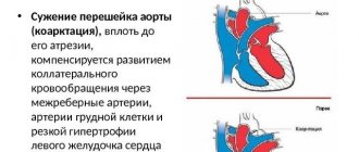
- Aortic arch. This is a small section of the main vessel, which is the basis of the entire circulatory system of the shoulder girdle and head. These vessels supplying the shoulders and head form a kind of arc that connects the descending and ascending sections of the main vessel.
- Thoracic (descending) department. Vessels that are located from the subclavian artery on the left and up to the diaphragm.
- Abdominal section. The area from the diaphragm to the bifurcation of the main vessel - the aorta.
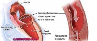
The pathology itself (aneurysm/expansion) is an increase in the diameter of the vessel by 1.5 times or more. In this state, the walls of the dilated vessel are no longer as elastic as possible, which significantly affects the speed of blood flow in the body and blood pressure. All dilations (aneurysms) are usually classified according to the zone of localization, the structure of the vessel walls, the shape and reasons for the formation of the pathology. Thus, depending on the location of the expansion, the following types of aneurysm are distinguished:
- Enlargement of the aortic root.
- Aneurysm of the ascending part of the vessel from the sinotubular ridge to the aortic arch itself.
- Arc expansion.
According to the ICD, the pathology code is I71-I71.9. This interval includes all subtypes of vessel dilatation.
General characteristics of the violation
With an aortic aneurysm, doctors diagnose a significant dilation of the vessel like a sac or spindle. This pathology can form in any part of the vessel. And based on the fact that it is through the aorta that blood is distributed to all organs with the help of increased pressure, the pathology is quite dangerous. Expansion of the lumen of the main vessel is an irreversible pathology.
Important : according to statistics, approximately 38% of cases occur in the expansion of the abdominal aorta, 24% in its ascending section and 18% in the arch.
Prognosis and complications
The long-term prognosis for patients with a thoracic aortic aneurysm depends on other medical problems, such as heart disease, high blood pressure, and diabetes. The size of the aneurysm is the main factor predicting its rupture. The survival rate for an aneurysm larger than 6 cm is 38-64% . Aneurysm rupture is the most common cause of death in patients who refuse surgery.
After rupture, the patient dies within 6 hours. Surgery significantly increases the survival rate.
Possible complications after surgery include:
- Stroke;
- Kidney damage;
- Infection;
- Heart attack;
- Arrhythmia;
- Paralysis;
- Death soon after surgery occurs in 5% of patients.
Complications from stenting also include damage to the blood vessels supplying the lower extremities, which will require another surgery.
Reasons for the development of pathology
Only a cardiologist is involved in diagnosing the enlargement of the aorta of the heart and treating this pathology. At the same time, doctors identify the following as the main reasons for the formation of pathology:
- past inflammatory and infectious processes;
- atherosclerosis (cholesterol plaques) of the main vessel;
- vascular injuries during operations on the cardiovascular system;
- congenital connective tissue dysplasia;
- congenital valve defect in a child;
- hypertension;
- congenital aneurysm in a newborn baby;
- genetic pathologies such as Marfan syndrome, etc.
Important : during pregnancy, a woman’s body develops a process of increased blood release into the aorta, which can also cause the development of vessel dilation. In addition, addiction to nicotine and alcohol can provoke vascular disease.
Types of extension
As mentioned above, all aneurysms are classified according to their localization zone. Below are the most common pathologies.
Enlargement of the abdominal aorta
One of the most common pathologies. In most cases, it is the result of blunt trauma to the abdominal area or smoking. In this case, patients often become men in the age group 75+. The danger of an aneurysm is that it always ruptures instantly and with almost no pain. However, if the rupture occurs precisely in the abdominal part, the patient feels a cutting pain in the abdomen or lower back.
If the rupture goes unnoticed, then with a high degree of probability the patient will die from internal blood loss.
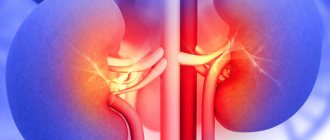
When the abdominal aorta expands, the patient may feel pain in the kidneys, pancreas, ureters, and intestines. If the dilated section of the vessel puts pressure on the ureter, this can provoke hydronephrosis. If the duodenum is compressed, the patient will experience stagnation of food in the intestines.
Important : a clear sign of such a vascular pathology is the constant felt pulsation of the vessel in the navel area.
Dilatation of the aortic arch
This area of the main vessel poses the greatest risk for aneurysm development. The fact is that in this area there is a sharp change in the direction of blood flow. That is, its speed, pressure and turbulence change. As a result, dilation of the vessel lumen may develop. Most often, an arch aneurysm manifests itself in the form of a characteristic dry cough and shortness of breath, hoarseness and dull soreness in the area of the shoulder blades, and constant aneurysmal pulsation in the wrist area.
Dilatation of the descending aorta
In this case, the expansion may have a sac-like or spindle-shaped form. Both the vessels of the thoracic and the vessels of the abdominal region can be affected. The cause of the development of a descending aneurysm is most often a cholesterol plaque. This type of pathology is detected by x-ray of the organs and vessels of the chest. Otherwise, the pathology is asymptomatic. If there are symptoms, then most often it is a burning and constant pain in the upper abdomen.
Symptoms
Thoracic aortic aneurysms grow slowly and are usually asymptomatic, making them difficult to identify. Some aneurysms will never rupture. Most small aneurysms do not progress or increase slowly in size, less than 1.2 centimeters per year. It is important to note that some aneurysms show a steady increase in diameter, which increases the risk of rupture. It is difficult to predict how quickly an aortic aneurysm will increase in diameter.
Some patients with an aortic aneurysm experience:
- soreness or pain in the abdomen or chest;
- backache;
Any part of the aorta that extends from the heart and runs in the abdomen can undergo aneurysmal change. If this occurs in the upper part of the aorta, it is called a thoracic aortic aneurysm. Most often, an aneurysm forms in the lower part of the aorta and is called an abdominal aortic aneurysm. Rarely, aneurysmal change can affect both the thoracic and abdominal aortas; this type of aneurysm is called thoracoabdominal.
You should see your doctor if you have symptoms of a thoracic aortic aneurysm.
If someone in your family has been diagnosed with an aortic aneurysm, Marfan syndrome, other connective tissue diseases, or a bicuspid aortic valve. Your doctor may recommend an ultrasound to detect an aortic aneurysm.
Diagnostics
In most cases, aortic dilatation occurs without obvious signs and symptoms. Vascular pathology is most often detected accidentally during the diagnosis of secondary diseases or during a preventive examination. If a doctor suspects dilation of a main vessel in the human body, he prescribes the following diagnostic measures to the patient:
- Radiography. Moreover, X-rays are used in relation to the section where dilation of the vessel is suspected (thoracic region or abdominal organs).
- Echocardiography. Most often used for aneurysm of the ascending section.
- CT or MRI of the thoracic/abdominal aorta as indicated.
- Angiography to assess vascular function.
Important : Often an aneurysm can masquerade as other pathological conditions, which leads the attending physician astray. That is why differentiation of aortic enlargement from tumors and other formations in the lungs or abdominal organs is required.
Risk factors
- Age.
Thoracic aortic aneurysm most often occurs in people over 60 years of age. - Tobacco smoking.
Smoking is one of the main risk factors for the formation of thoracic aortic aneurysm. With increasing smoking experience, the risk of aneurysm formation increases. - Arterial hypertension.
High blood pressure damages the blood vessels in the body and thereby increases the risk of developing an aortic aneurysm. - Atherosclerosis.
Increased levels of cholesterol and other substances that can damage the lining of blood vessels also increase the risk of aneurysms. - Floor.
Aortic aneurysms occur more often in men than in women. However, women with an aortic aneurysm have a higher risk of rupture than men. - Race.
Aortic aneurysm occurs more often in white people than in people of other races. - Family history.
If someone in the family has had cases of aortic aneurysm, then his blood relatives have an increased risk of developing an aneurysm. These people tend to form aneurysms at a younger age and have a higher risk of rupture.
Clinical picture
If we consider the signs of dilation of the main vessel, then most often the pathology is asymptomatic. If we are talking about pain, then it is usually pulsating and localized in the area of the aneurysm.
In turn, the symptoms of different types of aneurysm look like this:
- Dilatation of the abdominal aorta. Possible heaviness in the abdomen, vomiting and constipation, belching and decreased bowel function. During palpation, the doctor can feel a pulsating compaction.
- Expansion of the ascending department. Characterized by pain in the sternum (lungs, heart). In this case, the patient may experience swelling of the upper body, including the face. Dizziness, shortness of breath and tachycardia are possible.
- Expansion of the aortic arch. The patient may exhibit bradycardia (decreased heart rate), dry cough, and drooling. If compression of the aorta occurs in the area of the lungs and bronchi, then quite frequent pneumonia is possible.
Features of treatment
Treatment tactics for aortic enlargement are selected depending on the shape of the aneurysm, its location and size. With a slight expansion of the lumen of the vessel, the patient is simply observed in dynamics. A number of drugs are prescribed as maintenance drug therapy:
- antihypertensives to reduce blood pressure;
- venotonics, strengthening the walls of blood vessels;
- reducing the concentration of cholesterol in the blood;
- anticoagulants to prevent blood clots;
- vitamin complexes to normalize metabolic processes in the myocardium.
Important : all medications are prescribed only by the attending cardiologist. Folk remedies in the treatment of aneurysm are not effective.
If the lumen of the aorta in the abdominal region exceeds 4 cm, and in the thoracic region - 6 cm, then the patient is indicated for surgical intervention. Also, surgery is prescribed for a patient whose lumen has increased by 0.5 cm in six months.
The principle of surgical intervention for aortic enlargement is to remove the dilated (relaxed) section of the vessel and either sutured it or install a prosthetic stent. The operation can be performed either open or endoscopic.
Treatment of abdominal aortic aneurysm
The presence of an abdominal aortic aneurysm in a patient is an indication for surgical treatment, especially if the size of the protrusion increases by more than 0.4 cm per year.
The main operation for an abdominal aortic aneurysm is aneurysmectomy (excision of the aneurysmal sac) followed by plastic surgery of the removed portion of the blood vessel with a prosthesis made of Dacron or other synthetic material. Surgical intervention is performed through laparotomy (an incision in the abdominal wall). If the iliac arteries are also involved in the pathological process, then bifurcation aorto-iliac prosthesis is performed. Before, during and on the first day after surgery, the pressure in the cardiac cavities and the magnitude of cardiac output are monitored using a Swan-Gantz catheter.
Surgery to remove an abdominal aortic aneurysm is called an aneurysmectomy.
Contraindications to performing elective surgery for abdominal aortic aneurysm are:
- acute cerebrovascular accidents;
- fresh myocardial infarction;
- end-stage chronic renal failure;
- severe degree of cardiac and respiratory failure;
- widespread occlusion of the iliac and femoral arteries (partial or complete blockage of blood flow through them).
If an abdominal aortic aneurysm ruptures, the operation is performed for life-saving reasons on an emergency basis.
Abdominal aortic aneurysm ranks 15th on the list of diseases leading to death.
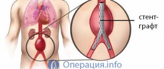
Currently, vascular surgeons prefer minimally invasive methods for treating abdominal aortic aneurysm. One of them is endovascular prosthetics of the area of pathological expansion using an implantable stent graft (a special metal structure). The stent is installed so that it completely covers the entire length of the aneurysmal sac. This leads to the fact that the blood stops putting pressure on the walls of the aneurysm, thereby preventing the risk of its further enlargement, as well as rupture. This operation for abdominal aortic aneurysm is characterized by minimal trauma, a low risk of complications in the postoperative period, and a short rehabilitation period.
Prevention
To avoid such an unpleasant disease as an aneurysm, you need to take your health seriously. In particular, from a young age, strengthen blood vessels by giving up tobacco and alcohol. It is very important after 45 years to constantly monitor blood pressure. And if there are problems with it, then even before the age of 45. Regular monitoring by a cardiac surgeon is also considered prevention for patients who are at risk.
You should always remember that a seemingly harmless problem is fraught with a very great danger. The fact is that the dilated aorta threatens the patient with sudden rupture at any moment, which can cause instant death. That is why it is worthwhile to approach the treatment of pathology adequately and with complete seriousness. Remember, delay can cost your life.