A local protrusion of the wall of the popliteal artery is called an aneurysm. It occurs after injury, surgery, inflammation or atherosclerosis. It manifests itself as pain, poor circulation, and the appearance of a pulsating formation. A complication is thrombosis, less often - rupture with the development of bleeding. Treatment requires surgery to remove, bypass, or install an arterial prosthesis.
What is a popliteal artery aneurysm?
The occurrence of aneurysmal dilatation of the artery is always associated with a violation of the structure of the vascular wall; it can be limited or widespread, in the form of a linear segment or a cavity or sac. Aneurysms can form inside, near the vessel or between its walls, and they always communicate with its lumen.
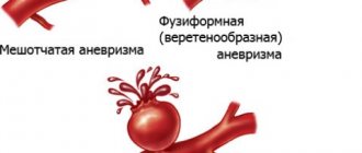
Among all such formations of peripheral arteries, the popliteal is the most typical location, almost 95% of patients are men over 60 years of age. The predominant form is fusiform; as a rule, both arteries are affected. The size of the expansion varies from 1 to 7 cm, most often it is within 2 - 3 cm.
Both large and small aneurysms can pose a circulatory risk if thrombosis occurs, or if part of a blood clot breaks off and blocks small vessels (embolism). Rupture is much less common and there are virtually no deaths once bleeding begins. The consequence of a wall tear is lameness.
We recommend reading the article about carotid aneurysm. From it you will learn about the pathology, its types and causes of development, symptoms of aneurysm, diagnostic methods and surgery. And here is more information about femoral artery aneurysm.
Vascular lesions of the liver. Hepatic artery aneurysm
Hepatic artery aneurysm
or its branches are a relatively rare lesion. Suspicion of it arises in cases of hemobilia or in the presence of a ring-shaped shadow at the site of the hepatic artery (it appears when lime salts are deposited in the wall of the aneurysm). A large aneurysm can cause depression on the upper contour of the duodenal bulb.
Arteriovenous fistula in the liver
is formed mainly as a result of puncture and biopsy of liver tissue. Recognition is based on arteriography data, in which blood discharge into the portal veins is observed.
Portal vein aneurysm
- an extremely rare lesion. Recognized only by angiographic examination. May be a congenital condition or develop in association with portal hypertension. It is located along the main trunk of the vein or deep in the liver.
Among the birth defects
refers to hypoplasia of the portal vein.
It is usually combined with preservation of patency of the umbilical vein and is accompanied by liver atrophy (this entire complex of pathological conditions is known as Cruvelier-Baumgarten disease).
A tangle of dilated veins in the area of the anterior abdominal wall and umbilicus suggests either Cruvelier-Baumgarten disease or Cruvelier-Baumgarten syndrome.
Arteriography and portography
make it possible to exclude cirrhosis of the liver, identify a narrow portal vein, a patent umbilical vein and a tangle of dilated vedae in the navel area.
Congenital cavernomatous transformation
The portal vein is rare. In most cases, cavernomatous transformation is a consequence of pylephlebitis and portal vein thrombosis.
Initially, with thrombosis, obstruction of the portal vein and a network of collaterals in the area of the splenic portal and along the splenic vein are determined.
Subsequently, instead of the shadow of the portal vein, splenoportography reveals a whole network of small tortuous vessels through which the contrast agent from the spleen enters the liver.
Hepatic artery
compensatory expanded. With incomplete thrombosis, the splenic vein is dilated, and the portal vein is narrow and has an uneven outline. With complete thrombosis, the shadow of the portal vein is interrupted, and the contrast agent fills the collaterals.
In the case of recanalization of a thrombus, a narrow channel is found along the portal vein, sometimes with longitudinal clearing along the axis of the vein. In most patients with phlebothrombosis, an enlarged spleen is detected.
In rare cases, portal vein thrombosis
This is evidenced by rounded shadows of calcifications visible on conventional radiographs along the portal vein.
With phlebosclerosis of the portal vein
lime deposits in its walls look like delicate parallel strips.
In the literature of recent years, there are many descriptions of the appearance of gas
in the intrahepatic branches of the portal vein.
As a rule, it occurs as a result of gangrenous or phlegmonous damage to the intestinal wall with the participation of gas-forming microorganisms. Therefore, prognostically this is usually an unfavorable sign. However, there are descriptions of a similar syndrome that arose as a result of irrigoscopy in patients with ulcerative or granulomatous colitis.
In such cases, the appearance of gas in the portal vessels was not accompanied by clinical symptoms.
Gas accumulations
cause the appearance on radiographs of narrow branching light stripes against the background of the shadow of the liver, mainly in the cranial parts of the organ.
Occlusive lesions of the hepatic veins
varied. Some of them develop as a result of thrombosis due to endophlebitis or polycythemia. Such primary venous lesions are commonly called Chiari disease.
There are occlusive lesions associated with a congenital anomaly (narrowing of the veins, the presence of membranes in them, etc.). Secondary obstructions of the hepatic veins are observed much more often with tumors originating from the liver or kidney, or with abdominal trauma.
Such secondary occlusive lesions are combined under the term Budd-Chiari syndrome.
If there is a membrane
and in the hepatic segment of the inferior vena cava, the only diagnostic method is inferior cavography. The membrane causes a thin light stripe that crosses the shadow of the vein.
Fibrous cords and expansively growing tumors cause a narrowing of the vein while maintaining the clarity of its outline.
Thrombosis of the inferior vena cava is radiologically manifested by single or multiple filling defects in its shadow (marginal defects, bizarre clearing along the axis of the vein).
With complete blockage of the inferior vena cava
X-ray images reveal shadows of collateral vessels.
Outflow of contrast agent
goes along the paravertebral and further azygos and semi-gypsy veins. With malignant tumors, filling defects with uneven contours are visible, up to the “amputation” of the vein.
Changes in the hepatic veins
detected using hepatovenography. The normal x-ray picture of these veins is disrupted, their curvature, narrowing, and their intermittency due to thrombosis are detected.
With thrombosis of the hepatic vein in one of the liver lobes, atrophy of this lobe and hypertrophy of the other lobe occur.
These changes can be detected by arteriography, but since they outwardly resemble the angiographic picture of a hepatoma or cirrhotic lesion, it is necessary to perform hepatography—direct injection of a contrast agent into the liver parenchyma.
This technique allows you to find out the cause of the postsinusoidal block
and establish the nature, location and extent of damage to the hepatic veins.
In Chiari disease, multiple tortuous collateral veins are identified, forming an X-ray “pattern” in the form of a web. The hepatic lesions are not visible, but portal vessels and lymphatic vessels are often visible.
When the veins are blocked in one lobe, the outflow of the contrast agent occurs in the branches of the portal vein, through which it enters the other lobe in a roundabout way.
Through intraparenchymal administration
contrast agent can recognize thrombotic, neoplastic lesions and membranes in the hepatic and inferior vena cava.
– Return to section table of contents “
diagnosis of diseases.”
Table of contents of the topic “Liver diseases.”: 1. Cholesterosis. Diagnosis of cholesterosis. 2. Adenomyomatosis. Lipomatosis. Hyalinocalcinosis. 3. Anomalies and paralysis of the diaphragm. Liver in systemic diseases. 4. Liver for gastrointestinal diseases. Liver for dysentery and ascariasis. 5. Biliary tract after surgery. Postcholecystectomy syndrome. 6. Diagnosis of postcholecystectomy syndrome. Bile ducts after cholecystectomy. 7. Diagnosis of cholecystoduodenostomy. Diagnosis of cholecystojejunostomy. 8. Blood flow in a normal liver. Study of liver blood flow. 9. Percutaneous transhepatic portography. Diagnosis of percutaneous transhepatic portography. 10. Lesions of liver vessels. Hepatic artery aneurysm.
Source: //medicalplanet.su/diagnostica/382.html
Reasons for development
The most common factor in the formation of an aneurysm is atherosclerosis, which leads to thinning of the artery wall and is accompanied by blockage of its lumen. The progressive course of the disease often causes thromboembolic complications requiring emergency surgical care.
Other causes of damage to the popliteal artery include:
- injuries - fractures, wounds, blows, operations, they contribute to the appearance of pulsating hematomas - false aneurysms near the site of arterial damage;
- arrosion (corrosion of the wall) - a tumor or infectious process;
- congenital structural anomaly, failure of connective tissue;
- arteritis of a bacterial or fungal nature, phlebitis.
Vascular atherosclerosis
What you need to know about the disease
More often, this pathology occurs as a complication of other aneuric vascular deformations (abdominal or iliac arteries), but can also be diagnosed as an independent disease.
With an aneurysm of the popliteal artery, dissection of the vascular wall occurs at the level of the popliteal fossa. The artery wall loses its elasticity (splits) at the site of damage and becomes deformed, forming a protrusion and expansion of the vessel. The vascular lumen can increase almost 2 times.
Aneurysms are characterized primarily by a disturbance in the speed of blood flow: at the site of deformation it slows down, often causing stagnation and thrombus formation. Slowing blood flow leads to the fact that the tissues below the site of pathological expansion receive less blood and experience oxygen starvation.
It is customary to distinguish two types of aneurysms:
- true ones, which appear under the influence of unfavorable factors;
- false, arising as a complication of punctures or surgery.
Both types manifest similar symptoms and are equally dangerous for the patient. The distinction between true and false aneurysms is important only for the angiosurgeon when determining treatment tactics.
People at risk
It has been established that aneurysms are most often diagnosed in patients with the following risk factors:
- smoking;
- chronic alcoholism;
- addiction;
- hypertonic disease;
- diabetes;
- unhealthy diet with excess animal fats;
- obesity;
- knee injuries or surgery.
Sports joint injuries
Causes
Popliteal artery aneurysms account for about 1% of all surgical vascular diseases and often occur in both legs. The main reason is the congenital weakness of the artery wall, which contributes to their pathological expansion. The majority of patients (95%) are elderly men with an average age of approximately 71 years. The exact reasons for the development of dilation in the popliteal artery are unknown, but there is a clear connection with atherosclerotic changes in the vessel wall. Sometimes the pathology develops as a result of injuries to the popliteal region, dislocations or fractures. Patients with multiple aneurysms in different arteries should have general tissue weakness. The exact nature of this is still unclear. The tendency of the popliteal artery to pathological expansion is associated with frequent flexion and extension of the vessel due to movements in the knee joint.
Symptoms of pathology
The disease may not manifest itself for a long period of time. Small formations are found by chance when examining the vessels of the extremities for other pathologies. One of the features of the popliteal artery is its location at the site of active movements, so such aneurysms tend to grow. As it increases, patients may experience leg pain, numbness, and tingling.
Signs of circulatory problems include:
- fatigue when walking;
- cold feet;
- pale skin with a cyanotic tint;
- convulsive muscle twitching;
- decreased tissue nutrition in the form of dry skin, dermatitis, ulcers.
Causes of hepatic artery aneurysm
The most common cause of hepatic aneurysm development is liver trauma; non-traumatic aneurysms can be caused by systemic inflammatory diseases, cholelithiasis and cholangitis. One possible cause is atherosclerosis with degeneration of the vascular wall of the hepatic artery.
Injuries and ruptures of the liver are one of the most common causes of intrahepatic aneurysm. Moreover, an aneurysm can develop any time after the injury. Since the disease manifests itself only during complications, diagnosis is either accidental or delayed.
Thrombosis and other complications
In the cavity of the aneurysm, the direction of blood movement changes - from linear to turbulent. This leads to the rapid formation of blood clots that gradually or suddenly cut off blood flow. A sign of the onset of thrombosis is unbearable pain in the leg; it practically does not decrease after the use of painkillers, and only increases over time.
In the future, the following are added to the pain syndrome:
- “stocking” type loss of sensitivity; Thrombosis of the extremities
- marbled leather pattern;
- sudden cold snap and paleness of the feet and legs;
- decreased muscle strength;
- pain on palpation of muscles;
- swelling of the lower leg;
- impaired mobility in the joints.
Progressive arterial insufficiency is dangerous for the development of gangrene of the lower extremities.
Complications.
The most severe complication of this disease is the rupture of an aneurysm with the development of gastrointestinal bleeding and hemorrhagic shock, leading to death. Blood breakthrough can occur into the abdominal cavity (43%), into the lumen of the stomach and duodenum (11%), into the bile ducts (41%) or into the portal vein (5%) [30]. The risk of rupture with an aneurysm diameter greater than 2 cm reaches 50%, and overall mortality due to rupture approaches 70% [15]. An increase in the diameter of the aneurysm when choosing non-surgical management tactics was noted in 27% of cases and amounted to up to 0.8 cm over three years [17]. In addition, it was noted that the risk of rupture of aneurysms resulting from non-atherosclerotic changes is much higher. Due to the high incidence of spontaneous rupture, surgical treatment is indicated for aneurysms larger than 2 cm in diameter [31].
Diagnostic methods
To identify an aneurysm, sometimes an examination is sufficient: a pulsating formation protruding above the skin is identified, upon auscultation of which a systolic murmur can be detected. Instrumental diagnostics helps to clarify the size of the aneurysm, the presence of a blood clot, and distinguish it from similar diseases. For this use:
- Ultrasound of the vessels of the lower extremities with Doppler scanning;
- angiography in combination with x-ray control;
- or MRI.
Before the operation, a complete assessment of the regional blood circulation is required; when performing bypass surgery with one’s own vein, the site of its intended extraction is examined.
Ultrasound of blood vessels of the lower extremities
Diagnostics
At the slightest suspicion of an aneurysm, you should consult a doctor to schedule an examination. Every person must remember that untimely detection of any disease can cause the development of pathological changes occurring in the body. Vascular aneurysm is a disease that requires strict control, high-quality diagnosis and proper treatment. The examination is carried out by a highly specialized specialist - a vascular surgeon.
For diagnostic examination the following are used:
- Ultrasound Doppler. This diagnosis combines standard ultrasound and Doppler scanning. With its help, you can see the movement of blood through the vessels, measure the speed of blood flow, determine the size of the vessels, and also identify possible blockage of the veins.
- Angiography. It is an x-ray examination and helps determine the correct functioning of the vascular system.
- MRI. Thanks to magnetic resonance imaging, the specialist receives a three-dimensional image, which, if desired, can be zoomed in to determine the condition of the vessels.
In some cases, the doctor may order a general blood and urine test. This is required in order to obtain an overall picture of the development of the aneurysm.
Treatment of popliteal artery aneurysm
Watchful waiting and constant monitoring of the patient are acceptable for small aneurysms (up to 2 cm) and the absence of clinical manifestations. This point of view is not supported by all surgeons, because even small formations can lead to acute thrombosis. Most patients are advised to have the aneurysm removed as soon as possible. If a blood clot is detected, surgery is performed on an emergency basis.
Surgical options
Two types of surgical interventions can be used to treat vascular pathology:
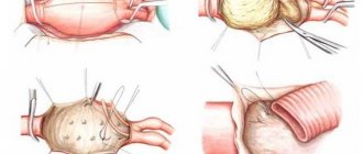
- Resection of the aneurysmal sac and replacement of part of the vessel with a synthetic prosthesis or one’s own vein. Most often, the aneurysm is ligated and a shunt is placed around it.
- Endosurgical method with installation of an intra-arterial endoprosthesis using a special catheter. This isolates the aneurysm wall from blood flow, reducing the risk of rupture and thrombosis.
Resection of the aneurysmal sac and replacement of part of the vessel with a synthetic prosthesis
Recovery after
On the first day after open aneurysm removal, patients are recommended to remain in bed. During endovascular operations, you can walk immediately after completion, but do not put excessive strain on your leg. Your doctor may prescribe compression stockings to reduce swelling and reduce the risk of blood clots.
During the rehabilitation period, the following recommendations must be followed:
- normalize body weight;
- do not consume fatty meats, offal, and fats;
- drink enough clean water (in the absence of edema);
- take medications to normalize blood clotting and cholesterol levels;
- control blood pressure and correct indicators using antihypertensive drugs;
- practice walking daily with a gradual increase in distance;
- treat the postoperative wound; if signs of inflammation appear, immediately contact a surgeon;
- During night sleep, give your legs an elevated position.
It usually takes at least a month to fully restore the functions of the lower limb, although pain and circulatory problems disappear almost immediately. To speed up the healing process and prevent the formation of blood clots, dosed physical activity is necessary. At the same time, both lack and excess of loads worsen the results of the operation.
Diagnosis and treatment of the disease
This disease should not be left to chance; at the first symptoms, it is better to see a doctor.
Then you will have to do a series of instrumental studies, including duplex ultrasound scanning and arteriography. These methods allow you to see the location of the arterial aneurysm and determine its size. In case of an asymptomatic course of the disease and with a slight increase in the lumen of the vessel, a wait-and-see approach is recommended, which consists of periodic monitoring of the condition of the artery using ultrasound scanning. With the possible development of complications, in particular thrombosis or rupture of an aneurysm, treatment is exclusively surgical.
The basic treatment method is surgery, which prevents potentially dangerous complications, in particular blockage of blood circulation in the lower limb and the formation of blood clots. In medicine, there are several methods of surgical intervention:
- Open surgery involves cutting through the soft tissue to gain direct access to the blocked artery. After which the aneurysm is bypassed, i.e. a vessel from another part of the body or an artificial implant is taken and sewn in, bypassing the aneurysm.
- Endovascular (closed) surgery is a minimally invasive procedure with a minimum of traumatic manipulations. A catheter is inserted through a small incision in the skin, then a cylindrical stent made of wire is installed. It serves to strengthen the walls of the vessel, preventing their stenosis (fusion), the formation of blood clots and rupture of the aneurysm. After such an operation, the patient recovers quickly and does not stay in the hospital for long.
Folk remedies to combat the problem
In the presence of an aneurysm, all conservative treatment methods, including herbal medicine, are completely ineffective, so if surgery is indicated, then there is no point in relying on traditional medicine. Infusions and decoctions of herbs can be beneficial for preventing vascular pathology and slightly slow down the growth of formations in small sizes.
For this purpose, you can use plants that:
| Action | Plant species |
| Regulate blood cholesterol levels | azure blue, clover, alfalfa, dioscorea; |
| Improve blood circulation | golden mustache, lemon balm, mint, horsetail; |
| Strengthens vascular walls | rue, horse chestnut; |
| Reduce the risk of blood clots | licorice, Kalanchoe juice, hop cones, sage; |
| Normalize blood pressure | mistletoe, sophora, calendula, hawthorn. |
To improve peripheral blood flow, you can use a collection of equal parts of dandelion root, rosemary sprigs and horsetail herb. A tablespoon of the mixture is poured into a thermos and poured a glass of boiling water overnight. You need to drink 80 ml three times a day half an hour before meals. Treatment is carried out for at least a month.
Causes of aneurysm formation
This pathology appears in cases where the vascular wall loses its tone and can no longer compensate for the internal blood pressure. Therefore, the reasons for the development of the disease include the following factors that cause weakening of the walls of arteries and veins:
- Genetics. When certain genes are mutated, the connective fibers that make up the blood vessels are weakened. Such mutations are inherited.
- Atherosclerosis. This inflammatory process leads to the formation of atherosclerotic plaques. They locally increase the pressure inside the vessel and cause the appearance of a bulge.
- Injuries. A simple blow to the area of a vessel can greatly weaken its walls and provoke an aneurysm.
- Infectious processes. They can be caused by infections and fungi. Tuberculosis and syphilis are considered the most dangerous in terms of aneurysm formation.
- Lack of nutrients in the body. Copper deficiency has a particularly strong effect on the condition of blood vessels. It is involved in the synthesis of an enzyme that accelerates the formation of collagen and elastin precursors. If there is a deficiency of the latter, the vascular walls will become fragile and flabby. This will promote the formation of bulges.
There are factors that are unable to independently cause an aneurysm, but significantly increase the risk of developing this pathology. This is heredity and high blood pressure.
Prevention of pathology
The formation of an aneurysm and its complications can be prevented by eliminating the main risk factors - smoking, alcohol abuse, and excess weight. To reduce blood cholesterol levels, a diet is required, which is based on:
- vegetables - salads with vegetable oil, boiled or steamed;
- cereals (buckwheat, oatmeal) for side dishes, casseroles, first courses;
- fresh fruits, berries or freshly squeezed juices from them;
- lean meat and fish, boiled or baked;
- fermented milk drinks (kefir, yogurt) homemade using starter cultures, low-fat cottage cheese;
- first courses without meat, steamed meatballs can be added before serving;
- drinks – green tea, rose hips, chicory.
The minimum duration of exercise therapy or walking should be 30 minutes per day. Sports such as swimming, cycling, skating, and skiing are useful. If you have a predisposition to vascular diseases of the lower extremities, it is not recommended to lift weights or exercise in the gym with weights.
We recommend reading about aortic aneurysm surgery. You will learn about the indications for surgery, what surgical interventions are performed, as well as rehabilitation and consequences after them. And here is more information about occlusion of the femoral artery.
Popliteal artery aneurysm is the most common type; it is most often diagnosed in older men. The course of the disease is asymptomatic for a long time; as it increases, pain and numbness appear in the legs. Among the complications, the most serious is thrombosis; with severe arterial insufficiency, gangrene develops.
If an aneurysm larger than 2 cm in size or signs of arterial blockage are detected, surgery for removal, bypass or endovascular replacement is indicated. To prevent the disease, correction of diet, lifestyle, and taking recommended medications and herbal remedies is required.
Iliac artery aneurysm mortality
The iliac artery is one of the largest (second only to the aorta) blood vessels. This is a paired vessel, its length is 5-7 centimeters, and its diameter is 11-13 millimeters.
The arteries begin at the bifurcation of the aorta, which is located at the level of the fourth lumbar vertebra.
And in the area of the articulation of the iliac bones and the sacrum, the arteries split into the internal and external iliac arteries.
Structure and functions of the artery
The iliac arteries are the largest in the human body, with the exception of the aorta, from which they emerge. In turn, these arteries also break up into smaller ones, which also break up into branches.
The internal artery divides into the iliopsoas, middle rectal, lateral, inferior and superior gluteal, sacral, as well as obturator, internal genital and inferior vesical branches.
They deliver blood to the inner walls of the pelvic cavity and to the organs.
The external artery also supplies blood to the pelvic cavity and passes into the femoral artery in the lower extremities. The femoral artery divides into branches that supply the thigh, foot and lower leg. In men, the iliac artery supplies blood to the membranes of the testicle, thigh, bladder and penis.
Iliac artery aneurysm
One of the dangerous diseases, an iliac artery aneurysm, can be completely asymptomatic at first, and only when it reaches a large size does it begin to cause discomfort.
The aneurysm itself is a protrusion of the vessel wall with the formation of a kind of sac. The artery wall begins to gradually lose elasticity and is replaced by connective tissue.
The causes of an aneurysm have not been fully established; it may be trauma, atherosclerosis or hypertension.
A ruptured aneurysm is a dangerous condition that can lead to gastrointestinal bleeding, decreased blood pressure and heart rate, and collapse.
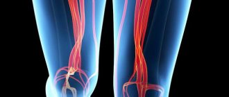
If the blood supply in the area of the aneurysm is disrupted, this can lead to thrombosis of the arteries of the leg, femoral artery and pelvic vessels.
Circulatory disorders are accompanied by pain and dysuric disorders.
Diagnosis of an aneurysm of this artery can be carried out in different ways, for example, using ultrasound, computed tomography or magnetic resonance imaging, duplex scanning or angiography.
Occlusion of the iliac arteries
Occlusion, like iliac artery stenosis, in most cases occurs as a consequence of arterial atherosclerosis, thromboangiitis obliterans, aortoarteritis, and fibromuscular dysplasia.
Stenosis of the iliac artery leads to the development of tissue hypoxia and disruption of tissue metabolism. Oxygen starvation of tissues contributes to the accumulation of under-oxidized metabolic products and metabolic acidosis.
And the increase in blood viscosity, which is inevitable in such a state, leads to the formation of blood clots.
There are different types of iliac artery occlusion:
- nonspecific aortitis,
- mixed form of arteritis, aortitis and atherosclerosis,
- iatrogenic occlusions,
- post-embolic occlusions,
- post-traumatic occlusions.
According to the nature of the lesion, chronic occlusion of the iliac arteries, thrombosis and stenosis are distinguished.
When treating occlusion, conservative and surgical methods are used. Conservative treatment includes pain relief, normalization of blood clotting, relief of vascular spasms and expansion of collaterals. Surgical treatment involves resection of the affected area with replacement with a transplant, opening of the artery with removal of plaques, sympathectomy, or a combination of different methods.
Popular articles on the topic: iliac artery aneurysm
The term “aneurysm” describes a saccular expansion of a vessel or heart, caused by exposure to damaging factors and leading to various disorders, and above all to significant problems with blood circulation.
Many doctors know what this condition is, but not everyone understands its etiology, clinical manifestations, and does not fully master the correct diagnostic and clinical algorithms for managing patients with abdominal ischemic syndrome (AIS).
The history of the study of atherosclerosis goes back more than three centuries; more than one generation of scientists has devoted their lives to this problem. Despite this, in the process of studying the pathogenesis and morphogenesis of atherosclerosis, more hypotheses and assumptions arose than answers.
On November 16, a very important event for our country took place in Kyiv - the opening of the 1st Congress of Vascular and Endovascular Surgeons of Ukraine.
Systemic vasculitis (SV) is a group of diseases characterized by primary damage to the walls of vessels of various sizes, such as focal inflammation and necrosis, and secondary involvement in the pathological process of organs and tissues of the vascular zone.
Currently, the problem of diabetes mellitus (DM) has become a problem of cardiovascular pathology: the fate and prognosis, ability to work and quality of life of the patient are determined by cardiovascular disorders. According to WHO experts, duration.
The relevance of discussing the problem of tactics and strategies for the behavior of a doctor in the presence of acute abdominal pain in a patient is beyond doubt.
Acute gastrointestinal bleeding can be a complication of a number of diseases; according to various authors, their frequency is 50-150 cases per 100 thousand population per year.
An aneurysm is an expansion of part of the aorta by more than 3 cm and a protrusion of its wall. The disease develops as a result of degenerative changes in vascular tissues and in 95% of cases is caused by atherosclerotic lesions.
The cause of the disease can be genetic predisposition, bruises and injuries, poor lifestyle (smoking, excess weight).
The risk group includes:
- elderly (over 55 years of age) people who suffer from acute pain in the abdomen or lower back, regardless of the presence or absence of a palpable pulsating formation;
- middle-aged patients who have suffered any trauma to the abdomen, spine, etc.;
- persons who received blunt abdominal injuries (bruises) as a result of an accident, as a result of which they develop a traumatic aneurysm.
Rice. 1. Abdominal aorta in normal condition
Symptoms of the disease
- Periodic or constant pain or discomfort in the abdominal area at rest and/or when eating or exercising;
- Pain in the side or lower back, possible spread of pain to other areas (buttocks, legs, groin). The nature of the pain can be different - aching or throbbing;
- Feeling of pulsation in the abdomen;
- Pain in the calf muscles when walking when the aneurysm spreads to the arteries of the lower extremities;
- Darkening or blueness of the fingers, their soreness, cold feet - with thrombosis of the aneurysmal section of the aorta and arteries of the lower extremities, which can lead to gangrene of the limb and, subsequently, amputation;
- Increased blood pressure that is not reduced by medications;
- Current symptoms of the inflammatory nature of the aortic aneurysm are weight loss, fever;
- The disease progresses over a long time and may be asymptomatic;
Rice. 3. Abdominal aortic aneurysm. Computed tomography of the abdominal aorta
Most abdominal aortic aneurysms are diagnosed as incidental findings, during examination or when performing ultrasound of the abdominal organs, blood vessels, CT (computed tomography) or MRI (magnetic resonance imaging).
When an abdominal aortic aneurysm is detected, it is determined whether there is a tear in the wall or a dissection, the size of the aneurysm and its exact location - a more detailed examination is performed (aortography, computed tomography of the abdominal cavity with contrast of the aorta and arteries of the legs (CT or MRI).
Blood tests: a general blood test with an assessment of the leukocyte formula, a biochemical blood test, with an assessment of whether there are disorders associated with the liver and kidneys, and a coagulogram is also performed.
Conservative and surgical treatment methods
Diagnostics provides all the necessary information to make a decision on the method of treatment. During the study, the size of the aneurysm is determined. If its diameter exceeds 5 cm, there is a high probability of rupture, internal bleeding and death.
Conservative treatment
If the diameter of the aneurysm is relatively small (small aneurysm, without the threat of dissection or rupture), conservative (medicinal) treatment is prescribed with mandatory dynamic monitoring of the abdominal aortic aneurysm. It assumes:
- transition to a healthy lifestyle (quitting smoking, alcohol, controlling cholesterol levels);
- taking medications to lower blood pressure (for hypertension);
Indications for use and methods of surgical treatment
If the diameter of the aneurysm is more than 5 cm or it increases by more than 5 mm per year, surgical treatment is indicated for patients.
The standard method is open surgery or aortic replacement.
Its essence is prosthetics (removal of the affected area of the aorta and replacing it with an artificial prosthesis): the aorta is clamped, the aneurysm is excised and a vascular prosthesis is installed in its place.
To access the aorta, a 7-15 cm long incision is made on the abdomen. The operation lasts 3-4 hours under general anesthesia.
When the aneurysm spreads to the iliac arteries (arteries of the lower extremities), bifurcation prosthetics is performed with removal and suturing of the prosthesis on the hips (in the groin areas), the so-called “pants” prosthesis.
Rice. 5. Prosthetics of the abdominal aorta and iliac arteries
Endovascular prosthetic method (stenting of the abdominal aorta) in which the damaged area of the aorta is replaced without an incision. The prosthesis (stent) is “delivered” using a catheter, which is inserted into the area of the inguinal fold. The movement of the catheter is monitored using x-rays.
As a result, the aneurysm ceases to participate in the main blood flow, as a result of which the likelihood of its rupture is significantly reduced.
Source: //serdce-moe.ru/zabolevaniya/anevrizma/anevrizma-podvzdoshnoj-arterii-letalnost
Symptoms of the disease
At the initial stages of development, aneurysms do not manifest themselves in any way. They can only be detected during a medical procedure. That's great rarity. Most often, the problem is discovered when a vessel ruptures and the patient’s life is threatened.
Symptoms depend on the location of the disease. For example, if the disease affects the blood vessels of the brain, the patient may experience the following symptoms:
- Headaches of unknown etiology.
- Sudden dizziness. Problems with coordination of movements.
- Speech dysfunction.
- Double vision.
If the aneurysm ruptures, hemorrhage occurs, leading to a subarachnoid hematoma. The patient experiences classic symptoms of a hemorrhagic stroke: muscle stiffness, loss of vision and hearing, convulsions, paralysis of the limbs.
Arterial aneurysms
All arterial aneurysms are accompanied by circulatory disorders in the periphery. The severity of symptoms depends on the type of damage, the magnitude of the expansion, the size of the aneurysmal sac, and the caliber of the vessel.
The appearance of terminal aneurysms leads to a complete blockage of blood flow along the main highways. The tissues of the limbs receive nutrition only through collaterals.
Lateral aneurysms with small sacs have virtually no effect on blood circulation.
If the collateral blood supply is sufficient, then ischemic disorders do not occur in the tissues of the extremities. If there is insufficient blood supply, foci of necrosis may appear on the patient’s arms and legs, followed by their transition to gangrene when an infection occurs.
Combined disorders
Combined aneurysms rarely lead to severe ischemic disorders in the extremities. Only one variety can cause gangrene. We are talking about a retrograde aneurysm.
Combined violations pose a different danger. They lead to the discharge of arterial blood into the venous bed, which significantly increases the load on the heart. This leads to myocardial hypertrophy and heart failure. The rate at which symptoms of heart disease appear depends on the volume of discharge and the distance from the zone of mixed expansion to the heart.
In the area of traumatic aneurysms, symptoms such as swelling and pulsation in the affected area occur.
Common symptoms of aneurysms include:
- Swelling of the limbs.
- Trophic ulcers.
- Numbness of legs and arms.
- Increased sensitivity to low and high temperatures.
- Dyspnea.
- Insomnia.
- Fatigue.
- Tachycardia.
All types of such pathology must be treated. Otherwise, serious complications may develop that can make a person disabled or cause his death.