general description
Thrombosis and arterial embolism is a disease that occurs when the lumen of the arteries is acutely blocked by a thrombus or embolus, leading to disruption of blood flow in the tissues.
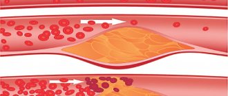
When the lumen of the vessel is blocked by a thrombus, further blood flow through the vessel stops, muscles and tissues experience oxygen starvation. In the absence of timely treatment, tissue death and the formation of gangrene occur. The most typical sites for localization of thrombi and emboli are the sites of vessel branching (bifurcation), since it is there that the diameter of the vessel decreases.
| The thrombus blocks the lumen of the vessel |
There are several possible sources of arterial thromboembolism:
1. Heart (most common source):
- for diseases characterized by rhythm disturbances;
- with aneurysm of the left ventricle;
- when endocarditis occurs (infectious and other forms);
- after undergoing operations with prosthetic heart valves.
2. Vessels with the presence of atherosclerotic plaques:
- with aortic aneurysm;
- in places of ulceration of atherosclerotic plaques.
| Thromboembolism of the left common iliac artery at the origin of the aorta |
Causes of the disease
Acute subclavian vein thrombosis is a rare pathology that occurs due to the formation of a blood clot in the vessels. Vulnerable to the disease are people who play sports and patients who take injectable drugs for a long time. Common reasons influencing the development of the disease are:
- occlusion of the brachial vein due to physical activity and excessive stress;
- genetic predisposition of blood cells to clotting and the formation of blood clots;
- prolonged administration of medications through the catheter system with subsequent clot formation.
Return to contents
Symptoms of acute thrombosis (embolism) of the arteries of the limb
According to the classical classification according to Savelyev V.S., which fully reflects the clinical picture of the disease, there are 3 degrees.
I degree:
- IA - the appearance of numbness in the distal parts of the limb and coldness of the feet and hands.
- I B - the addition of pain syndrome (pain is constant).
II degree:
- II A - impaired sensitivity and active movements in the limb (lack of tactile sensitivity in the fingers and toes).
- II B - complete absence of active movements in the limb (passive movements are preserved).
III degree:
- III A - subfascial edema of the limb occurs.
- III B - muscle-joint contracture is formed (the presence of limb contracture indicates the death of the limb and the development of gangrene, which, in turn, requires surgical treatment in the amount of amputation of the limb).
Localization options
The formation of blood clots is a natural process for the human body, since it is blood clots that clog damaged vessels, stopping bleeding. However, if a blood clot forms in a healthy artery, this cannot be called a favorable process.
The resulting clot interferes with blood circulation, and as a result of separation, it can clog the vessels of vital organs, which will lead to more serious pathologies.
Although blood clots can form in any artery, there are a few vessels that are most often at risk.
Brachial artery thrombosis
Pathology with this localization is different in that in most cases it is asymptomatic, and the patient is unaware of its presence for a long time until the last stage of development occurs.
When the blood clot reaches a significant size and significantly interferes with blood circulation in the affected area, the upper part of the arm gradually becomes numb, becomes cold to the touch, the pulse in it slows down, and the skin acquires a bluish tint. In this case, you must definitely consult a doctor for help.
In most cases, surgical intervention is performed, since conservative therapy is often unproductive, and delay can lead to complete loss of the limb.
Aortic thrombosis
The aorta is the largest artery in the human body, from which all the others branch. A blood clot formed in this vessel may not be dangerous if it reaches a small size and is diagnosed promptly. However, there is another option, in which the clot grows so large that it completely blocks the lumen of the vessel, causing instant death.
Thrombosis of the superior mesenteric artery
In another way, this pathology is called blockage of mesenteric vessels. Symptoms of the disease include weight loss, abdominal pain and bowel movements.
Most often, this type of thrombosis occurs in weakened elderly people with a tendency to cardiovascular diseases. Obstruction of the superior mesenteric artery leads to acute oxygen deprivation and necrosis, followed by death.
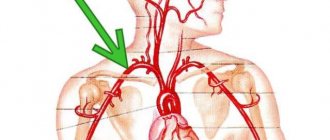
Thrombosis of the subclavian artery
This pathology leads to impaired blood circulation in the upper limb and brain. The left side is most often affected because it is directly connected to the aortic arch.
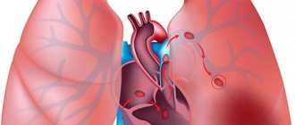
Coronary artery thrombosis
The coronary arteries deliver oxygen to the elements of the heart, so the condition of these vessels is important for its smooth operation. Coronary thrombosis is dangerous because its symptoms are similar to those of other heart diseases, making diagnosis difficult.
With this pathology, the patient may feel pain in the chest, lack of air, breathing rhythm, heartbeat may be disrupted, and skin may become pale. The formation of a blood clot in a coronary artery often causes myocardial infarction.
Arterial thrombosis is no less dangerous than venous thrombosis, since embolism can occur as a result of the formation of a blood clot and its rupture.
This is a condition in which a blood clot moves through the bloodstream from one organ to another, and it is impossible to predict where it will go. In some cases, it is possible to avoid negative consequences if the clot clogs a vessel near which there are bypass paths for blood flow (collateral vessels). However, in most cases the prognosis for patients is poor.
A white blood clot, which consists of platelets, most often forms in the artery.
Diagnostics
1. Consultation with a vascular surgeon and examination. After consultation with a specialist, urgent hospitalization in a hospital for surgical treatment is necessary.
2. Instrumental diagnostic methods:
- Doppler ultrasound of arteries.
- Angiography is the most informative diagnostic method, allowing you to decide on further treatment tactics.
- Computed tomography with contrast injection.
In most cases, an examination by a specialist is enough to make an accurate diagnosis and decide on further treatment tactics.
Treatment of acute thrombosis (embolism) of the arteries of the limb
The first priority in therapy is the intravenous administration of Heparin (to reduce the risk of thrombosis of distal areas), antispasmodics (to relieve vasospasm) and antiplatelet agents (Reopoliglyukina, Trentala).
Treatment is aimed at eliminating the true cause of the disease (endocarditis, etc.).
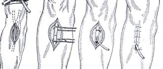
Surgical treatment is most often performed under local anesthesia, less often under general anesthesia or epidural anesthesia. The choice of surgical intervention depends on the location of the occlusion of the vessel lumen. Direct or indirect thrombectomy is performed.
| Indirect thrombectomy with a Fogarty probe. An arteriotomy is performed, a probe is inserted through the hole in the artery, the balloon is inflated and pulled out along with the blood clots | Direct thrombectomy. An artery is isolated at the site of the thrombosed area, an arteriotomy is performed, and the blood clot is removed using instruments within the wound |
Essential drugs
There are contraindications. Specialist consultation is required.
- Heparin sodium (direct anticoagulant). Dosage regimen: intravenous bolus - 80 units per 1 kg of body weight, initial infusion rate - 18 units per 1 kg of body weight per hour (not less than 1250 units/hour). Next, dose selection is carried out by determining the APTT. The duration of administration of sodium heparin should be 5-7 days. (with a gradual transition to NACG). When transferring a patient to NACG, the latter are prescribed 4-5 days in advance. before the expected withdrawal of UFH. The administration of UFH is stopped only when the INR value reaches 2.0-3.0.
- Streptokinase (fibrinolytic agent). Dosage regimen: intravenous infusion of 250 thousand - 500 thousand IU over 15-30 minutes, then 100 thousand IU/hour for 12-72 hours; intravenous infusion of 1500 thousand IU is possible over 2 hours.
- Urokinase (fibrinolytic agent). Dosage regimen: intravenous infusion of 4400 IU/kg over 10-20 minutes, then 4400 IU/kg per hour for 12-24 hours; intravenous infusion of 3 million IU over 2 hours is possible.
- Reopoliglucin (improving microcirculation, antiaggregation agent). Dosage regimen: 400 ml intravenously (for at least 30-60 minutes) 1-2 times a day.
- Pentoxifylline (improves microcirculation, vasodilator). Dosage regimen: intravenously in the form of infusions 2 times a day, morning and afternoon. A single dose (per 1 infusion) is 200 mg of pentoxifylline (2 ampoules of 5 ml) or 300 mg of pentoxifylline (3 ampoules of 5 ml) in 250 ml or 500 ml of 0.9% sodium chloride solution or Ringer's solution.
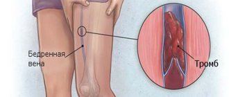
Thrombosis of the axillary vein: how dangerous?
Thrombosis is a fairly serious disease, which is characterized by various types of problems leading to temporary disability. However, with thrombosis of the upper extremities, the prognosis is much more favorable than in other places.
Thrombosis of the subclavian vein can occur as a result of playing sports or significant stress on the upper shoulder girdle due to the specifics of work. It is described as Paget's syndrome. If thrombosis of the right subclavian vein is diagnosed, then the person is often right-handed and loads this particular arm. Such diseases lie in wait for fairly young men.
Incidence (per 100,000 people)
| Men | Women | |||||||||||||
| Age, years | 0-1 | 1-3 | 3-14 | 14-25 | 25-40 | 40-60 | 60 + | 0-1 | 1-3 | 3-14 | 14-25 | 25-40 | 40-60 | 60 + |
| Number of sick people | 0 | 0 | 0.5 | 0.5 | 0.5 | 15 | 25 | 0 | 0 | 0.5 | 5 | 5 | 25 | 35 |
Symptoms
| Occurrence (how often a symptom occurs in a given disease) | |
| Numbness of one, two or all limbs | 100% |
| Pain in the hand spreading to the fingers | 90% |
| Constant pain in legs | 90% |
| Paleness of fingers | 80% |
| Cold weather, chilly fingers | 80% |
| Coldness, chilly toes | 80% |
| Weakness in the leg (right or left) | 50% |
| Weakness in the arm (right or left) | 50% |
Preventive actions
Any illness or disease is easier to prevent than to use therapy. This also applies to thrombophlebitis of the shoulder. Preventive measures include exercise, swimming, cycling and walking. It is not recommended to wear high-heeled shoes for a long time. Taking cranberry tinctures also helps. St. John's wort and rose hips. Carrying out therapy in the primary stages of the disease is much simpler, therefore, at the slightest sign of pathology, you should immediately consult your doctor.