You can live with an aneurysm for years
I was diagnosed with an aneurysm. I would like to know more about this disease. What are the causes of its occurrence and how to treat it?
An aneurysm is a protrusion of the wall of an artery (less commonly a vein) as a result of its thinning or stretching. In this case, an aneurysmal sac appears, which can compress nearby tissues. Depending on the location, aneurysms can be arterial or venous. When an artery and vein are simultaneously damaged, an arteriovenous aneurysm may develop. Based on their shape, they distinguish between saccular and fusiform, false and true aneurysms. Cerebral aneurysm is the most dangerous and common form of this disease. Complications after the rupture of such an aneurysm are comparable to the consequences of a stroke. Aortic aneurysm is an equally dangerous disease in which the lumen of the aorta expands 2 times compared to normal. More often, aortic aneurysm occurs in men suffering from atherosclerosis. The peculiarity of a thoracic aortic aneurysm is that it can develop within 20 years after the injury. An abdominal aortic aneurysm is asymptomatic. However, a very thin person may feel a pulsation when he puts his hand on his stomach. If this aneurysm compresses the roots of the spinal cord, the pain becomes unbearable. With a peripheral vascular aneurysm (blood vessels in the extremities), a person may experience severe pain in the legs and arms. With a cardiac aneurysm, a sac-like protrusion of the heart wall appears. Acquired cardiac aneurysm occurs in 5–20% of people who have had a myocardial infarction. It can develop either immediately after a heart attack or several months after it. Most often, an aneurysm is congenital and can be caused by a disease of the vessel wall. Moreover, after birth this defect is invisible, and the baby develops completely normally. Acquired aneurysm is more common in people over 50 years of age. In young people, it usually occurs after injuries received in car accidents or while playing extreme sports. Diseases that thin blood vessels also lead to aneurysm: hypertension, atherosclerosis, syphilis (at a late stage). The risk of developing this insidious disease also appears when infected blood clots form. Spreading to the vascular wall, the infection leads to the formation of an aneurysm. You can live with an aneurysm for years and not notice any signs of illness. In the meantime, the aneurysm will imperceptibly grow, threatening to rupture or dissection at any moment. The manifestations of an aneurysm depend on its location or size. Symptoms usually occur when adjacent tissues are compressed. The most common complication of an aneurysm is its rupture. Ruptures of aneurysms of the heart, aorta and large arteries are especially dangerous, as this results in severe bleeding, which often causes death. When an aneurysm ruptures, hemorrhage occurs, which leads to a serious condition. When an aneurysm ruptures, a person feels pain and his blood pressure begins to drop rapidly. Since an aneurysm rarely manifests itself, it is often discovered accidentally during an X-ray examination or a routine medical examination. Ultrasound allows you to determine the size, shape and location of the aneurysm. Using aortography (an X-ray image of the aorta after the injection of a dye), it is possible to assess the condition of the vessels in the area of the aneurysm and its size. If an aneurysm is detected, you must constantly be under close medical supervision. The operation is not performed for all aneurysms. If it is small, then the doctor can only observe and wait. When an aneurysm reaches life-threatening sizes, surgical treatment is necessary. This involves removing the damaged portion of the vessel and replacing it with a plastic prosthesis or a fragment of a blood vessel taken from another part of the body. Sometimes an aneurysm that sticks out like a bag is clamped at the base with a special clip. If an aneurysm ruptures, emergency surgery is performed. To prevent an aneurysm, it is first necessary to normalize the level of blood pressure and cholesterol in the blood, since they are the main risk factors for the occurrence of an aneurysm. To do this, you need to eat foods low in cholesterol, exercise, quit smoking and lose excess weight.
Cardiac aneurysm after a heart attack: types, diagnosis, surgery, prognosis
Myocardial infarction is a complication of heart pathologies. It provokes the appearance of other problems in the functioning of the organ. These include post-infarction cardiac aneurysm, in which protrusion of the heart wall occurs. The pathology is quite dangerous and requires timely treatment. If help is not provided, complications will lead to the death of the patient.
Types of aneurysms
Depending on the time of appearance, aneurysms are:
- Spicy. Their development is observed during the first two weeks after a heart attack. It is difficult to predict the behavior of such a formation. The wall of the aneurysm is just beginning to become covered with collagen fibers and can quickly increase in size or rupture.
- Subacute. Such formations appear within 3-8 weeks after the attack. The tissue components are strengthened. Therefore, the risk of rupture is reduced.
- Chronic. They appear two months after a heart attack. The formation slowly increases in size, but blood clots form inside it and it leads to heart rhythm disturbances.
Most often, a left ventricular aneurysm is observed after a heart attack. This is due to high pressure in this part of the organ, which causes the wall of the heart to bulge.
Aneurysms can be small, medium-sized, or giant. The latter lead to changes in heart volume. They can reach the shape of the left ventricle. It is by the size of the formation that a forecast can be made. The larger it is, the sadder the consequences will be.
Causes
Aneurysms occur when the walls of the heart weaken. The internal pressure in the organ is increased. Therefore, under pressure, the weakened wall forms a bulge.
The ventricles have higher pressure than the atria, so formations appear in them most often. At the same time, the tissue cells of the heart wall gradually die, and the pressure increases more and more, especially during physical exertion.
These problems are usually observed in various diseases, so an aneurysm is considered a complication of the pathological process, and not a separate disease. The problem may result from:
- A previous attack of myocardial infarction. This cause causes aneurysm in 90% of cases. In this condition, the heart muscle suffers from an acute lack of oxygen, which causes the death of normal cells, but does not recover after this. In their place, a focus of connective tissue is formed that cannot contract. An aneurysm may appear at this site. It is usually located on the wall of the left ventricle, reaches a size of 5-7 cm, and often grows quickly and ruptures.
- Diseases of infectious origin. Infections do not directly cause aneurysm. But they give impetus to the development of the pathological process. Viruses or bacteria enter the heart through the blood and cause inflammation, which is accompanied by cell death. After the inflammation is eliminated, connective tissue forms at its source. An aneurysm can form months or even years after infections.
- Congenital disorders. Such formations appear during the first weeks after the birth of the child. This is due to defects that occurred during intrauterine development. If cells no longer divide correctly in a certain area of the organ, then these areas become thinner. When the baby is in the uterus, blood pressure is still low, but with birth, blood circulation increases and aneurysms form under pressure. A problem may arise if the mother smoked, drank alcohol, or had measles or rubella during pregnancy. This leads to disruption of the normal process of cell division with all the ensuing consequences.
- Surgical interventions on the heart. Often, if an elderly person or child has been operated on due to a heart defect, a bulge appears in this place. This happens if the tissues on which the suture was placed do not heal well, scars appear on them, pressure increases and the number of heartbeats increases. Therefore, complications may occur even if the operation was successful.
- Injuries in which the heart wall was damaged.
- Systemic inflammatory diseases. They cause myocarditis with cardiosclerosis. Most often this occurs with rheumatism. Large inflammatory lesions increase the risk of aneurysm formation.
- Exposure to radiation. Under normal conditions, a person cannot receive a large dose of radiation. This can happen during radiation treatment of malignant tumors.
Symptoms indicating the occurrence of aneurysms
The clinical presentation of the condition may vary. How the problem will manifest itself depends on where the bulge is located and how much it has enlarged. Many patients experience no symptoms after a heart attack. Only a small part of patients complain of:
- Painful sensations in the heart area. The aneurysm itself does not hurt, since it has no nerve endings. Pain occurs in the area around the scar. The symptom is usually localized behind the sternum on the left. It has a paroxysmal nature and appears as a result of physical activity, drinking alcohol or smoking.
- Weakness. The muscles and nervous system suffer from insufficient oxygen and do not work at full strength.
- Heart rhythm disturbances. The attacks appear from time to time and quickly stop. It can be caused by stress and physical activity.
- Shortness of breath. In this case, breathing rhythm is periodically disrupted. An aneurysm is characterized by a slowdown in gas exchange in the alveoli, which causes shortness of breath.
- Pallor of the skin. The symptom occurs due to insufficient blood supply to the skin, since the heart is not able to pump the required amount of blood. Because of this, the hands and feet also become cold and the sensitivity of the skin is impaired.
- Cough. This symptom is uncommon. It manifests itself if the aneurysm reaches a large size. The mass compresses the lung and irritates it, causing a dry cough. But fever, wheezing in the lungs and sputum production are not observed.
- Feeling of heartbeat. In a normal state, a person does not feel his heart beating. With an aneurysm, this is associated with disturbances in heart rhythm and increased contractions due to an increase in the volume of the left ventricle.
Also read: Types of complications after myocardial infarction
It is important to report any discomfort after a heart attack to your doctor. This will help to detect an aneurysm in a timely manner.
Diagnostic methods
Diagnosing an aneurysm after myocardial infarction is not an easy task. This is due to the absence of complaints in most patients. Large formations are easiest to notice. But to identify small ones, special examinations are needed. The problem definition consists of:
- Physical examination. At the same time, blood pressure is measured, the chest is felt, and the function of the organ is listened to with a stethoscope. If after this there is a suspicion of an aneurysm, other studies are prescribed.
- Electrocardiography. The device registers the movement of a bioelectric impulse to the myocardium. An experienced doctor can determine the disorder based on the results of this study alone. This is an accessible and informative procedure.
- Echocardiography. This is an accurate diagnostic method that will help confirm the presence of a formation in the heart. The procedure provides accurate information about the condition of the heart.
- Myocardial scintigraphy. During the study, an isotope of thallium is injected into the blood. It accumulates in the walls of the heart and creates a contrast by which an aneurysm is easily identified.
- X-rays. This method is rarely used, since it can only detect large formations.
In addition to these procedures, blood and urine tests may be prescribed to identify concomitant disorders.
Treatment methods
In most cases, they prefer to treat an aneurysm surgically, since no medication will help get rid of it. Conservative therapy is carried out to eliminate symptoms and prevent complications.
The operation is prescribed if:
- heart failure increases rapidly;
- severe arrhythmia develops that is difficult to treat;
- blood clots form;
- false aneurysms were detected;
- education has collapsed. In most cases, the problem ends in the death of the patient.
If a person is diagnosed with an aneurysm, he can only be cured with the help of surgery, as well as alleviate the symptoms of the problem and prevent complications by using medications.
Surgical treatment involves a complex procedure in which the doctor must open the chest. After this, the blood flow is stopped and connected to a special device that temporarily performs the functions of the heart.
When the blood pumping process is stopped. The surgeon begins to remove the formation. He excises areas with pathological tissue, removes blood clots, if any, and firmly sutures the heart wall. In some cases, synthetic materials are used to support it.
To restore normal blood circulation, coronary artery bypass surgery may be necessary. This will reduce the risk of another heart attack.
The procedure takes quite a long time and is performed under general anesthesia. But even a successful operation does not provide a 100% guarantee of survival. 8% of cases end in death.
TC with the help of medications usually reduces the load on the left ventricle and prevents the formation of blood clots. If the examination shows that there are no blood clots and the formation is not growing, then maintenance treatment is carried out. It is prescribed to elderly people who may not be able to undergo surgery. Conservative treatment includes the use of:
- Beta blockers. They are necessary to stabilize the heart rate and weaken heart contractions.
- Thrombolytics. Drugs in this group prevent platelets from sticking together and thin the blood, which helps prevent blood clots.
- Diuretics. They help reduce pressure in the arteries. The lower it is, the less likely it is that the formation will rupture.
- Nitrates. Medicines dilate the coronary vessels, allowing more blood to flow to the myocardium, which reduces pain.
The drug and its dosage should be determined only by the attending physician.
Possible complications
If the aneurysm is not treated correctly, the risk of another heart attack and rupture of the formation increases. These consequences are fatal.
After excision of the aneurysm:
- blood accumulates in the pericardium;
- left ventricular output decreases;
- heart rhythm is disturbed;
- blood clots form.
But these complications can be combated with special medications, and there is no risk of aneurysm rupture and heart attack.
Forecast
A cardiac aneurysm after a heart attack may have a different prognosis. Most often it is unfavorable, but the situation can be improved with surgical treatment. But this procedure also has certain risks. Elderly people may develop other life-threatening complications after anesthesia.
The prognosis of a left ventricular aneurysm after a heart attack will be poor if the patient refuses surgery and the formation continues to increase in volume.
How long a patient with this diagnosis will live depends on the size, location of the aneurysm, and the general condition of the body.
Prevention
To avoid the development of complications and alleviate the symptoms of the problem, the patient should:
- stop smoking;
- avoid alcoholic drinks;
- reduce the intensity of physical activity;
- follow a diet: patients are usually advised to give up salty, spicy, fatty foods, coffee, and tea.
With these measures, you can avoid rupture of the formation in the first few weeks after a heart attack. The patient must adhere to these rules throughout his life or until surgery, when the formation is eliminated.
To prevent an aneurysm from appearing, diseases of the cardiovascular system should be treated promptly and prevent the development of a heart attack.
Source: https://KardioPuls.ru/bolezni/razryv/anevrizma-serdtsa-posle-infarkta/
General characteristics of the disease
Aneurysm is an insidious disease, in many cases it does not manifest itself symptomatically. Most often it is discovered accidentally during a medical examination of a person for other diseases.
It can be congenital or acquired as a result of the development of diseases such as arterio- or atherosclerosis. In addition, an aneurysm may be the result of trauma, mycotic or syphilitic vascular damage. An aneurysm most often appears in the aortic area.
There are true and false aneurysms. All layers of the blood vessel wall are involved in the formation of the true one, which occurs with syphilis and atherosclerosis. False aneurysms appear as a result of trauma to blood vessels, in which blood flows into the tissue.
After some time, the walls of an aneurysm form around this area with blood, and “aneurysmal shock” is formed, as a result of which the walls of the vessels begin to gradually bulge, which leads to compression of the surrounding organs.
Many doctors call an aneurysm nothing more than a “time bomb” that can “explode” at any moment.
According to theory, an aneurysm can be located in absolutely any artery, however, practice shows that the aorta, the largest artery, is most often affected (the disease in this case is called aortic aneurysm) and the arteries of the brain (the disease is cerebral aneurysm).
The aorta is divided into two sections: abdominal and thoracic; respectively, an abdominal aortic aneurysm and a thoracic aortic aneurysm are distinguished.
Read also
Diagnosis and treatment of high monocyte levels in a child
Symptoms indicating the occurrence of aneurysms
The clinical presentation of the condition may vary. How the problem will manifest itself depends on where the bulge is located and how much it has enlarged. Many patients experience no symptoms after a heart attack. Only a small part of patients complain of:
- Painful sensations in the heart area. The aneurysm itself does not hurt, since it has no nerve endings. Pain occurs in the area around the scar. The symptom is usually localized behind the sternum on the left. It has a paroxysmal nature and appears as a result of physical activity, drinking alcohol or smoking.
- Weakness. The muscles and nervous system suffer from insufficient oxygen and do not work at full strength.
- Heart rhythm disturbances. The attacks appear from time to time and quickly stop. It can be caused by stress and physical activity.
- Shortness of breath. In this case, breathing rhythm is periodically disrupted. An aneurysm is characterized by a slowdown in gas exchange in the alveoli, which causes shortness of breath.
- Pallor of the skin. The symptom occurs due to insufficient blood supply to the skin, since the heart is not able to pump the required amount of blood. Because of this, the hands and feet also become cold and the sensitivity of the skin is impaired.
- Cough. This symptom is uncommon. It manifests itself if the aneurysm reaches a large size. The mass compresses the lung and irritates it, causing a dry cough. But fever, wheezing in the lungs and sputum production are not observed.
- Feeling of heartbeat. In a normal state, a person does not feel his heart beating. With an aneurysm, this is associated with disturbances in heart rhythm and increased contractions due to an increase in the volume of the left ventricle.
Abdominal aortic aneurysm
The abdominal aorta carries blood to the lower part of the body. When the aorta has a weakened area, it begins to either bulge or expand. This is how an abdominal aortic aneurysm appears. It is very dangerous and poses a real threat to human health. When it ruptures, internal bleeding begins, which can be fatal.
Another unpleasant “surprise” of an abdominal aortic aneurysm is the possibility of the formation of blood clots that can break away from the walls of the vessel and block the lumen of smaller vessels. The result is arterial thrombosis, causing severe pain and serious complications. One of them is the possibility of losing a limb.
Causes
Aneurysms occur when the walls of the heart weaken. The internal pressure in the organ is increased. Therefore, under pressure, the weakened wall forms a bulge.
The ventricles have higher pressure than the atria, so formations appear in them most often. At the same time, the tissue cells of the heart wall gradually die, and the pressure increases more and more, especially during physical exertion.
These problems are usually observed in various diseases, so an aneurysm is considered a complication of the pathological process, and not a separate disease. The problem may result from:
- A previous attack of myocardial infarction. This cause causes aneurysm in 90% of cases. In this condition, the heart muscle suffers from an acute lack of oxygen, which causes the death of normal cells, but does not recover after this. In their place, a focus of connective tissue is formed that cannot contract. An aneurysm may appear at this site. It is usually located on the wall of the left ventricle, reaches a size of 5-7 cm, and often grows quickly and ruptures.
- Diseases of infectious origin. Infections do not directly cause aneurysm. But they give impetus to the development of the pathological process. Viruses or bacteria enter the heart through the blood and cause inflammation, which is accompanied by cell death. After the inflammation is eliminated, connective tissue forms at its source. An aneurysm can form months or even years after infections.
- Congenital disorders. Such formations appear during the first weeks after the birth of the child. This is due to defects that occurred during intrauterine development. If cells no longer divide correctly in a certain area of the organ, then these areas become thinner. When the baby is in the uterus, blood pressure is still low, but with birth, blood circulation increases and aneurysms form under pressure. A problem may arise if the mother smoked, drank alcohol, or had measles or rubella during pregnancy. This leads to disruption of the normal process of cell division with all the ensuing consequences.
- Surgical interventions on the heart. Often, if an elderly person or child has been operated on due to a heart defect, a bulge appears in this place. This happens if the tissues on which the suture was placed do not heal well, scars appear on them, pressure increases and the number of heartbeats increases. Therefore, complications may occur even if the operation was successful.
- Injuries in which the heart wall was damaged.
- Systemic inflammatory diseases. They cause myocarditis with cardiosclerosis. Most often this occurs with rheumatism. Large inflammatory lesions increase the risk of aneurysm formation.
- Exposure to radiation. Under normal conditions, a person cannot receive a large dose of radiation. This can happen during radiation treatment of malignant tumors.
Cerebral aneurysm
The disease can develop due to congenital changes in the walls of blood vessels. In addition, an aneurysm is found in people who have genetic disorders in the body, which include: connective tissue diseases, polycystic kidney disease, and circulatory disorders.
A cerebral aneurysm can be a consequence of a head injury, appear due to a tumor, infection, high blood pressure and other vascular diseases. Drug use and smoking are also causes of cerebral aneurysm.
This disease is also dangerous; it can result in brain hemorrhage, damage to the nervous system, stroke or death. There is a risk of formation and development of several aneurysms, which further increases the risk of the disease.
General characteristics of cardiac aneurysms and its structures: what is it and how does it manifest?
Aneurysmal dilatations of the heart are a group of diseases that differ in form, mechanism of occurrence and course.
Most aneurysms are detected in people over 50 years of age. Features of the pathology are an erased course, a connection with other heart diseases and a significant impact on the blood supply to internal organs.
What it is?
A cardiac aneurysm is a limited expansion of one of its cavities, caused by anatomical insufficiency of the walls of its chambers and not participating in contractions. More than 90% of formations develop in the left ventricle. The diameter ranges from 2 mm to 20 cm. Read more about left ventricular aneurysm here.
The ICD-10 code for cardiac aneurysms is I25.3.
Differences from vascular disease
Cardiac aneurysms have the following differences from vascular aneurysms:
- Most of them are associated with myocardial infarction;
- Rarely remain asymptomatic (except for those localized in the interatrial septum);
- Represented by a thin layer of fibrous or scar tissue;
- With a progressive course, they cause changes in the entire cardiac hemodynamics, and not just an increase in the chamber.
Causes
Common Causes of Heart Aneurysm in Adults:
The disease may be congenital. Causes :
- Defects in endothelial structural genes;
- Muscular dysplasia;
- Congenital heart defects;
- Prematurity;
- Large fruit;
- Pathology of pregnancy in the mother.
Causes in children:
- Left ventricular diverticulum;
- Dysplasia and systemic connective tissue diseases;
- Viral myocarditis;
- Chickenpox;
- Adenoviral infection;
- Potassium and magnesium deficiency.
Development mechanism
Congenital cardiac aneurysms develop under the influence of defective structural proteins : instead of a three-layer heart wall, newborns develop a thin layer of connective tissue that is not capable of contraction. Under the influence of blood pressure, the defective area is pressed and begins to bulge in the direction of the lowest pressure.
Acquired forms are a consequence of traumatic damage to the heart . Immediately after the injury, delicate connective fibers form in the lesion, which are gradually replaced by scar tissue. The scar is not capable of elastic expansion, so it becomes thinner and bulges under the influence of blood flow, forming a protrusion.
After a heart attack
Post-infarction cardiac aneurysms are classified as post-traumatic. After the formation of necrosis, the dead area of the myocardium undergoes lysis and destruction, which leads to local softening of the cardiac fibers.
After this, new connective fibers are formed in the lesion, devoid of muscle layer and elasticity. Under the influence of heart contractions, the wall of the formed scar becomes thinner and protrudes.
The prognosis is unfavorable. The danger is expressed in the possibility of rupture, turning into cardiac tamponade.
How long do people live with a cardiac aneurysm after a heart attack: life expectancy is reduced by 5-7 years due to developing heart failure.
Acute, subacute and chronic forms
- The acute variety is typical for the first two weeks after the action of the traumatic factor.
It can be true or false, in shape - sac-like or spindle-shaped (you will learn about all types and forms of aneurysms here). The course is acute. The ischemic area swells, increases in size and causes pain behind the sternum. Possible fever. In complicated cases, acute heart failure develops. Treatment is aimed at stabilizing vital functions - breathing and circulation. - The subacute form is typical for 3-8 weeks of the disease.
The course is erased, painful attacks are periodic, of medium intensity. The affected area is limited, and scar tissue begins to form. Treatment is aimed at reducing pain and maintaining cardiac output and blood pressure. - The chronic stage begins at 9 weeks.
The protrusion is represented by a fibrous, thinned sac, in the wall of which muscle fibers can be detected. Thrombosis is characteristic. The course is determined by chronic heart failure. Treatment is aimed at compensating the function of the left ventricle and preventing the appearance of edema.
Diagnostic methods
Diagnosis is based on medical history and visualization of the lesion. When conducting a comprehensive examination, the following results are observed:
- Survey . Complaints are nonspecific; if the aneurysm size is up to 3 mm, there are no complaints. Patients indicate a heart attack, chest pain, heaviness in the chest, forced sitting, swelling, abdominal enlargement;
- Objective examination . An increase in the boundaries of relative dullness of the heart (detected by percussion), systolic murmur above the projection of the protrusion (detected by auscultation);
- Radiography . Enlarged cardiac shadow with displacement of the lung on the affected side; parietal hematoma;
- ECG . Displacement of the heart axis to the right or left depending on the location of the lesion, extrasystoles, tachycardia, fibrillation, decreased voltage of the R wave, pathological Q wave, displacement of the ST segment below the isoline;
- Ultrasound of the heart . Identification of the exact location of the aneurysm, dyskinesia of the affected area, decreased cardiac output and left ventricular shock force;
- Color mapping allows you to determine the filling of the aneurysm with blood and thrombus formation;
- Cardiac catheterization . Confirmation of a rupture of the heart wall by the free passage of the catheter;
- Ventriculography . Determination of the exact location, shape and size of the lesion;
- CT (MRI) . Determination of the stage of myocardial infarction, local edema, thickness of scar tissue, parietal hematoma.
Symptoms by localization
Let's consider the symptoms and diagnostic signs of cardiac aneurysms (including on the ECG) depending on the location in the heart muscle.
| Aneurysm of the right or left ventricle of the heart . The frequency per 100,000 population is 14-16 cases. | ||
| Clinic | Distinctive features | Diagnostic results |
| Chest pain, loss of consciousness, forced positioning, swelling, shortness of breath, pale or blue skin | In 95% of cases there is a connection with a heart attack, a progressive course, rapid onset of cardiopulmonary failure | Shoe-shaped heart (x-ray), “Frozen ECG” - ST segment elevation for more than 6 weeks, deep Q wave, tachyarrhythmia; local edema and thinning of the ventricular wall (CT), dys- or akinesia of the affected area (ultrasound) |
| Aneurysm of the right or left atrium . The frequency per 100,000 population is 4-5.5 cases. | ||
| Clinic | Distinctive features | Diagnostic results |
| Missing or erased. Complaints of shortness of breath, irregular heart function, palpitations | Long-term erased course, thrombus formation is characteristic | Enlarged atrial arch (radiography), extrasystoles, fibrillation, biphasic P wave (ECG), dyskinesia, mural thrombi (ultrasound) |
| Aneurysm of the sinus of Valsalva . The frequency per 100,000 population is 0.1-0.5 cases. | ||
| Clinic | Distinctive features | Diagnostic results |
| Painful attacks caused by coronary artery ischemia, loss of consciousness | Attachment of secondary aortic insufficiency, myocardial infarction | Pathological protrusion at the aortic root (x-ray), myocardial ischemia (decreased ST segment, Q wave on ECG), coronary insufficiency (coronary angiography) |
| Aneurysms of the cardiac septa (intergastric, interatrial) in adults and children. The frequency per 100,000 population is about 12 cases. | ||
| Clinic | Distinctive features | Diagnostic results |
| If the IVS is affected - asymptomatic, IVS - swelling, pain behind the sternum, abdominal enlargement, shortness of breath | Benign course with lesions of the IVS, with lesions of the IVS - early development of right ventricular failure | Atrial extrasystoles, atrial fibrillation and flutter, biphasic R (ECG), dyskinesia (ultrasound), local edema, thrombus formation (CT), rupture (catheterization) |
False cardiac aneurysms
False protrusions form secondary to injury or myocardial infarction . They represent a rupture of the wall (usually the left ventricle) with the formation of a hematoma limited to the pericardium. Patients complain of shortness of breath, pressing pain in the chest, and palpitations.
Methods for confirming the diagnosis - ultrasound, CT, MRI (detection of a hematoma limited to the pericardium and the ventricular wall). Surgical treatment is aimed at removing the hematoma and suturing the rupture site.
Choice of treatment tactics
How to treat a cardiac aneurysm is determined by the doctor based on the patient’s complaints and the size of the lesion. If the condition is satisfactory and the size of the formation is small, conservative treatment is carried out . Groups of drugs used:
- Adrenergic blockers;
- Antiarrhythmic drugs;
- Diuretics;
- Symptomatic therapy (pain medications, nitroglycerin, cardioprotectors).
Indications for surgery for cardiac aneurysms:
- Presence of complaints;
- Fainting;
- Threat of rupture;
- Risk of developing a false aneurysm;
- Combination with heart defects;
- Right ventricular failure;
- Decreased stroke volume and cardiac output.
Types of surgical treatment:
- Aneurysm resection;
- Suturing the wall defect;
- Thrombectomy;
- Removal of part of the heart chamber;
- Septoplasty according to Cooley.
If a newborn or older child is sick
When diagnosing a newborn, preference is given to expectant management , which is associated with high risks of surgical intervention. Conservative therapy is carried out aimed at eliminating symptoms and preparing for surgery. Doses of funds are calculated based on the child’s weight. Upon reaching 5-7 years of age, the issue of surgical intervention is decided.
In children, treatment is aimed at eliminating protrusion and correcting associated defects . In the absence of complaints, therapy is conservative. If shortness of breath, edema and pulmonary hypertension develop, the protrusion is excised and the heart wall is sutured.
Why it’s dangerous: possible consequences
The danger is expressed in the threat of rupture with heavy bleeding. In the absence of timely diagnosis, death occurs in 95-98% of patients.
Consequences without treatment:
- Cardiopulmonary failure;
- Thrombosis;
- Thromboembolism in the vessels of the systemic circulation;
- Arrhythmia;
- Cardiac tamponade;
- Ventricular fibrillation.
Complications after treatment are observed in 3-5% of cases:
- Traumatic injury of the myocardium;
- Arrhythmia;
- Small output syndrome;
- Seams coming apart.
How to live with a heart aneurysm?
When making a diagnosis, it is necessary to register with a cardiologist. Periodic medical examinations and dynamic monitoring help prevent complications (ultrasound, ECG). Drinking alcohol, smoking and professional sports should be excluded. Fried, salty foods, semi-finished products, foods with transgenic fats are removed from the diet, and sweet foods and baked goods are limited.
FAQ
- Can you join the army with a heart aneurysm? Aneurysm of the sac is not a contraindication to military service. In other cases, untreated patients are released from the army, those treated are assigned to the reserve category.
- Do they give you disability? With the development of cardiopulmonary failure, thromboembolism, and pulmonary hypertension, disability is established. The category is determined individually.
- How does the quality of life change? The quality of life decreases with the development of heart failure, arrhythmia, fibrillation, and thrombus formation.
Practical recommendations for parents if their child is sick
- Register your child with a cardiologist.
- Observe his mobility, appetite, and general condition daily.
- Control weight gain.
- Prevent seasonal infections (take multivitamins, magnesium, potassium).
- Limit physical activity.
- Avoid injury.
- If shortness of breath, cyanosis, or fainting occurs, seek help immediately.
Cardiac aneurysms are a group of pathological conditions that occur primarily or against the background of cardiac pathology.
Protrusion of one of the walls of the heart leads to hemodynamic disturbances and is manifested by a variety of nonspecific complaints. Treatment is determined by the diameter of the formation, age and complaints of the patient .
Most patients are indicated for surgical correction in combination with therapy for concomitant cardiac diseases.
Source: https://oserdce.com/sosudy/anevrizmy/serdca
Symptoms of an aneurysm
The symptoms of the disease depend on the presence of complications of the aneurysm, the type of development and its location. Symptoms of an aneurysm may not appear for several years. According to statistics, 25 percent of patients suffering from an aneurysm initially mistook it for a migraine.
Asymptomatic cerebral aneurysm is usually discovered incidentally. When not ruptured, it causes sensations such as compression of the brain and cranial nerves, which in turn leads to recurring headaches. As a result of the disease, vision is impaired, strabismus may appear, the sense of smell is partially lost, and sometimes patients suffer from epileptic seizures. When an aneurysm ruptures, internal bleeding occurs with associated symptoms.
An abdominal aortic aneurysm may also be asymptomatic. But in some cases, symptoms of an aneurysm appear - the patient feels throbbing and pressing pain in the abdomen, chest, between the shoulder blades, in the lower back, sides, buttocks, and legs. Sometimes there is cyanosis of the fingers and a change in the color of the skin of the hands.
A thoracic aneurysm is characterized by deep, throbbing pain in the chest, sometimes radiating to the shoulder. Shortness of breath, pain and discomfort when swallowing, and cough are also observed. Fever and even weight loss are possible.
When an aneurysm ruptures, a person experiences severe pain, leading to shock. Shock is expressed in impaired respiratory function, rapid heartbeat, lack of response to questions asked, and loss of the ability to move.
If you experience sudden pain in the head, abdomen or chest, or if the aneurysm symptoms described above occur, you should immediately consult a doctor.
Provided timely diagnosis, aortic aneurysm responds well to treatment, and its rupture most often leads to death.
Cardiac aneurysm - what kind of disease it is, treatment, symptoms and diagnosis on MED Zoon
Cardiac aneurysm is a disease characterized by thinning and protrusion of the myocardial wall, lack of contractility in this area of the heart muscle. As a rule, an aneurysm forms in the wall of the left, sometimes in the area of the right ventricle.
In newborns, protrusions of the interatrial and interventricular septa are more common. Acquired cardiac aneurysm is one of the common complications of myocardial infarction; the disease is diagnosed in 10-35% of patients in the post-infarction period.
Men over forty are at risk.
Cardiac aneurysms are classified according to many criteria: the cause and course of the disease, cellular structure and mechanism of formation, shape and size. A detailed classification of cardiac aneurysms is given in the table.
Types of cardiac aneurysms
| Classification sign | Type of aneurysm | Decoding |
| Cause of the disease | congenital | are formed due to disorders of intrauterine development of the fetus |
| acquired | in the vast majority of cases occur after a myocardial infarction | |
| Flow | spicy | form within one to two weeks after a heart attack |
| subacute | are formed from the third to the eighth week after a heart attack | |
| chronic | occur later than the eighth week after myocardial infarction | |
| Cellular structure | muscular | made up of muscle fibers |
| fibrous | the walls of the aneurysm are formed by fibrous tissue | |
| fibromuscular | include muscle and fibrous tissue | |
| Education mechanism | true | have a three-layer structure, consist of dead scar tissue |
| false | are formed when the myocardium ruptures and the formation of a cavity limited by the pericardium (the outer lining of the heart) | |
| functional | areas of viable myocardium that protrude during ventricular contraction | |
| Form | diffuse | have a flat shape |
| saccular | characterized by the presence of a “neck”, which, when expanded, forms a sac-like cavity | |
| delaminating | are formed due to rupture of the endocardium (the inner lining of the heart) |
Cardiac aneurysms can come in different sizes. There are significant aneurysmal protrusions - more than eight centimeters, medium - from three to seven centimeters in size, and small (hidden) aneurysms - less than three centimeters.
Why is a cardiac aneurysm dangerous?
A cardiac aneurysm is dangerous due to the formation of blood clots. Echocardiography often reveals blood clots in a distended cavity. A blood clot can break off at any time and lead to serious consequences.
Common complications of cardiac aneurysm include:
- ischemic stroke - a violation of cerebral circulation with damage to brain tissue;
- thromboembolism (blockage) of the pulmonary artery;
- kidney infarction - an acute circulatory disorder leading to the death of organ tissue;
- gangrene – necrosis of body tissues, accompanied by their rotting;
- repeated myocardial infarction is an acute disturbance of blood circulation in the heart muscle.
In addition to the risk of blood clot rupture, a cardiac aneurysm contributes to the development of heart failure with characteristic symptoms: shortness of breath, interruptions in heart function, chest pain, and swelling.
How long do people live with a cardiac aneurysm?
Life expectancy with a cardiac aneurysm depends on several factors: the size of the protrusion, location, rate of increase, concomitant diseases and the age of the patient. People with small aneurysms live for years without surgical treatment, but under the constant supervision of a cardiologist.
In cases where the size of the aneurysm is so large that the disease threatens a person’s life, surgery is necessary; without it, death is inevitable.
An aneurysm is not a death sentence, but in no case should you let the situation take its course; only an experienced cardiologist can decide on the choice of treatment tactics.
Causes of cardiac aneurysm formation
The causes of congenital and acquired cardiac aneurysm differ. In adult patients, in 95-97% of cases, pathology develops after extensive myocardial infarction. Less commonly, the formation of a cardiac aneurysm is caused by trauma, injury and infection (rheumatism, tuberculosis, syphilis, infective endocarditis).
Aggravating factors are:
- high blood pressure;
- chronic heart failure;
- post-infarction cardiosclerosis is a pathology characterized by the growth of scar tissue in the myocardium.
Among the causes of congenital cardiac aneurysms, researchers identify:
- disruption of the formation of connective tissue during intrauterine development of the fetus;
- genetic predisposition;
- infections suffered by a pregnant woman;
- negative environmental factors.
Congenital cardiac aneurysm is quite rare and accounts for slightly more than 1% of all anomalies of intrauterine fetal development.
Symptoms of a cardiac aneurysm
Symptoms of a cardiac aneurysm depend on the course of the disease.
Characteristic signs of acute cardiac aneurysm are:
- severe pain in the chest that appears suddenly and lasts from several hours to several days;
- a feeling of lack of air, turning into suffocation;
- pronounced pallor of the skin;
- blue discoloration of mucous membranes;
- with the development of pulmonary edema, coughing, wheezing appear, breathing becomes bubbling, and copious sputum is produced in the form of foam.
With chronic cardiac aneurysm, the symptoms are different.
Most often, patients complain about:
- minor periodic pain in the heart area;
- heartbeat;
- weakness and dizziness;
- shortness of breath;
- swelling.
The listed symptoms of a cardiac aneurysm can also occur with other diseases of the cardiovascular system. Additional research methods help make an accurate diagnosis.
Diagnosis of the disease
Laboratory diagnostic methods are not very informative for cardiac aneurysm, therefore they are used only as auxiliary studies.
The main diagnostic methods for this disease are:
- electrocardiogram (ECG) - shows changes characteristic of an aneurysm (pathological O wave and persistent ST segment elevation in the precordial leads);
- echocardiography (ultrasound of the heart) – allows you to see the location, shape and size of the aneurysm;
- contrast ventriculography - a picture of the heart with the introduction of radiopaque substances;
- magnetic resonance imaging (MRI) – layer-by-layer images of the heart;
- coronary angiography – allows you to determine the nature, location and size of the aneurysm.
Treatment of cardiac aneurysm
The choice of treatment method is selected individually depending on the size of the protrusion, the rate of increase and the age of the patient.
In adults
drug therapy includes taking medications aimed at treating heart rhythm disturbances and restoring blood circulation to the organ.
Adult patients are prescribed medications for:
- reducing blood viscosity and cholesterol levels;
- stabilization of blood pressure;
- preventing attacks of arrhythmia;
- reducing swelling.
indications for surgical treatment are:
- large size of the aneurysm;
- increasing heart failure;
- thromboembolism;
- myocardial rupture;
- progressive mitral regurgitation (dysfunction of the mitral valve of the heart).
The main goal of surgery is removal of the aneurysm followed by reconstruction (restoration) of the heart.
in children
When diagnosing an aneurysm of the interatrial septum in a child, which is small in size and does not affect the quality of life, conservative treatment methods are used:
- taking medications that help improve the structure of connective tissue;
- taking medications to strengthen the heart muscle;
- antiarrhythmic drugs;
- physical therapy and massage.
congenital left ventricular aneurysm is a direct indication for surgery. if the protrusion does not increase in size and does not pose a threat to life, surgery is performed after one year.
From the moment of birth until the operation, the child is under constant supervision of a cardiologist.
Surgical treatment of congenital aneurysm is carried out in different ways: complete removal (resection) of the protrusion and verticuloplasty - correction (restoration) of the structure of the heart.
in newborns
the choice of treatment tactics for cardiac aneurysm in newborns is determined by the severity of the pathology. in the absence of a threat to life, the question of surgery is postponed until one year.
small aneurysms do not require surgical treatment, however, children with such pathology are registered with a cardiologist and undergo regular examinations.
surgery to remove a cardiac aneurysm in newborns is performed only in emergency cases when there is a clear threat to the baby’s life. The surgical technique and timing are determined individually by a medical council.
There are a number of contraindications for surgical treatment of cardiac aneurysm in children: severe heart and kidney failure, cancer, severe diabetes, intolerance to anesthesia.
how to live with a heart aneurysm
Cardiac aneurysm is a serious disease that requires a person to follow certain rules of life. However, if you follow your doctor’s recommendations and undergo regular monitoring with a cardiologist, the risk of blood clot formation and separation is significantly reduced.
For patients with cardiac aneurysm, it is recommended to:
- maintain a daily routine;
- eat a balanced diet;
- exclude strong physical activity;
- engage in physical therapy;
- undergo a heart examination at least twice a year;
- take maintenance medications as prescribed by your doctor.
Following simple rules and taking care of your health will help maintain quality and increase life expectancy.
Disease prognosis
The prognosis for cardiac aneurysm depends on many factors. First of all, it depends on the size and location of the protrusion, the state of the cardiovascular system, concomitant pathologies, and the person’s age. If the aneurysm is small in size and you see a doctor in a timely manner, the prognosis for the disease is favorable.
An aggravating prognostic factor is the addition of heart failure. Lack of treatment for extensive post-infarction aneurysms also significantly worsens the prognosis.
Uncomplicated flat cardiac aneurysms occur most easily; saccular protrusions are often complicated by intracardiac thrombosis.
Source: https://zoon.ru/med/anevrizma-serdca/
Diagnosis of the disease
Before treating an aneurysm, a thorough diagnosis is necessary. Currently, there are several methods for diagnosing an aneurysm: contrast X-ray examination of the vessel (angiography) is carried out to determine its condition, the nature of the blood flow and the size of pathological changes.
Using computed tomography angiography and magnetic resonance angiography, images of blood vessels are obtained and the characteristics of blood flow in them are assessed.
Doppler ultrasound (ultrasound examination of blood vessels) allows you to see a picture of the location of the vessel in the volume, assess its condition from different angles, and determine the degree of the pathological process.
Diagnostics
Pain is a useful but late symptom.
Patients may feel a pulsating mass in the middle of the abdomen with their fingers. Using a stethoscope placed in the middle of the abdomen, doctors usually hear a noise caused by blood flowing through the aneurysm. However, in obese people, even large aneurysms cannot be detected.
Rapidly growing aneurysms that rupture always cause pain and a painful sensation that is transmitted to the abdominal cavity upon palpation (touch). Skin X-ray studies show whether there is calcium deposits on the wall of the aneurysm, but this procedure does not provide much information.
If an aneurysm is identified, ultrasound examinations may be performed over several months to determine the extent of its growth. A CT scan of the abdomen, especially if done after an injection of radioactive isotopes, can better determine the size and shape of the aneurysm. MRI is also effective, but is not always available as quickly as ultrasound or CT.
Treatment of aneurysm
Modern medicine uses only one method of treating an aneurysm - surgery. During the operation, the affected vessel is removed, and an artificial one is sewn in its place.
Conduct open and closed operations. When open in the abdominal area, the surgeon makes an incision through which the expanded part of the abdominal aorta is removed and a prosthesis is placed in its place.
The second method of treating an aneurysm is endoprosthetics. During this procedure, a small incision is made in the patient's groin, through which a prosthesis is inserted and placed in the cavity of the aneurysm.
This operation is performed according to indications only in a small number of patients. This is explained by the fact that after it there is a possibility of a new aneurysm forming, and this will lead to a repeat operation.
As a rule, prostheses are not rejected by the human body, in most cases do not require replacement and serve until the end of the patient’s life.
Treatment methods
In most cases, they prefer to treat an aneurysm surgically, since no medication will help get rid of it. Conservative therapy is carried out to eliminate symptoms and prevent complications.
- heart failure increases rapidly;
- severe arrhythmia develops that is difficult to treat;
- blood clots form;
- false aneurysms were detected;
- education has collapsed. In most cases, the problem ends in the death of the patient.
If a person is diagnosed with an aneurysm, he can only be cured with the help of surgery, as well as alleviate the symptoms of the problem and prevent complications by using medications.
Surgical treatment involves a complex procedure in which the doctor must open the chest. After this, the blood flow is stopped and connected to a special device that temporarily performs the functions of the heart.
When the blood pumping process is stopped. The surgeon begins to remove the formation. He excises areas with pathological tissue, removes blood clots, if any, and firmly sutures the heart wall. In some cases, synthetic materials are used to support it.
To restore normal blood circulation, coronary artery bypass surgery may be necessary. This will reduce the risk of another heart attack.
The procedure takes quite a long time and is performed under general anesthesia. But even a successful operation does not provide a 100% guarantee of survival. 8% of cases end in death.
TC with the help of medications usually reduces the load on the left ventricle and prevents the formation of blood clots. If the examination shows that there are no blood clots and the formation is not growing, then maintenance treatment is carried out. It is prescribed to elderly people who may not be able to undergo surgery. Conservative treatment includes the use of:
- Beta blockers. They are necessary to stabilize the heart rate and weaken heart contractions.
- Thrombolytics. Drugs in this group prevent platelets from sticking together and thin the blood, which helps prevent blood clots.
- Diuretics. They help reduce pressure in the arteries. The lower it is, the less likely it is that the formation will rupture.
- Nitrates. Medicines dilate the coronary vessels, allowing more blood to flow to the myocardium, which reduces pain.
The drug and its dosage should be determined only by the attending physician.
An aneurysm may not show symptoms for many years.
Aneurysm is a tricky disease that hides for a long time. It forms in the wall of a blood vessel, usually in a part of an artery that is weakened for some reason. Blood flowing under pressure is pushed towards the weakened part of the vessel. When the pressure rises dangerously, the aneurysm can rupture and cause massive bleeding. Find out how aneurysms occur. How to recognize the symptoms of cerebral aneurysm and aortic aneurysm?
Aneurysms are localized bulges and distortions formed in the walls of arteries, and less commonly in veins. The aneurysm's arterial wall location becomes thinner and more susceptible to injury, with the risk of rupture, massive bleeding, and (in the case of an aortic aneurysm) overall fatal outcome.
An aneurysm is an abnormality in the structure of the wall of a large blood vessel, usually an artery. This is a local bulge in the wall of the artery, which at this point becomes thinner and more susceptible to damage. Depending on the location, aneurysms of the aorta, heart, femoral artery, popliteal artery, renal artery and cerebral aneurysms (cerebral arteries) are distinguished. An aneurysm, especially when it ruptures, poses a serious threat to health and life.
An aneurysm can appear anywhere, but most often occurs in the aorta and blood vessels in the brain.
Read also
Cocoa butter for hemorrhoids: methods of use and effectiveness
There are pseudo- and true aneurysms. A true aneurysm occurs when there is a total expansion of the lumen of the vessel while maintaining the continuity of its walls.
An aneurysm may not cause any symptoms (asymptomatic). It is usually recognized when it reaches a large size or when it breaks. An aneurysm may also be diagnosed incidentally during imaging. The symptoms that accompany aneurysms are closely related to the site of their origin, that is, the vessel around which the aneurysm was created.
Types of Aneurysms
Aneurysms can develop anywhere along the aorta. Three-quarters of aortic aneurysms develop in the abdominal aorta (the part of the aorta that runs through the abdomen), and the rest develop in the thoracic (aortic) aorta.
Aneurysms can also develop in the arteries behind the knee and in the arteries that supply blood to the heart (coronary arteries). In adults, aneurysms often occur in areas where there are arterial branches (for example, when the abdominal aorta is divided into the right and left iliac arteries) or in areas where more stress is present (the popliteal artery). Aneurysms can be round or cylindrical in shape. Most of them are cylindrical.
Damage to the myocardium with subsequent destruction of the wall due to pressure is considered an unfavorable variant of the pathology. Ideally, it should be detected before a rupture occurs.
Causes of aneurysms
An aneurysm can develop slowly at a weakened point in the artery wall that bends due to the force of the blood flowing through it. However, the exact cause that causes the formation of aneurysms has not yet been determined. It is known, however, that certain factors increase the risk of their occurrence.
Risk factors include:
- hypertension (high blood pressure) that persists for many years, causing damage and weakening of blood vessels;
- atherosclerosis, which weakens the walls of blood vessels; trauma, including chest trauma;
- polycystic kidney - increases the risk of developing a cerebral aneurysm; an infection that attacks and weakens part of a blood vessel;
- a place in the wall of a blood vessel with a weakened structure is assessed as a congenital defect (aneurysm).
Causes of aneurysm after a heart attack
In most patients, the previous condition was extensive myocardial infarction with transmural (all layers of the wall) necrosis. In this case, an aneurysm can occur both in the first week and several months after the onset of the disease.
The most common place of formation is the apex and anterior wall of the left ventricle. The destroyed muscle tissue is highly extensible and, during the period of heart contraction, protrudes outward or into the lumen of the right ventricle if the formation is located on the septum.
Factors that can provoke the development of an aneurysm after a heart attack include:
- elderly patients, diabetes mellitus, exhaustion (the process of scarring of the infarction zone is disrupted);
- early physical activity;
- severe form of arterial hypertension;
- the occurrence of repeated infarction, especially in patients with cardiomyopathy, myocardial dystrophy, cardiosclerosis;
- late start of treatment of the disease;
- atypical, including painless forms;
- absence of collateral (bypass) blood flow during the first severe attack of angina.
Read more about the complications of myocardial infarction here.
Brain aneurysm
A brain aneurysm, also called an intracranial aneurysm or cerebral vascular aneurysm, is a cerebrovascular disease.
This occurs when one of the layers that make up the artery becomes weakened somewhere. Blood flowing under pressure begins to push out the weakened part of the artery, resulting in a characteristic bulge. This dominance increases over time and threatens to break.
It is not explained why brain aneurysms occur only in some people. Among the causes of cerebral aneurysm, all congenital anomalies in the formation of the arterial wall and atherosclerosis, as well as inflammation in the vascular walls, are mentioned.
A cerebral aneurysm most often occurs at the site of arterial bifurcation on distal branches or at the origin of individual arteries. An aneurysm is formed as a bulge in the wall of an artery that resembles the shape of a sac—which is why the most common aneurysms are called saccular aneurysms (80% of all aneurysms). Fusiform aneurysms are less common and are located primarily on the basilar and middle cerebral arteries.
A brain aneurysm may not show any symptoms. Only in some cases, a large aneurysm can put pressure on neighboring structures and cause headaches and various neurological problems. Some aneurysms produce nonspecific symptoms, such as dilation of one pupil, double vision, or drooping eyelids. However, most often patients become aware of the existence of an aneurysm during its rupture, that is, subarachnoid hemorrhage.
When an aneurysm ruptures, a sudden and very severe headache occurs. This may be accompanied by symptoms such as neck stiffness, photophobia, nausea and vomiting. Some patients lose consciousness or have speech difficulties. In extreme cases, a ruptured brain aneurysm can lead to coma and even death.
Aneurysms occur over a long period of time, so they are most often found in the fourth, fifth, and sixth decades of life, and they are present sporadically in newborns.
There are two methods of treating cerebral aneurysm (brain blood vessels). The first involves inserting a clip for the neck of the aneurysm, which requires opening the patient's skull. The second method does not require such an invasive procedure - neuroradiological treatment, or embolization, involves filling the aneurysm with springs through a catheter that is inserted through the femoral artery into the groin.
The doctor decides on the treatment method in consultation with the patient. When choosing a treatment method, the location of the aneurysm, its size, shape, neck width and adjacent vessels should be taken into account. The risk of aneurysm rupture increases with size. The larger the aneurysm (it can get larger over time), the greater the chance of it breaking and causing hemorrhage.
Diagnostic methods
Signs that can be detected when examining a patient include:
- inspection and palpation - asynchronous pulsation in the 3rd - 4th intercostal space outside the apical impulse zone (yoke symptom);
- Auscultation – murmur during systole with relative mitral insufficiency;
- radiography - pathological protrusion of the left ventricular arch, expansion of the borders of the heart, pulmonary congestion;
- ECG – “frozen” symptoms of a heart attack, no improvement (ST recovery);
- EchoCG - an aneurysm is determined, its size, the presence of blood clots, as well as hemodynamic parameters - ejection fraction, final ventricular volumes in systole and diastole can be examined;
- coronary angiography – blockage of the coronary artery and changes in blood flow behind it;
- ventriculography and MRI - help to establish the size, shape, location, changes in the heart muscle outside the aneurysm, thrombosis of the ventricular cavity;
- PET of the heart (positron emission tomography) – needed to determine the “sleeping” myocardium in adjacent areas;
- EPI (electrophysiological study) is indicated in the presence of arrhythmia.
Aortic aneurysm (abdominal/thoracic)
The aorta is the main artery in the body. The left ventricle of the heart sends blood to it, and further branches deliver it to all organs and tissues.
The diameter of the aorta is not a constant value. Diameter is affected by age, gender, body size and blood pressure. Although the diameter of the aorta naturally increases with age, it is a slow expansion process. However, in some cases, excessive dilatation of the aorta occurs, which exceeds the norm by more than 50% for a given episode - then we talk about an aortic aneurysm.
For changes in the aortic wall that cause it to become weaker and less flexible, this usually results in atherosclerosis. Other, less common causes include degenerative changes in the vascular walls during genetic disorders (eg, Marfan syndrome), post-inflammatory changes during autoimmune diseases (Takayasu disease), inflammation in diseases such as syphilis, endocarditis, sepsis, and post-traumatic changes.
Aortic aneurysms are divided due to their location. Eat:
- Abdominal aortic aneurysms: This is the most common location of aneurysms. Occurs in the descending portion below the diaphragm;
- aneurysms of the thoracic aorta: occur in the ascending part, the aortic arch or the initial part of the descending aorta - up to the height of the diaphragm;
- Thoracoabdominal aneurysms: they occur at the border of both areas.
Aortic aneurysm most often remains asymptomatic for a long time. However, in light of the dilated aorta, blood does not flow properly, creating the risk of blood clots forming in the aneurysm. Fragments of the clot can become detached and travel with the blood, causing blockages, which are often the first symptom of an aneurysm. This can lead to stroke, ischemia of the lower extremities or intestines, kidney infarction, and blue fingers.
Symptoms of a thoracic aortic aneurysm include constant pain, bone pain, and back pain. Less common are dysphagia, hoarseness, cough, and shortness of breath. In the case of an ascending aortic aneurysm or aortic arch, there may be symptoms of aortic regurgitation or symptoms of the superior vena cava (shortness of breath, swelling and redness of the face and neck).
Symptoms of an abdominal aortic aneurysm: Usually there are no symptoms, they may only appear with large aneurysms. The pattern may be constant, with crushing pain around the navel, abdomen or lumbar region.
Read also
Removal of hemorrhoids using the Longo method: features, advantages and disadvantages of the procedure
Large aneurysms usually cause pain. They also pose a high risk of rupture. When this occurs, the patient may experience chest laceration or abdominal pain, rapidly worsening the general condition and increasing symptoms of shock. You may lose consciousness.
Treatment for aortic aneurysm involves pharmacological treatment, which is a chronic treatment that slows the rate at which aneurysms grow. Surgical treatment is a radical method of removing the diseased segment of the aorta and implanting a prosthesis, that is, an artificial segment of the aorta.
An extremely dangerous pathology is a cardiac aneurysm after a heart attack.
One of the severe complications of a heart attack is aneurysmal protrusion of the wall in the necrosis zone. Signs of its development are: circulatory failure, thromboembolism, arrhythmia. Treatment requires surgery; without it, patients die from cardiac decompensation or ruptured aneurysm.
Causes of aneurysm after a heart attack
In most patients, the previous condition was extensive myocardial infarction with transmural (all layers of the wall) necrosis. In this case, an aneurysm can occur both in the first week and several months after the onset of the disease.
The most common place of formation is the apex and anterior wall of the left ventricle. The destroyed muscle tissue is highly extensible and, during the period of heart contraction, protrudes outward or into the lumen of the right ventricle if the formation is located on the septum.
Factors that can provoke the development of an aneurysm after a heart attack include:
We recommend reading the article about post-infarction cardiosclerosis. From it you will learn about the causes and symptoms of the pathology, types of post-infarction cardiosclerosis, possible complications, diagnosis and treatment.
Read more about the complications of myocardial infarction here.
Signs and symptoms
When the ventricle contracts, the blood partially passes into the aneurysmal sac, which reduces cardiac output and contributes to the manifestations of heart and vascular failure:
- dyspnea,
- severe weakness
- dizziness,
- fainting states,
- cough,
- attacks of suffocation,
- swelling in the legs,
- liver enlargement.
Due to the turbulent movement of blood in the cavity of the aneurysm, blood clots are formed - parietal thrombi. They can be destroyed, and their parts move along the arterial bed. Such emboli lead to blockage of blood vessels in the extremities, brain, lungs, intestines, and kidneys, causing gangrene, organ infarctions (including secondary myocardial necrosis), and stroke.
If an aneurysm occurs in the acute or subacute period of the disease, then its wall contains insufficiently formed scar tissue, which can lead to cardiac rupture, pericardial hemorrhage, tamponade and cardiac arrest.
How long do people live with post-infarction aneurysm without treatment?
The formation of a protrusion of the heart wall is an unfavorable variant of the course of myocardial infarction. If the operation is not performed in time, then within 2 - 3 years from the moment of aneurysm formation, patients die from acute coronary or cardiac decompensation, thrombosis. Factors that increase the likelihood of death include:
- progression of circulatory failure;
- resumption of angina attacks during treatment;
- relative mitral valve insufficiency due to dilation of the left ventricular cavity;
- severe types of rhythm disturbances;
- large size of the aneurysm, sac-like or mushroom-shaped.
A more benign course is observed in asymptomatic and small formations formed after 2 months from the onset of a heart attack.
Watch the video about an aneurysm and the danger it poses:
Classification of education
Depending on the location of the aneurysm, there may be:
- ventricular (mainly left);
- septal (interventricular septum).
There are dangerous formations in the shape of a bag, a mushroom, and multilayer ones (one inside the other); flat ones, having the configuration of a bowl on the inside, are considered more durable. The structure of the aneurysm wall can be muscle or connective tissue fibers; more often they are combined in various proportions.
Complications can occur in the acute (up to the 2nd week), subacute (up to the 8th) periods, and chronic ones form after the 2nd month from the attack.
True ventricular aneurysms contain all three layers of the myocardium, while false ones appear when muscle tissue ruptures; they are limited only by the epicardium and the pericardial sac; the probability of rupture in this case is extremely high.
During a heart attack, a functional variant may also appear - viable cells are in a dormant state (zone of the hibernating heart), they do not contract during the systole phase, but protrude under blood pressure. Once blood flow to the area is restored, the aneurysm disappears.
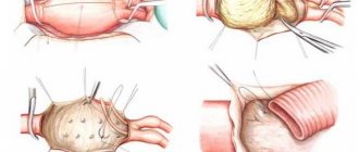
Surgery as the only treatment option
Drug treatment can be prescribed in exceptional cases - with contraindications to surgery (danger of anesthesia, dead cells around the aneurysm, severe mitral regurgitation), asymptomatic small aneurysm. In all other situations, surgical intervention is indicated.
The patient is connected to an artificial respiration apparatus, after opening the aneurysm sac, it is cleared of blood clots, excised, leaving about 2 cm of scar tissue, and then stitched with a linear or purse-string suture.
For larger lesions, a patch is placed over the sutures.
Along with the removal of an aneurysm, coronary artery bypass surgery is sometimes performed to restore blood flow in the area of the infarction, or surgery on the heart valves.
Prognosis after for the patient
The performed operation significantly reduces the mortality rate of patients. But since it is performed on an open heart, and the volume of the ventricle decreases after suturing, complications are possible in the postoperative period:
- heart and respiratory failure;
- low cardiac output (hypotension, syncope, collapse);
- arrhythmia;
- bleeding;
- stroke;
- renal failure.
The five-year survival rate after aneurysm resection is about 75%, and the ten-year survival rate is approximately 35%. In most patients, the cause of death is a repeated acute violation of the coronary circulation.
We recommend reading the article about surgery for aortic aneurysm. From it you will learn about indications for surgery, types of operations, rehabilitation period and prognosis for patients.
And here is more information about repeated myocardial infarction.
Post-infarction aneurysm is formed with extensive and transmural lesions of the myocardium. Most often it occurs in the left ventricle. Leads to heart failure, vascular thrombosis, and rhythm disturbances. When it appears in the acute and subacute periods, it has a weak wall, which threatens heart rupture.
The most accurate diagnosis is made using echocardiography, ventriculography and MRI. Treatment requires surgery - excision of tissue with subsequent restoration of the integrity of the walls of the ventricle.
Source: https://CardioBook.ru/anevrizma-serdca-posle-infarkta/
Time bomb
Every year, 50,000 people worldwide are diagnosed with aortic aneurysm At the same time, there are other types of such pathology that are no less common and deadly.
itself is a bulging of the wall of an artery (in some cases a vein) due to thinning or excessive stretching. The consequence of this is the so-called. an aneurysmal sac that compresses nearby tissues. Moreover, it can be not only acquired, but also congenital.
The main problem of pathology is that it does not manifest itself in any way for the time being. And often the whole situation ends in the rupture of an aneurysm, which most often leads to death.
What types of pathology are there, what causes it, and who is at risk?
How long do people live with post-infarction aneurysm without treatment?
The formation of a protrusion of the heart wall is an unfavorable variant of the course of myocardial infarction. If the operation is not performed in time, then within 2 - 3 years from the moment of aneurysm formation, patients die from acute coronary or cardiac decompensation, thrombosis. Factors that increase the likelihood of death include:
- progression of circulatory failure;
- resumption of angina attacks during treatment;
- relative mitral valve insufficiency due to dilation of the left ventricular cavity;
- severe types of rhythm disturbances;
- large size of the aneurysm, sac-like or mushroom-shaped.
A more benign course is observed in asymptomatic and small formations formed after 2 months from the onset of a heart attack.
Watch the video about an aneurysm and the danger it poses:
Different types
When talking about an aneurysm, they often mean aortic aneurysm . At the same time, there are quite a lot of different types of this pathology.
Thus, one of the first to be called is a cerebral aneurysm .
Here, both single and multiple defects can be observed, which naturally aggravates the situation. Such neoplasms may not manifest themselves for a long time, but as they grow, they often give nonspecific symptoms. You can recognize the presence of such a problem by:
- Visual impairment
- The appearance of hallucinations
- Coordination and balance problems
- Failure of one limb
- Speech disorders
- Headaches
This is due to the fact that the aneurysm compresses tissue and brain structures. Due to the fact that it takes on some of the blood, a person may experience neurological disorders and even a stroke. When a vessel ruptures, half of the victims die from hemorrhage.
An abdominal aortic aneurysm is a defect that affects the largest vessel in the human body. Its walls are very dense and can easily separate into components. If there is such a problem, a person will be tormented by:
- Burning
- Chest pain
- Pulsation in the stomach area
- Cold feet
When such an aneurysm ruptures, acute abdominal pain and vomiting are observed. If the blood flows too quickly, then within a few minutes the person will feel an unpleasant tingling sensation in the legs. A complication in this case may be a stroke.
Aneurysm of peripheral vessels causes increased thrombosis. The thrombus can break away from the wall of the aneurysm and move into the vessels of vital organs: heart, brain, lungs, kidneys. Symptoms vary depending on the location of the defect.
Cardiac aneurysm , as a rule, becomes a consequence of a heart attack and other cardiac problems. This is due to the fact that the wall of the heart, having experienced such a strong load, cannot withstand the pressure. As a result, it begins to stretch. Indicates the presence of such a problem:
- Weakness
- Edema
- Stagnation of mucus in the lungs
- Dyspnea
- Increased heart rate
Pathology cannot be underestimated. For example, in the United States alone, every year as many people die from this defect as from AIDS: about 24,000.
Acquired aneurysm is often considered to be the lot of the elderly. But young people are also susceptible to it: they develop it due to injuries received in car accidents or during extreme sports.
Types of aneurysms
Depending on the time of appearance, aneurysms are:
- Spicy. Their development is observed during the first two weeks after a heart attack. It is difficult to predict the behavior of such a formation. The wall of the aneurysm is just beginning to become covered with collagen fibers and can quickly increase in size or rupture.
- Subacute. Such formations appear within 3-8 weeks after the attack. The tissue components are strengthened. Therefore, the risk of rupture is reduced.
- Chronic. They appear two months after a heart attack. The formation slowly increases in size, but blood clots form inside it and it leads to heart rhythm disturbances.
Most often, a left ventricular aneurysm is observed after a heart attack. This is due to high pressure in this part of the organ, which causes the wall of the heart to bulge.
Aneurysms can be small, medium-sized, or giant. The latter lead to changes in heart volume. They can reach the shape of the left ventricle. It is by the size of the formation that a forecast can be made. The larger it is, the sadder the consequences will be.
Possible complications
The main danger of an aneurysm is that it can rupture. This often ends in death, but it can also lead to complications that will be extremely serious. Thus, when a rupture occurs, damage to the central nervous system and the development of a hemorrhagic stroke may occur.
In addition, it is worth understanding that aneurysm often recurs. Since it does not produce any symptoms, in most cases the relapse occurs unexpectedly. Most often, with relapses, a cerebral hemorrhage occurs.
Treatment
Treatment of cardiac aneurysm occurs in a complex manner using pharmacological agents of different groups. At the same time, drug treatment does not always have positive dynamics, which requires surgical therapy. The treatment process is accompanied by the administration of the following types of medications:
- Antianginal drugs. They can eliminate attacks of chest pain (unstable angina). For this purpose, drugs from the Nitrates group are prescribed (Nitrong, Nitroglycerin, Nitromint). Coronarolytics are also used: Papaverine or Validol. Calcium channel blockers (Amlodipine, Felodipine) have a vasoconstrictor effect.
- To prevent the formation of blood clots in the lumen of blood vessels, cardiac Aspirin, Rovocor, Lovastatin (statin group) is prescribed.
- To correct and stabilize blood pressure, pharmacological agents from the group of ACE inhibitors (Captopril, Lisinopril or Fosinopril) are used.
The lack of the desired effect or a significant increase in the size of the aneurysm suggests the use of surgical therapy. In this case, polymer surgery techniques, resection of affected areas, or coronary artery bypass grafting may be used. Aneurysm of the heart muscle is a dangerous complication, which in most cases has a disappointing prognosis. This is due to the fact that the pathology in most cases is recorded in an advanced form. In addition, it occurs against the background of other diseases of the internal organs. A relatively favorable prognosis will be only if the disease is detected at an early stage of development.
Aneurysms of other arteries
Aneurysms can also occur in other arteries, such as:
- popliteal,
- femoral,
- coronary,
- laryngeal,
- sleepy.
Most of these aneurysms are the result of congenital (inherited) wall weakness or an atherosclerotic process. Remaining aneurysms are the result of injury, infection, or inflammation of the artery wall. Infections usually start elsewhere in the body, most commonly in the heart valves (infective endocarditis).
Most dilatations of the popliteal and femoral arteries do not cause symptoms, but blockage of blood in the area of the aneurysm leads to the formation of clots (thrombi), which can become detached from the vessel wall and become emboli, which can block arteries located distal to the foot or leg.
Femoral, coronary, and carotid aneurysms rarely rupture. The doctor may find a pulsating mass in the area of the aneurysm. Ultrasound or CT can confirm the diagnosis. For popliteal aneurysms larger than 2.5 cm in diameter, the options are open surgery or endovascular stent grafting. Typically, femoral and carotid aneurysms are removed surgically.
Aneurysms can occur in the arteries of the brain (cerebral arteries). Rupture of cerebral aneurysms can cause bleeding in the brain tissue (intracerebral hemorrhage), leading to stroke. Cerebral artery aneurysms are dangerous and require early treatment. Treatment always involves surgery.
The disease that develops most often after myocardial infarction is a cardiac aneurysm. A cardiac aneurysm can also be defined as a congenital condition of the heart muscle. It develops as a complication of traumatic, infectious, postoperative disorders.
The aneurysm stands out above the wall of the heart. The most common location for cardiac aneurysm after a heart attack is the anterior wall and left ventricle of the heart.
Another important cause is hypertension - due to increased pressure in the left ventricle.
The clinical picture of aneurysm heart disease is accompanied by the following symptoms:
- strong heartbeat,
- shortness of breath and pain in the heart and chest,
- attacks of angina.
When nerves are damaged and there is a lack of oxygen in the muscle tissue, weakness forms. The fact is that it is the blood that carries oxygen, and in a situation where an aneurysm develops, a volume that does not extend beyond the cavity of the aneurysm is removed from the bloodstream. After all, connective tissue does not have the ability to contract. Another characteristic sign is arrhythmia. It is provoked by emotional and physical stress. It passes quite quickly, but leaves behind the effect of a gallop rhythm.
Shortness of breath becomes a natural consequence of blood stagnation. The pressure inside the heart chambers increases. This affects the condition of blood vessels and their function. We are talking about the arteries that feed the lungs. This is why breathing function is inhibited. At the same time, pallor of the skin is recorded. It is observed on the upper and lower extremities and facial skin. It then spreads throughout the body. Patients feel that they are cold, and the sensitivity of the skin also decreases. This also leads to tissues not being oxygenated well enough.
A cardiac aneurysm suggests another characteristic symptom in the form of cough. It may not be accompanied by a sore throat, but it is paroxysmal and is a consequence of congestion in the blood vessels of the lung. It occurs if pressure is applied to the lung in the area where the protrusion is located. But this can be if it has reached an impressive size. Other symptoms include increased sweating and heaviness in the chest, poor coordination of movements, swelling in the face and limbs, and hoarseness.
The disease is characterized by a hemodynamic disorder due to changes in the shape and volume of the heart chamber and the accumulation of part of the blood in the aneurysmal sac. Pathological atrial pulsations and progressive heart failure are formed.
The diagnosis of cardiac aneurysm is made after a detailed interview based on patient complaints. Physical, instrumental and laboratory tests are carried out. From physical studies, auscultation (listening) of the heart reveals the following pathological changes:
- weakening of the first tone,
- increased second tone over the pulmonary artery,
- systolic murmur
- sometimes pericardial friction is heard.
According to instrumental methods, the most informative is the electrocardiogram (ECG). This study demonstrates a large transmural infarction with the presence of QS and QR waves and an arcuate constant height of the ST segment. The ECG registers an abnormal heart rhythm.
To diagnose cardiac aneurysms, laboratory tests are important. They reveal:
- leukocytosis (increased number of white blood cells),
- accelerated ESR (erythrocyte sedimentation rate).
Treatment for cardiac aneurysm necessarily requires surgery in most cases. Preparation for surgery and prevention of complications includes conservative therapy. The key indications for surgical treatment of cardiac aneurysm are:
- progressive heart failure,
- thromboembolism,
- arrhythmia,
- increase in angina attacks.
Surgical treatment of cardiac aneurysm is carried out in conditions of maintaining blood circulation through special equipment.
Diagnosis of cardiac aneurysm
In 50% of patients, pathological precordial pulsation is detected.
ECG signs are nonspecific - a “frozen” picture of acute transmural myocardial infarction is revealed, there may be rhythm disturbances (ventricular extrasystole) and conduction (left bundle branch block).
ECHO-CG allows you to visualize the aneurysm cavity, determine its size and location, and identify the presence of a parietal thrombus.
The viability of the myocardium in the area of chronic cardiac aneurysm is determined by stress echocardiography and PET .
Using chest x-ray, it is possible to detect cardiomegaly and congestive processes in the blood circulation.
ventriculography, MRI and MSCT are also used to determine the size of the heart aneurysm and detect thrombosis of its cavity.
For the purpose of differential diagnosis of the disease from coelomic pericardial cyst, mitral heart disease, mediastinal tumors, probing of the heart cavities and coronary angiography may be prescribed .
Thoracic aortic aneurysms
They are an extension of where the aorta passes through the chest cavity (chest). Involvement of the thoracic aorta may cause no symptoms or may cause pain, coughing, or wheezing in the chest.
If an aneurysm ruptures, the patient may feel a stabbing pain that starts high in the back and travels down the spine to the abdomen. Aneurysms are often detected by X-ray, magnetic resonance imaging, or CT scan performed for another case. The doctor may try to surgically remove the aneurysm before it ruptures.
The types of aneurysm in question are now more common than before due to the use of computed tomography to study other diseases. Most often, in a thoracic aortic aneurysm, the wall of the aorta degrades (a condition called cystic necrosis) and the part of the aorta that is close to the heart becomes enlarged. This increase can lead to insufficient functioning between the valves of the heart and the aorta, allowing blood to return to the heart when the valves are closed. This condition is called aortic regurgitation. About half of people with this form of aneurysm also have Marfan syndrome.
For the other half, the cause is unclear, although most have high blood pressure. Although rare, syphilis causes aneurysmal dilatation of the aorta near the heart.
Thoracic aneurysms can grow large without causing symptoms. The onset of symptoms is the result of aortic pressure on surrounding structures, depending on where the aneurysm is located.