Sudden changes in blood flow to the brain are classified as hemorrhagic (bleeding) and ischemic disorders. Such a division is important for the correct choice of therapy method.
The classic abbreviated name for the pathology in acute cerebrovascular accident is “ischemic stroke.” If hemorrhage is confirmed, then it is considered hemorrhagic.
In ICD-10, ACME codes may vary, depending on the type of violation:
- G45 is an established designation for transient cerebral attacks;
- I63 - recommended for statistical registration of cerebral infarction;
- I64 - an option used for unknown differences between cerebral infarction and hemorrhage, used when the patient is admitted in an extremely serious condition, unsuccessful treatment and imminent death.
The frequency of ischemic strokes exceeds hemorrhagic strokes by 4 times and is more associated with general human diseases. The problem of prevention and treatment is considered in programs at the state level, because 1/3 of patients who have suffered the disease die in the first month and 60% remain permanently disabled requiring social assistance.
Why does a lack of blood supply to the brain occur?
Acute ischemic cerebrovascular accident is often a secondary pathology and occurs against the background of existing diseases:
- arterial hypertension;
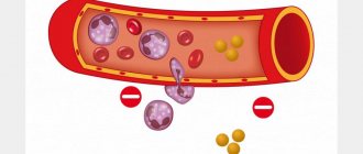
- widespread atherosclerotic vascular lesions (up to 55% of cases develop due to pronounced atherosclerotic changes or thromboembolism from plaques located in the aortic arch, brachiocephalic trunk or intracranial arteries);
- previous myocardial infarction;
- endocarditis;
- heart rhythm disturbances;
- changes in the valvular apparatus of the heart;
- vasculitis and angiopathy;
- vascular aneurysms and developmental anomalies;
- blood diseases;
- diabetes mellitus
Up to 90% of patients have changes in the heart and main arteries of the neck. The combination of these reasons sharply increases the risk of ischemia.
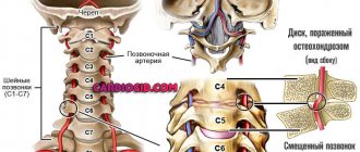
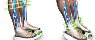
Possible compression of the vertebral artery by the processes of the vertebrae
Transient attacks are most often caused by:
- spasm of the arterial brain stems or short-term compression of the carotid and vertebral arteries;
- embolization of small branches.
The following risk factors can provoke the disease:
- elderly and senile age;
- excess weight;
- the effect of nicotine on blood vessels (smoking);
- experienced stress.
The basis of the influencing factors is the narrowing of the lumen of the vessels through which blood flows to the brain cells. However, the consequences of such a malnutrition may vary according to:
- stamina,
- localization,
- prevalence,
- severity of vessel stenosis,
- gravity.
The combination of factors determines the form of the disease and clinical symptoms.
An ischemic stroke can be considered recurrent if it occurs in the area of the blood supply of one vessel within 28 days after the initial manifestations of the first event. A recurrent stroke is called a stroke at a later date.
Pathogenesis of various forms of acute cerebral ischemia
Transient ischemic attack was previously called transient cerebrovascular accident. It is identified as a separate form because it is characterized by reversible disorders; the heart attack does not have time to form. Usually the diagnosis is made retrospectively (after the disappearance of the main symptoms), within a day. Before this, the patient is treated as if he had a stroke.
The main role in the development of hypertensive cerebral crises belongs to the increased level of venous and intracranial pressure with damage to the walls of blood vessels and the release of fluid and protein into the intercellular space.
Swelling of brain tissue in this case is called vasogenic
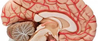
The feeding artery is necessarily involved in the development of ischemic stroke. The cessation of blood flow leads to oxygen deficiency in the lesion formed in accordance with the boundaries of the basin of the affected vessel.
Local ischemia causes necrosis of an area of brain tissue.
Depending on the pathogenesis of ischemic changes, types of ischemic strokes are distinguished:
- atherothrombotic - develops when the integrity of the atherosclerotic plaque is disrupted, which causes complete closure of the internal or external feeding arteries of the brain or their sharp narrowing;
- cardioembolic - the source of thrombosis is pathological growths on the endocardium or heart valves, fragments of a blood clot, they are delivered to the brain with the general blood flow (especially when the foramen ovale is not closed) after attacks of atrial fibrillation, tachyarrhythmia, atrial fibrillation in patients in the post-infarction period;
- lacunar - more often occurs when small intracerebral vessels are damaged in arterial hypertension, diabetes mellitus, is characterized by the small size of the lesion (up to 15 mm) and relatively minor neurological disorders;
- hemodynamic - cerebral ischemia with a general decrease in blood circulation speed and a drop in pressure against the background of chronic heart disease, cardiogenic shock.
In case of hemodynamic disturbances, blood flow in the vessels of the brain may decrease to a critical level and below
It is worth explaining the development of strokes of unknown etiology. This often happens when there are two or more reasons. For example, in a patient with carotid artery stenosis and fibrillation after an acute infarction. It should be taken into account that elderly patients already have stenosis of the carotid arteries on the side of the alleged disorder, caused by atherosclerosis, in the amount of up to half the lumen of the vessel.
Prevention, prognosis
The prognosis for hemorrhages and hematomas is unfavorable. The mortality rate reaches 70%, of which about 50% of deaths are associated with the removal of hematomas located between areas of the brain lobes. The reason for the high mortality rate is rapidly increasing swelling, compression, displacement of the brain, and repeated hemorrhages. The prognosis generally depends on the amount of blood shed, the volume of the hematoma, its location, and the age of the victim. About 80% of surviving patients become disabled.
The mortality rate for ischemic stroke is about 22%. The prognosis depends on the affected area and the speed of medical care. Preventive measures are aimed at timely detection of diseases and their treatment, especially for people at risk. These are people with high cholesterol, hypertension, diabetes, and cardiovascular diseases.
Secondary prevention measures are necessary for people at risk who have had a stroke. They include taking medications that normalize blood pressure, anticoagulants or antiplatelet agents, and medications that lower bad cholesterol levels. Secondary prevention includes planned surgical treatment for partial occlusion of important blood trunks.
According to statistics, people are more often diagnosed with ischemic stroke – about 80%. It is often called a cerebral hemorrhage, but this is incorrect; when blood escapes into the brain tissue, a hemorrhagic stroke develops. The death of nerve cells in the brain can lead to serious consequences, disability, and death.
Stages of cerebral infarction
The stages of pathological changes are distinguished conditionally; they are not necessarily present in every case:
- Stage I - hypoxia (oxygen deficiency) disrupts the process of permeability of the endothelium of small vessels in the lesion (capillaries and venules). This leads to the transfer of fluid and protein from the blood plasma into the brain tissue and the development of edema.
- Stage II - at the level of capillaries, pressure continues to decrease, which disrupts the functions of the cell membrane, the nerve receptors located on it, and electrolyte channels. It is important that all changes are reversible for now.
- Stage III - cell metabolism is disrupted, lactic acid accumulates, and a transition to energy synthesis occurs without the participation of oxygen molecules (anaerobic). This species does not allow maintaining the necessary level of life of neuronal cells and astrocytes. Therefore, they swell and cause structural damage. Clinically expressed in the manifestation of focal neurological signs.
What is the reversibility of the pathology?
For timely diagnosis, it is important to establish a period of symptom reversibility. Morphologically, this means preserved neuronal functions. Brain cells are in a phase of functional paralysis (parabiosis), but retain their integrity and usefulness.
The ischemic zone is much larger than the necrosis area; the neurons in it are still alive
In the irreversible stage, it is possible to identify a zone of necrosis in which cells are dead and cannot be restored. Around it there is an ischemic zone. Treatment is aimed at supporting adequate nutrition of neurons in this area and at least partially restoring function.
Modern research has shown extensive connections between brain cells. A person does not use all reserves and opportunities in his life. Some cells are able to replace dead ones and provide their functions. This process is slow, so doctors believe that rehabilitation of a patient after an ischemic stroke should continue for at least three years.
Signs of transient cerebral circulatory disorders
Clinicians include the following in the group of transient cerebrovascular accidents:
- transient ischemic attacks (TIA);
- hypertensive cerebral crises.
Features of transient attacks:
- the duration ranges from several minutes to a day;
- every tenth patient after a TIA develops an ischemic stroke within a month;
- neurological manifestations are not grossly severe;
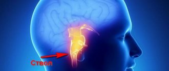
- mild manifestations of bulbar palsy (focus in the brain stem) with oculomotor disorders are possible;
- blurred vision in one eye combined with paresis (loss of sensation and weakness) in the limbs of the opposite side (often accompanied by incomplete narrowing of the internal carotid artery).
Features of hypertensive cerebral crises:
- the main manifestations are cerebral symptoms;
- focal signs occur rarely and are mild.
The patient complains of:
- sharp headache, often in the back of the head, temples or crown of the head;
- state of stupefaction, noise in the head, dizziness;
- nausea, vomiting.
People around note:
- temporary confusion;
- excited state;
- sometimes - a short-term attack with loss of consciousness, convulsions.
Transient disorders are not accompanied by any abnormalities in computed tomography and magnetic resonance imaging, since they do not have organic changes.
How to help during an attack
Poor blood circulation in the brain can lead to a stroke. During an exacerbation, it is important to competently provide assistance. This will avoid serious consequences and alleviate the suffering of the patient.
During an attack, it is necessary to measure blood pressure and count the pulse. If your heart function worsens, your blood pressure is high or very low, you need to call an ambulance. Offering any medications if they have not been prescribed by the attending doctor is strictly prohibited. It is not recommended to exceed the dosage of previously prescribed medications - this will not improve the situation, and can cause great harm.
Signs of a cerebral stroke
Ischemic stroke means the occurrence of irreversible changes in brain cells. At the clinic, neurologists distinguish periods of the disease:
- acute - continues from the onset of symptoms for 2–5 days;
- acute - lasts up to 21 days;
- early recovery - up to six months after the elimination of acute symptoms;
- late recovery - takes from six months to two years;
- consequences and residual effects - over two years.
Some doctors continue to distinguish small forms of stroke or focal ones. They develop suddenly, the symptoms do not differ from cerebral crises, but last up to three weeks, then completely disappear. The diagnosis is also retrospective. During the examination, no organic abnormalities were found.
Cerebral ischemia, in addition to general symptoms (headaches, nausea, vomiting, dizziness), manifests itself locally. Their nature depends on the artery that is “turned off” from the blood supply, the state of the collaterals, and the dominant hemisphere of the patient’s brain.
Let's consider the zonal signs of blockage of the cerebral and extracranial arteries.
We recommend reading: Symptoms of ischemic cerebral stroke
If the internal carotid artery is damaged:
- vision is impaired on the side of the blocked vessel;
- the sensitivity of the skin on the limbs and face on the opposite side of the body changes;
- paralysis or muscle paresis is observed in the same area;
- possible loss of speech function;
- inability to realize one’s illness (if the focus is in the parietal and occipital lobes of the cortex);
- loss of orientation in parts of one’s own body;
- loss of visual fields.
Narrowing of the vertebral artery at the level of the neck causes:
- hearing loss;
- nystagmus of the pupils (twitching when deviating to the side);
- double vision.
If the narrowing occurs at the confluence with the basilar artery, then the clinical symptoms are more severe, since cerebellar damage predominates:
- inability to move;
- impaired gesticulation;
- chanted speech;
- violation of joint movements of the trunk and limbs.
The possibility of developing compensatory collateral circulation is much higher when the patency of extracranial vessels is impaired, since there are connecting arteries for blood flow from the other side of the body.
If there is insufficient blood flow in the basilar artery, manifestations of visual and brain stem disorders (impaired breathing and blood pressure) occur.
If the anterior cerebral artery is damaged:
- hemiparesis of the opposite side of the body (unilateral loss of sensation and movement), often in the leg;
- slowness of movements;
- increased tone of flexor muscles;
- loss of speech;
- inability to stand and walk.
Blockage of the middle cerebral artery is characterized by symptoms depending on the damage to the deep branches (feeding the subcortical nodes) or long ones (approaching the cerebral cortex)
Obstruction of the middle cerebral artery:
- when the main trunk is completely blocked, a deep coma occurs;
- lack of sensitivity and movement in half of the body;
- inability to fix the gaze on an object;
- loss of visual fields;
- loss of speech;
- inability to distinguish the left side from the right.
Obstruction of the posterior cerebral artery causes:
- blindness in one or both eyes;
- double vision;
- gaze paresis;
- seizures;
- large tremor;
- impaired swallowing;
- paralysis on one or both sides;
- respiratory and blood pressure disturbances;
- brain coma
When the optic geniculate artery is blocked, the following appears:
- loss of sensation in the opposite side of the body, face;
- severe pain when touching the skin;
- inability to localize the stimulus;
- perverted perceptions of light, knocking;
- “thalamic hand” syndrome - the shoulder and forearm are bent, the fingers are extended at the terminal phalanges and bent at the base.
Impaired blood circulation in the area of the visual thalamus is caused by:
- sweeping movements;
- large tremor;
- loss of coordination;
- impaired sensitivity in half of the body;
- sweating;
- early bedsores.
The combination of damage to several branches causes complex syndromes of loss of sensitivity and false sensations in the limbs. The ability to diagnose ischemic changes depends primarily on the neurologist’s knowledge of the clinical manifestations of vascular disorders.
Probability of recovery
At the mere mention of this diagnosis, many who hear it feel, if not panic, then some kind of anxiety and internal discomfort. Indeed, the vast majority of the population associates this diagnosis with disability or even death.
Let's take a closer look to see if this is really the case.
There are many cases of recovery, if not complete, then almost complete.
In fact, the state of affairs is such that in the same neurological department a person can be treated for stroke, physical activity is limited only by doctor’s orders and bedridden, unable to move independently even within the hospital ward.
In the first case: the hospital patient walks calmly without support or auxiliary objects. He can even walk up stairs without handrail support. Speech is preserved, completely oriented in time and space. Coordination of movements is also not impaired. Externally, there are no signs of a serious illness. The loss of neurological function is minimal and its manifestations can only be detected by neurological examination.
In the second case: a person cannot move independently; strength is only in the left arm and leg, coordination of movements in them is impaired. He is in a hospital bed. He can only turn slightly in bed to one side. Raising the head end of the bed causes dizziness. The speech is not intelligible; only certain fragments of it are understandable. Verbal communication - responds with gestures and facial expressions, selectively - to individual questions.
As you can see, the difference between stroke cases can be enormous. Moreover, both in its acute period - the first 21 days, and a year after it occurred.
This difference is due, first of all, to the size of the lesion in the substance of the brain. This is one of the most important factors influencing the depth of impairment of neurological consequences.
The size of the dead tissue as a result of a hemispheric stroke is no more than 20-30 mm. in diameter and localized outside the zone of passage of large nerve tracts (pyramidal, optic radiation) are favorable in relation to the extent of neurological dysfunction and their recovery
Lesions larger than 30-40 mm in diameter, localized in areas where large nerve tracts pass or in the brain stem area, have an unfavorable prognosis in terms of the depth of neurological disorders and recovery from them.
The location of the stroke site plays a major role in recovery. More pronounced symptoms of brain damage will occur when the lesion is localized near the nerve pathways or in their area, even if they are small in size. This also applies to the brainstem localization of stroke. With equal sizes of dead nervous tissue, the depth of loss of function will be greater when localized in the region of the trunk.
This happens due to the high density of nerve conductors located here. The danger of this localization is due to the location in this area of a large number of vital nerve centers, including those responsible for blood circulation, breathing, digestion and other vital functions of the human body.
In what cases can acute stroke be suspected?
The above clinical forms and manifestations require careful examination, sometimes not by one, but by a group of doctors of different specialties.
Cerebrovascular accident is very likely if the patient exhibits the following changes:
- sudden loss of sensation, weakness in the limbs, face, especially one-sided;
- acute loss of vision, the occurrence of blindness (in one eye or both);
- difficulty in pronunciation, understanding words and phrases, composing sentences;
- dizziness, loss of balance, impaired coordination of movements;
- confusion;
- lack of movement in the limbs;
- intense headache.
Additional examination allows us to establish the exact cause of the pathology, the level and location of the vessel lesion.
Symptoms of the disease
The overall picture of the disorder depends on its type, but several general signs of the disease can be identified.
- pain in certain areas of the head;
- numbness in the limbs and face;
- problems with coordination of movements;
- dizziness, sharp and unpleasant;
- weakness in the upper and lower extremities, most often this symptom manifests itself on one side of the body;
- vision problems, loss of fields, inaccurate picture, splitting of objects;
- problems with speech, unclear words spoken by the patient.
All signs may not be present, which aggravates the situation with diagnostic accuracy.
Purpose of diagnosis
Diagnosis is important for choosing a treatment method. To do this you need:
- confirm the diagnosis of stroke and its form;
- identify structural changes in brain tissue, focal area, affected vessel;
- clearly distinguish between ischemic and hemorrhagic forms of stroke;
- based on pathogenesis, establish the type of ischemia for starting specific therapy in the first 3–6 in order to get into the “therapeutic window”;
- assess indications and contraindications for drug thrombolysis.
It is practically important to use diagnostic methods on an emergency basis. But not all hospitals have enough medical equipment to operate around the clock. The use of echoencephaloscopy and cerebrospinal fluid studies yields up to 20% errors and cannot be used to resolve the issue of thrombolysis. The most reliable methods should be used in diagnosis.
Foci of softening on MRI allow differential diagnosis of hemorrhagic and ischemic strokes
Computed and magnetic resonance imaging allows you to:
- distinguish a stroke from space-occupying processes in the brain (tumors, aneurysms);
- accurately determine the size and location of the pathological focus;
- determine the degree of edema, disturbances in the structure of the ventricles of the brain;
- identify extracranial locations of stenosis;
- diagnose vascular diseases that contribute to stenosis (arteritis, aneurysm, dysplasia, vein thrombosis).
Computed tomography is more accessible and has advantages in studying bone structures. And magnetic resonance imaging better diagnoses changes in the parenchyma of brain tissue and the size of edema.
Echoencephaloscopy can only reveal signs of displacement of the median structures with a massive tumor or hemorrhage.
During ischemia, cerebrospinal fluid rarely shows slight lymphocytosis with increased protein. Most often no change. If the patient has a hemorrhage, blood may appear. And with meningitis - inflammatory elements.
Ultrasound examination of blood vessels - Dopplerography of the arteries of the neck indicates:
- development of early atherosclerosis;
- stenosis of extracranial vessels;
- sufficiency of collateral connections;
- the presence and movement of an embolus.
Duplex sonography can determine the condition of the atherosclerotic plaque and artery walls.
Cerebral angiography is performed if technically possible for emergency indications. Typically, the method is considered more sensitive in identifying aneurysms and foci of subarachnoid hemorrhage. Allows you to clarify the diagnosis of pathology identified on tomography.
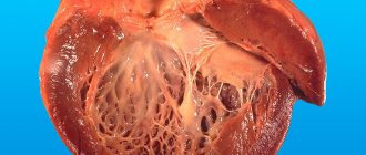
Cardiac ultrasound is performed to detect cardioembolic ischemia in heart disease.
It is mandatory to study blood clotting: hematocrit, viscosity, prothrombin time, level of platelet and erythrocyte aggregation, fibrinogen.
Causes, symptoms of hemorrhagic stroke
The most common causes leading to hemorrhagic stroke are:
- hypertonic disease;
- smoking;
- aneurysms;
- anomalies, pathologies of cerebral vessels;
- long-term use of blood thinning medications;
- addiction;
- alcoholism;
- hemophilia;
- inflammatory processes in cerebral vessels;
- decreased platelet levels.
Examination algorithm
The examination algorithm for suspected acute stroke proceeds according to the following plan:
- examination by a specialist in the first 30-60 minutes after the patient’s admission to the hospital, examination of the neurological status, clarification of the medical history;
- taking blood and studying its coagulability, glucose, electrolytes, enzymes for myocardial infarction, and the level of hypoxia;
- if it is not possible to conduct MRI and CT, do an ultrasound of the brain;
- spinal puncture to exclude hemorrhage.
Diagnostic methods and tools
Distinguishing this pathology from a stroke is sometimes problematic, therefore, before the start of the treatment process, several methods are used to determine the disease.
- detailed collection of data from the anamnesis, including interviewing the patient and his relatives;
- carrying out a blood test, determining cholesterol and coagulability levels;
- identification of causal factors of a pathological nature that influence the disease;
- Ultrasound of cerebral vessels, which makes it possible to identify signs of underlying diseases;
- MRI - the event is aimed at conducting a detailed study of the affected area.
It is also necessary to send the patient for consultation to other specialists. This could be a vascular surgeon, neurologist, cardiologist.
Treatment
The most important importance in the treatment of cerebral ischemia belongs to the urgency and intensity in the first hours of admission. 6 hours from the onset of clinical manifestations is called the “therapeutic window”. This is the time for the most effective use of the thrombolysis technique to dissolve a blood clot in a vessel and restore impaired functions.
Regardless of the type and form of stroke, the following are carried out in the hospital:
- increased oxygenation (filling with oxygen) of the lungs and normalization of respiratory function (if necessary, through transfer and mechanical ventilation);
- correction of impaired blood circulation (heart rhythm, blood pressure);
- normalization of electrolyte composition, acid-base balance;
- reducing cerebral edema by administering diuretics and magnesium;
- relief of agitation and seizures with special antipsychotic drugs.
A semi-liquid diet is prescribed for the patient's nutrition; if swallowing is impossible, parenteral therapy is prescribed. The patient is provided with constant care, prevention of bedsores, massage and passive exercises.
Rehabilitation begins from the first days
This allows you to get rid of negative consequences in the form of:
- muscle contractures;
- congestive pneumonia;
- DIC syndrome;
- pulmonary embolism;
- damage to the stomach and intestines.
Thrombolysis is a specific therapy for stroke of ischemic type. The method allows you to preserve the viability of neurons around the necrosis zone, returning all weakened cells to life.
You can read more about the indications and methods of thrombolysis in this article.
The administration of anticoagulants begins with Heparin derivatives (in the first 3–4 days). Drugs of this group are contraindicated for:
- high blood pressure;
- peptic ulcer;
- diabetic retinopathy;
- bleeding;
- impossibility of organizing regular monitoring of blood clotting.
After 10 days they switch to indirect anticoagulants.
Drugs that improve metabolism in neurons include Glycine, Cortexin, Cerebrolysin, Mexidol. Although they are not listed as effective in the evidence-based medicine database, their use leads to improvement in the condition.
Decompression craniotomy is performed in case of increasing edema in the brain stem area
Patients may need symptomatic treatment depending on the specific manifestations: anticonvulsants, sedatives, painkillers.
Antibacterial agents are prescribed to prevent kidney infection and pneumonia.