Violation of the functional activity of blood vessels is a common problem in cardiological practice. Mainly of organic origin, defects lead to the impossibility of adequate blood circulation in the tissues. Is it dangerous.
Aortic thickening is not a diagnosis. We are talking about a random clinical finding obtained as a result of an instrumental study. Objectively, it represents hypertrophy (proliferation) of tissue throughout the entire thickness of the vessel structure.
Contrary to possible misconception, this is not good. Rigidity (decrease in elasticity) affects the vessel’s ability to withstand enormous loads.
It is possible and even probable that many dangerous complications will develop: from an aneurysm to an instant rupture with massive bleeding and death in a matter of seconds.
If the aorta is compacted, this means that there is a third-party pathology; the deviation is not primary, it is always caused by another disease.
The vast majority of cases are associated with tuberculosis, arterial hypertension and atherosclerosis. Accordingly, therapy for the underlying condition is indicated.
Development mechanism
The basis of the pathological process is destruction of the aortic wall or a constant increased impact on the structure.
In the first case, we are talking about infectious-inflammatory, less often autoimmune processes. As a result of the constant violation of the anatomical integrity of the endothelium and other tissues, severe scarring occurs.
Epithelization leads to a gradual overgrowing of the lumen with dense connective tissue and its narrowing. The blood flow weakens, and the pressure in the local area becomes higher due to the need to overcome a mechanical obstacle.
The second factor is approximately identical, in terms of creating excess load on the walls of blood vessels. The main etiological point is an increase in blood pressure and the constant preservation of tonometer readings at consistently high numbers.
Both options pose a colossal danger to human life and health.
Organic disorders are difficult to treat, and not every doctor is ready to take on such unpredictable cases. Therefore, the logical solution would be to carry out treatment in the early stages, when there are no anatomical abnormalities yet.
Causes
There are both fundamental factors and risk factors that do not directly determine the development of the pathological process. First group:
Current or previous tuberculosis
This is a dangerous infectious disease. It is provoked by mycobacterium, also called Koch's bacillus. Can exist for a long time without symptoms. The clinic is already formed at pronounced stages.
Damage to the aorta is a complication, the prevalence is up to 25% of the total number of cases. Timing does not matter; involvement is possible at the very first stages, which affects the overall forecast.
The walls of the vessel are destroyed and scarred, the lumen narrows, and the process progresses independently. Recovery is possible only with comprehensive treatment of tuberculosis in a hospital setting.
The total duration of supervision is 6-12 months. Hospital stage - no more than 3. The rest of the period is spent on outpatient care. Throughout the rest of life, the patient should be monitored for relapses.
Syphilis
Danger does not come immediately. This sexually transmitted infection can be transmitted non-sexually, but the likelihood of this option is extremely low.
Urgent treatment of the condition is required. Because aortic damage occurs in approximately 30% of situations.
Thickening of the aortic walls does not occur immediately; it is a later complication. There are known cases of the development of a delayed phenomenon after 10-20 years from the conditional cure of syphilis.
Therefore, such patients are recommended to undergo at least echocardiography every 6 months and closely monitor their well-being.
Aortitis
Inflammatory process in the corresponding vessel. Relatively rare. Again, as a consequence of an infectious disease.
It is a complication of herpes, damage by pyogenic flora, and septic myocarditis. In rare cases, it develops as a result of autoimmune pathologies. For example, rheumatism or vasculitis.
Inflammatory diseases need to be treated in a timely manner, because the likelihood of complications is significant.
Aortitis can be treated with antibiotics or immunosuppressants, depending on the etiology of the process. In any case, hospitalization in a cardiac hospital is indicated.
Hypertension
Stable increase in blood pressure. It occurs for many reasons, which themselves require identification and qualified assessment by doctors.
At the first stage of deviation, when the disease is still unstable in its manifestations, there is a chance for normalization. As the condition progresses, the risks of impairment increase. The third degree of hypertension cannot be treated.
As mentioned, hardening of the aorta of the heart is the result of degeneration, excessive stress on the walls, or a combination of these factors.
In the absence of therapy, there is a high probability of vessel rupture. Sooner or later this will happen. Specific medications are prescribed to lower blood pressure (ACE inhibitors and others, as indicated).
Age-related changes
Violation of the functional activity, anatomical characteristics of the connective tissue that makes up the aorta and its walls. Mainly appears to be the result of age-related changes.
Variations are possible. The same phenomena are observed in diabetes mellitus, genetic pathologies, anomalies of intrauterine development with spontaneous features.
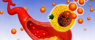
Atherosclerosis
There are two types.
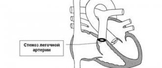
- The first is a narrowing of the vessel, stenosis. Occurs in smokers, alcoholics, drug addicts, and people with arterial hypertension. It is a complication and requires urgent help. If the process is stable, surgery may be required if conservative methods do not have an effect.
- The second option is occlusion or blockage of the vessel with a cholesterol plaque. The result of a disorder of lipid metabolism in the body. It is less dangerous because the condition develops over months or even years.
Gradually, the formation becomes calcified and becomes covered with mineral salts. Such an atherosclerotic plaque cannot be dissolved with statins. Surgical treatment is required.
Therapy methods
The disease, detected at an early stage, requires constant dynamic monitoring.
If the disease does not progress, an effective therapeutic method for the patient can be a radical revision of his lifestyle: switching to a healthy diet and eliminating all bad habits. Moderate physical activity and long walks in the fresh air will be extremely beneficial. An important factor promoting recovery is protecting the patient from all kinds of stress and anxiety. All these actions have both therapeutic and preventive significance. A good addition to the above measures can be a course of drug treatment prescribed by the attending physician. Progression of the disease is an indication for surgical intervention. If there is significant damage to the heart valves, they resort to prosthetics or plastic surgery. If the disease has led to significant destruction of the heart muscle structures, a donor organ transplant may be required. It is possible to treat aortic thickening with folk remedies only with the consent of the treating specialist. The most popular methods of alternative medicine include the practice of vegetarianism or raw food diet, the use of herbal medicine, acupuncture massage and treatment with natural juices. When including folk remedies in the general treatment regimen, it is very important to take into account existing contraindications and the high likelihood of developing an allergic reaction to the components of the drugs used
A subject index to common diseases of the cardiovascular system will help you quickly find the material you need.
In the absence of pronounced manifestations of fibrosis, treatment is not required. It is enough for the patient to be observed by a cardiologist and periodically undergo ultrasound of the heart. The exception is people who have concomitant diseases (arrhythmia, hypertension, ischemia). In their case, it will be necessary to use therapeutic agents, depending on the pathological process.
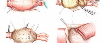
Significant changes that provoked severe stenosis of the valve ring are eliminated surgically. Its essence is in valve prosthetics or plastic surgery. Most of the operations are full-scale, that is, on an open heart, using a cardiopulmonary bypass device.
The use of traditional medicine must be agreed with the attending physician. In fact, it cannot help in the treatment of fibrosis; it will only reduce nervous tension and saturate the body with useful substances.
Prevention measures
The appearance of fibrous tissue is easier to prevent than to stop or treat. The following tips will help with this:
- prevent rheumatism;
- eliminate infectious diseases without causing complications;
- make a proper diet;
- walk in the fresh air more often;
- follow the recommendations of your doctor;
- undergo a full medical examination annually;
- strengthen the immune system;
- avoid physical overload;
- avoid stressful situations;
- exercise.
https://youtube.com/watch?v=4JHYtVF5n50
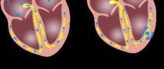
Fibrosis of the valve apparatus is characterized by the appearance of negative consequences, for example, acquired heart disease (stenosis of the valve ring or insufficiency of its leaflets). If the changes are caused by rheumatism, then the chance of their development is higher than due to ischemia or heart attack.
Atherosclerosis of the aorta provokes non-rheumatic types of defects much more often than myocarditis. It is extremely difficult to predict the likelihood of their formation. Doctors try to focus on the results of examinations and the course of the main pathological process that provokes changes in the heart in order to keep the situation under control.
Risk factors
And the corresponding groups with an increased likelihood of developing compaction:
Physical inactivity
Insufficient physical activity of a person. It is common both for bedridden patients confined to a wheelchair and for persons leading a sedentary lifestyle. Which is the majority these days.
Blood stasis develops, general hemodynamics are disrupted. A hardening of the aorta is a possibility, but not the only problem.
To reduce the risks of the process, at least an hour of walking or cycling is enough. More is possible, at the discretion of the person himself. If there are no contraindications.
Excessive physical activity
Leads to an increase in blood pressure, the release of inadequate amounts of cortisol, strerlide hormones and catecholamines.
This provokes a narrowing of the lumen of blood vessels, including the aorta. Blood flow is disrupted, and the load on structures increases. The body is forced to resist.
In general, the thickening of the aortic walls is a compensatory mechanism aimed at preventing rupture as a result of excess load.
Obesity
It is not increased body weight that has an effect, although it plays a role. Responsibility lies with disruption of normal lipid metabolism. Fats are deposited (deposited), there is no rapid catabolism, that is, breakdown.
The excess enters the bloodstream. Due to their structure, lipids are well attached to blood vessels. Then the process continues, the formation grows, the walls of the aorta thicken and lose elasticity.
The disorder can only be stopped with the help of drugs, statins. A change in diet is also indicated. Less fat and easily digestible carbohydrates, more protein, plant foods.
Excess weight causes an increase in blood pressure, acceleration of cardiac activity due to the high load on the muscles (just imagine who will have it easier: a person with empty hands or someone carrying a bag of potatoes).
Diabetes
Causes generalized disruption of all organs and systems. Including circulatory.
The process is twofold. On the one hand, there is a stable narrowing of the aorta and other structures, on the other, a violation of lipid metabolism. Atherosclerosis develops.
The likelihood of death from blood vessel rupture increases without treatment every month, if not daily. Therefore, timely diagnosis of endocrine disease is necessary.
Smoking
Long lasting. Against the background of constant consumption of tobacco, tar, and cadmium compounds, a physiological addiction of the body is formed. Stenotic atherosclerosis develops.
Even after giving up the bad habit, spontaneous regression of narrowing does not occur. Treatment is needed. Considering that many return to addiction, the likelihood of further development of pathology increases.
Finally, alcoholism and the use of psychoactive substances have an effect. Many drug addicts do not have time to live to see either a rupture or an aortic aneurysm. Death occurs from heart failure much earlier. Alcoholics have a better chance.
Reasons are being eliminated gradually. One at a time during the diagnosis.
Symptoms
There are no such manifestations. If the walls of the aorta are compacted, there is no clinical picture. Signs and disturbances in well-being arise later, after complications of the pathological process.
Organic changes, such as an aortic aneurysm, give the following moments:
- Pain syndrome. Localization and nature depend on the location of the violation. If the abdominal region is affected, there is discomfort in the abdomen, and in the chest there is a pressing, burning sensation. If the aortic arch is compacted, there is pain in the neck, turning into discomfort at the location of the heart.
- Dyspnea. Disruption of a natural physiological process.
Further manifestations depend on which area has undergone changes.
Abdominal:
- Nausea, vomiting, heartburn, belching, other dyspeptic symptoms. Such patients are falsely sent to gastroenterology.
- Increased gas formation in the intestines.
- Pulsating sensations in the abdomen.
Arc, descending section:
- Inability to swallow normally.
- Violations of voice, timbre and general ability to speak.
- Excessive saliva production.
- Weakness.
- Numbness of the upper and lower extremities. In exceptional cases, paresis and paralysis occur. This is the result of compression of the nerve roots.
Such conditions require urgent treatment; irreversible changes in the musculoskeletal system are possible.
Compaction of the aortic root and ascending section:
- Chest pain.
- Dyspnea.
- Abnormal heart rate, other arrhythmias.
- Cephalgia, vertigo.
- Loss of consciousness, fainting.
Classic signs of damage to cardiac structures.
Attention:
You cannot focus on symptoms when identifying the causes of poor health. These are unreliable clinical signs.
Diagnostics
It takes place on an outpatient basis, under the supervision of a cardiologist or vascular surgeon. The following activities are shown:
- Oral interview with a person. To get a rough picture.
- Anamnesis collection. Family history, addictions, past pathologies.
- Measurement of blood pressure and heart rate.
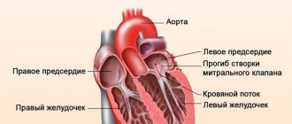
- Echocardiography. One of the first methods. Gives an idea of the state of the muscular organ itself and the ascending aorta, arch.
- Aortography. Targeted assessment of the largest vessel. It is used both to verify the diagnosis and to determine the effectiveness of the treatment.
- MRI. Used to clarify the localization of the pathological focus. Typically, tomography is prescribed before surgery as an informative way to visualize tissue.
- ECG. To identify functional disorders that inevitably arise at advanced stages of changes in the aorta.
- Ultrasound of the abdominal cavity.
- Plain chest x-ray.
Methods provide nuggets of valuable information. To get a complete picture, you need to carry out all these activities. They are not dangerous and painless. Therefore, it is not recommended to hesitate.
How to Diagnose the Problem
Aortic thickening is a change that can only be detected by instrumental diagnostic methods:
- Ultrasound of the problem area;
- X-ray of the chest (fluorography) and abdominal cavity;
- computer and magnetic resonance imaging (CT and MRI of the chest and abdominal cavities);
- contrast aorto-arteriography.
Methods for diagnosing aortic compaction
Treatment
Depends on the primary diagnosis. The goals of therapy are to prevent complications and eliminate the origin of the condition. Medicines are used.
Which ones are determined by the doctor based on the underlying disease:
- Antihypertensive. ACE inhibitors, calcium antagonists, centrally acting agents, beta blockers, mild diuretics.
- Statins. They dissolve cholesterol and prevent its further deposition on the walls of blood vessels.
- Thrombolytics, antiplatelet agents. They prevent blood from stagnating, improve its fluidity and rheological properties, and prevent the formation of dangerous clots.
- Antituberculosis drugs. Also medications for the treatment of syphilis.
Surgery is required when a threatening aneurysm forms due to calcification of the cholesterol plaque. At the discretion of the specialist after careful consideration of all options.
A change in diet is shown throughout life. Less fat, easily digestible carbohydrates, more protein and plant foods, walking, minimal physical activity, proper rest at night. Issues of lifestyle correction are also resolved by a specialist.
Prevention measures
Preventive measures can help reduce the risk of developing UAS. It is especially important to adhere to them for those who have diseases that provoke thickening - atherosclerosis, hypertension, etc.
Basic preventive measures:
- stop smoking completely;
- give up alcohol (use is possible, but in the most moderate quantities);
- eat right, follow a diet;
- visit your doctor regularly;
- walk more and play sports;
- follow the work and rest schedule, get enough sleep;
- avoid stress.
Forecast
In itself, compaction of the aorta is not a diagnosis, but just an instrumental finding. You can say anything concrete after assessing the root cause.
The following factors play a major role:
- Dynamics of deviation. Progression is associated with more severe general condition and outcome.
- The age of the person. Elderly people have a relatively unfavorable prognosis.
- Family history. If there was at least one representative in the family with the condition in question, it is necessary to carefully check every six months. In such individuals, the anomaly progresses rapidly. Therefore, delay reduces the chances of successful treatment.
- The presence of concomitant somatic pathologies. Among the especially dangerous ones are hypertension, diabetes mellitus, metabolic syndromes, and atherosclerosis.
- Possibility and necessity of radical treatment. If surgery is required, the prognosis is initially worse. Further, everything depends on the success of the therapy. The absence of such a need gives a better outcome, although it does not guarantee it.
In general, even an aneurysm, as one of the most severe complications, is associated with a survival rate of 85-90%.
Attention:
Heart failure and circulatory disorders in the brain reduce the patient's chances by 30-40%.
Signs when the aorta is hardened
You need to worry when the compaction begins to intensify - this can lead to disturbances in the circulatory system and the threat of dissection and rupture of the vessel, which will lead to dangerous bleeding.
Risk factors that may cause pathology to develop include:
- Smoking;
- Abuse of fatty foods;
- Atherosclerosis;
- Age over 50 years;
- Diabetes.
Diagnosis of seals is carried out both in a selected area and along the entire length - ultrasound of the aortic walls. The study may also include x-rays, CT and MRI of the abdominal and chest cavities. In the initial stages, the disease does not manifest itself in any way, but over time, it leads to the lumen of the blood vessels becoming narrower, and the nutrition of the internal organs may be impaired.
If the blood supply to the heart muscle is disrupted, myocardial infarction and angina attacks may develop. If damage to the blood vessels of the brain occurs, headache, irritability, fatigue, and dizziness appear. In the area of the peritoneum, compaction of the aorta manifests itself in the form of pain in the abdominal area, signs of gastrointestinal dysfunction, and sudden weight loss. In its most severe form, signs of acute peritonitis may occur. The abdominal aorta supplies blood vessels to the legs and, if they become blocked, lameness, cramps and severe pain appear.
Complications
Possible consequences:
- Thrombosis. The formation of a clot in one of the arteries, sometimes in the aorta itself. Causes local ischemia. When torn off and moved along the riverbed, it often blocks the pulmonary vessel and coronary structures, which immediately leads to death.
- Aneurysm. Wall protrusion. It is observed against the background of stable degeneration and dystrophy of aortic tissue.
- Rupture, delamination. Leads to death within a matter of seconds. There were cases to the contrary, but they were casuistic. There are hardly more than 0.5%.
Thickening of the aortic walls is not an independent disease. It is not considered a diagnosis and is not represented in the ICD-10 classifier. This is a consequence, an accidental find.
After eliminating the root cause, there is every chance to stop the progression and achieve results without affecting the quality and length of life.