Study of the heart and blood vessels
24,000—37,000 rubles
Cardiologists from the Stolitsa network of clinics will give you a complete medical examination of your heart and blood vessels within 2 days, from which you will learn:
- what condition is your cardiovascular system in?
- what you need to do to avoid the development of heart and vascular diseases,
- how to treat heart and vascular diseases identified during the study in order to prevent further development of the disease.
A medical examination of the heart and blood vessels will allow timely identification of risk factors and prevention of the development of diseases that sharply reduce the quality of life, such as:
- coronary heart disease (its consequences - heart pain, limitation of activity, threat of myocardial infarction),
- hypertension and symptomatic hypertension (headaches, hypertensive crises, threat of stroke),
- cerebral atherosclerosis (memory impairment, decreased performance, sleep disturbances, mental disorders),
- obliterating atherosclerosis of the vessels of the lower extremities (more often in men - chilly legs, pain in the calf muscles when walking, threat of gangrene and amputation).
CardioProfi (full examination) is a program for those who already have diseases of the cardiovascular system and need to assess the dynamics of the disease, the condition of the heart and blood vessels and carry out treatment adjustments.
CardioNorm is the optimal program for those who have initial signs of heart and vascular diseases or health problems (fatigue, headaches, palpitations, shortness of breath, and others).
View all cardiology programs
| Program | "Cardioprofi" | "CardioNorm" |
| Program cost (RUB) | 37 000 | 24 000 |
Initial consultation with a cardiologist.
| + | + |
| Repeated consultation with a cardiologist. | + | + |
| ECG with interpretation | + | + |
| ECG Holter monitor (24 hours) | + | + |
| BP-Monitor (24 hours) | + | |
| Bicycle ergometry | + | + |
| ECHO CG with Dopplerography | + | + |
| Ultrasound of the abdominal aorta | + | |
| Ultrasound of the thyroid gland | + | |
| Ultrasound of kidneys + adrenal glands | + | + |
| Triplex scanning of the vessels of the lower extremities | ||
| Triplex scanning of renal vessels | + | + |
| Triplex scanning of extracranial vessels | + | + |
| Clinical blood test, ESR | + | + |
| General urine analysis | + | + |
| Prothrombin index + clotting time | + | + |
| Rheumatic tests | + | |
| Thyroid hormones (T3f, T4f, TSH, AT TPO, AT TG) | + | |
| Biochemical blood test (urea, creatinine, electrolytes, uric acid, glucose, total bilirubin, direct bilirubin, ALT, AST, total cholesterol, LDL cholesterol, HDL cholesterol, triglycerides) | + | + |
| Consultation with an ophthalmologist (external examination, visometry, refractometry, tonometry, biomicroscopy, ophthalmoscopy) | + | + |
| Advanced markers of atherosclerosis (apolipoprotein A1, apolipoprotein B, lipoproptein (a), homocysteine, fibrinogen, ) | + | |
| Cardiologist’s conclusion based on examination results, recommendations, treatment adjustments | + | + |
View all cardiology programs
Make a choice in favor of professional medicine!
How is an examination by a cardiologist carried out and whether you need to prepare for it 03/24/2017
Preparing the patient for a visit to a cardiologist greatly facilitates the diagnosis process. So, when going to see a doctor, you need to make a complete list of complaints that caused concern and served as a reason for making an appointment with a cardiologist. It is important not to miss anything, because any change in well-being, even the most short-term, can be a symptomatic manifestation of a serious pathology of the cardiovascular system. Before consulting a cardiologist, you should try to remember under what circumstances the deterioration occurred (for no reason, after strong emotional stress, during physical exertion, etc.), for how long the alarming symptoms lasted (a couple of seconds, more than a minute, in during the day), how exactly the improvement occurred (after taking any drug, after tracking, on its own), with what frequency the deterioration occurred.
When preparing to go to a cardiologist, it is advisable to check with your closest relatives about whether they have any problems with the cardiovascular system. Cardiologists explain the significance of such information by the fact that it is the hereditary factor that quite often plays a key role in the development of heart pathologies, which is important to take into account when developing a treatment strategy. People who have previously undergone examination of the heart and/or blood vessels, were observed by another cardiologist, or have undergone a course of treatment aimed at eliminating a particular disease of the circulatory system are recommended to take with them to their appointment with a cardiologist all the medical documentation they have on this issue . So, you should prepare a medical card, certificates, extracts, laboratory test results, old cardiograms, ultrasound reports, etc. Of particular diagnostic value for a cardiologist will be the results of examinations completed over the past year. If the patient has taken or continues to take any medications, for a consultation with a cardiologist he should take a pre-compiled list of these medications, preferably indicating doses.
In preparation for visiting a cardiologist, at least 2 hours before the scheduled consultation time, it is strongly recommended to stop smoking and drinking caffeine-containing drinks, because otherwise the doctor will not be able to correctly assess the functional capacity of the cardiovascular system. Cardiologists at the Agapit clinic in Chernigovskaya metro station emphasize that ignoring such restrictions can significantly distort the real picture of the condition of the heart and blood vessels, which can lead to errors when making a diagnosis.
As a rule, an initial appointment with a cardiologist begins with collecting the patient’s life history and discussing the complaints that led to the need to seek medical help. This is usually followed by a physical examination, during which the cardiologist evaluates the color of the skin, checks for swelling, palpates the heart area, taps the chest, performs auscultation, measures blood pressure and carefully examines the pulse in both arms. Based on the data obtained during the examination, the cardiologist makes a preliminary diagnosis and determines a list of additional studies that will help confirm or refute the initial conclusion.
Basic instrumental methods for studying the heart
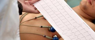
1. Electrocardiography.
This is the registration and further study of electrical impulses produced by the heart muscle during its work. In a healthy heart, electrical excitation travels through the conduction system and different parts of the heart in a certain sequence. Its violation can be a manifestation of various pathological conditions.
Fluctuations in the potential difference between the excited and non-excited parts of the muscle are recorded in the form of waves, resulting in a specific curve - the ECG itself. An ECG is recorded using a device called an electrocardiograph.
An ECG allows you to evaluate the nature of the heart rhythm and the electrical processes occurring in it. This is important for diagnosing various arrhythmias and other heart rhythm disturbances, coronary heart disease; ECG changes are also possible with myocarditis, metabolic and electrolyte disorders, non-cardiac diseases (pulmonary emphysema, pleurisy, obesity, etc.).
Electrocardiography can also be part of other research methods.
- Holter (24-hour) monitoring is a 24-hour ECG recording, used to monitor seriously ill patients or to identify periodically occurring pathological conditions, such as short-term arrhythmias.
- Bicycle ergometry - recording an ECG during and after physical activity. This method allows you to determine the patient's susceptibility to stress and is used in the diagnosis of coronary heart disease, in particular exertional angina.
2. Phonocardiography.
A method of graphically recording sounds - tones and noises - of the heart. The recording is made using a phonocardiograph, which is most often an additional attachment to an electrocardiograph, since PKG is necessarily recorded synchronously with electrocardiography. This method allows you to evaluate the sound symptoms of various heart diseases and more reliably identify pathological noises.
3. Echocardiography.
Ultrasound examination of the heart muscle. Currently, there are three modifications of this method of cardiac examination:
- one-dimensional echocardiography gives a projection of the heart in one plane, is carried out to assess the thickness of the heart walls and the size of the organ, the functioning of the valves, the condition of the heart during and after contraction;
- two-dimensional echocardiography allows you to obtain a three-dimensional image of the heart and is more informative;
- Doppler echocardiography is a study of intracardiac blood flow, used to assess the hemodynamics of the heart, diagnose defects and shunts.
4. X-ray methods for studying the heart.
Radiography (RG) and fluoroscopy are used to determine the size and shape of the organ and large pericardial vessels, and the presence of fluid in the pericardial cavity. This method involves irradiating the patient with X-rays, and therefore is carried out only according to strict indications when other research methods are not informative. X-rays are contraindicated in pregnant women.
A type of RG is computed tomography. This method allows you to obtain more information, since the image of the heart is simulated on a computer screen, but the radiation dose when using it is greater than when performing conventional X-ray.
5. Radionuclide methods for studying the heart
is the introduction of radioisotopes of metals into the bloodstream and subsequent assessment of their distribution. Various radioisotopes are used to determine the localization of myocardial infarction, accumulating in the lesion, others help assess the condition of the cavities of the heart and large vessels. During this study, the body is also exposed to radiation.
6. Magnetic resonance imaging.
Evaluation of a computer image of the heart obtained as a result of the resonance of hydrogen nuclei contained in cells when the body is exposed to a magnetic field. Allows you to evaluate not only structural changes in heart tissue, but also local chemical cellular disorders: acidosis, ischemia, swelling.
7. Angiocardiography.
Injection into the heart of a substance that has radiopaque properties. Using this method, many structural and physiological parameters of the heart muscle can be assessed. Angiocardiography is also used to clarify indications for heart surgery. It is always carried out simultaneously with cardiac catheterization (the catheter is inserted most often through the subclavian or femoral arteries), which also has a diagnostic value - it allows you to obtain blood samples from the cardiac cavities and assess the pressure inside the heart.
All these methods are not used simultaneously in one patient; for each one or more methods of studying the heart are selected depending on the condition of the body, age and diagnosed pathology.
Often, a person who first feels any complaints from the cardiovascular system, upon initial outpatient treatment, immediately receives treatment, bypassing a full cardiological and related examination, which should include both instrumental and laboratory diagnostic methods.
Depending on the prevalence of clinical symptoms, three main groups of patients can be divided:
1. Patients with complaints of pain in the heart area 2. Patients with high blood pressure 3. Patients with rhythm disturbances, interruptions in heart function
Various combinations of clinical symptoms are also possible (rhythm disturbances and heart pain against the background of high blood pressure).
The minimum examination should include:
- Examination by a cardiologist with detailed collection of complaints and physical examination (auscultation, percussion)
- ECG (12-lead ECG, long strip ECG, cardiotopography, ECTG-60, ECG with isometric stress)
- Ultrasound (ultrasound) of the heart, dopplerography of blood vessels, transesophageal ultrasound (often necessary for rhythm disturbances to exclude the presence of blood clots in the cavities of the heart), ultrasound of the kidneys, adrenal glands, thyroid gland
- 24-hour blood pressure and ECG monitoring (Holter monitoring)
- Load tests (velergometric test, treadmill test, informational and pharmacological tests)
- Consultations of related specialists (endocrinologist, gynecologist, ophthalmologist, gastroenterologist, neurologist, nephrologist, etc.)
- Laboratory tests: biochemical blood test (glucose, electrolytes, lipid spectrum, cholesterol, etc., cardiac enzymes), determination of the level of certain hormones (thyroid gland, brain natriuretic peptide).