Physiological characteristics of the female body
Women have a number of physiological characteristics that distinguish them from men.
- The former have a smaller heart size , myocardial thickness, systolic and minute blood volumes (MBV), and diastole duration. The increase in IOC in response to physical activity occurs mainly due to an increase in heart rate. This mechanism is wasteful and has less functionality of the heart.
- Increased elasticity and extensibility of ligaments , which leads to frequent injury.
- Higher excitability and reactivity of the nervous system is manifested by pronounced emotionality.
- The menstrual cycle, pregnancy, menopause - all these features change the level of estrogen and progesterone, which entails lability of mood, sleep, basal metabolism, and catecholamine metabolism.
Diff. diagnosis of angina pectoris and diseases of the lungs, pleura, mediastinum
Pneumonia
- Pneumonia. With pneumonia, there is an increase in body temperature, increased pain in the chest when inhaling, and on auscultation - wheezing in the lungs, crepitus. X-ray confirms the diagnosis.
- Chronic cor pulmonale. Chest pain is constantly felt in such patients, there is no irradiation to the arm or shoulder blade, and it is not reduced by taking nitroglycerin, but the use of bronchodilator therapy helps very well.
- Diseases of the esophagus. Burning pain in the chest is not related to exercise, but to meals: after eating and when swallowing, they intensify, and there will be no ischemia on the ECG. Recognition of pathology is facilitated by FGDS and fluoroscopy of the esophagus.
- Mediastenitis and mediastinal tumors. When pain syndrome appears in the chest due to this pathology, as a rule, there are already other signs of the disease: difficulty swallowing due to compression of the esophagus, swelling of the neck veins, breathing problems, thickening of the neck. Mediastenitis is accompanied by an increase in temperature and a sharp acceleration of ESR. Help with differential X-rays provide diagnostics.
Common reasons
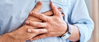
The sources of heart pain in women are varied. Today it is customary to divide possible etiological factors into two large groups:
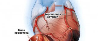
- Coronarogenic (true cardialgia) is a type of pain that is associated with organic pathology of the cardiovascular system, namely the coronary arteries. With their atherosclerosis, spasm, the heart receives an insufficient amount of oxygen from the blood, as a result of which the appearance of coronary disease is observed.
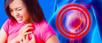
- Non-coronary - a category of pain that is not associated with impaired blood flow through the coronary vessels. The reasons are heart rhythm disturbances, psychoneurotic conditions, exacerbation of concomitant chronic diseases. Below is a photo with typical localization of pain points in the heart in women.
True cardialgia
The causes of true cardialgia are the following factors:
- Stable exertional angina. It implies the appearance of pain behind the sternum of a pressing, squeezing nature, which is associated with physical activity or stressful situations. It lasts no more than 15 minutes and is easily relieved by taking Nitroglycerin.
- Coronary syndrome. Retrosternal pain attacks are often more intense and react worse to Nitroglycerin. The peculiarity of this pathology is that the coronary vessels are not involved in the development of the disease, and the diagnosis is made by exclusion.
- Prinzmetal's angina (vasospastic) . An anginal attack occurs more often in the morning or at night.
- Unstable angina is a complex of symptoms that appeared for the first time (the last two months) or aggravation of the manifestations of existing angina (pain bothers more often and with less stress, sometimes even at rest, while sensitivity to nitrate drugs decreases).
- Myocardial infarction is a dangerous condition that threatens human life. Typical features: sharp paroxysmal pain in the chest of a burning, tearing nature with possible irradiation to the left arm, shoulder blade, leg, elbows. The attack lasts more than 15 minutes and is not relieved by nitro drugs. Accompanied by severe shortness of breath, sweating, general weakness, and rapid heartbeat.
- Dissecting aortic aneurysm . Pain in the chest occurs unexpectedly, at the time of physical activity, a rise in blood pressure, and radiates to the spine, upper limbs, and head. Signs of shock are observed with a transition to a state of imaginary well-being, when the pain subsides, blood pressure and heart rate stabilize. The migration of pain is characteristic: first it is localized in the heart area, then in the epigastrium, lower back, and lower limb.
- Pulmonary embolism . The attack occurs acutely, while the patient suffocates, the skin becomes bluish due to severe suffocation, and the heartbeat quickens; subsequently hemoptysis appears.
- Dry pericarditis is manifested by prolonged pain in the heart area, shortness of breath, and fever.
Extracardiac sources of pain
- Cervical and thoracic osteochondrosis , spondylosis. The pain is stabbing, sometimes shooting in nature, increases gradually and lasts for days, increases at night, decreases with walking, running, and taking NSAIDs. A special feature is a direct connection with body movements - rotation, abduction and raising of the arms. It intensifies when you inhale and touch the problem point. Often accompanied by cephalalgia and dizziness.
- Intercostal neuralgia . The pain is sharp, burning, and occurs along the rib.
- Left-sided plexitis is an inflammation of the brachial nerve bundle, which is accompanied by severe pain and numbness in the left arm. The sensations intensify with movements of the affected limb.
- Tietze syndrome is an aseptic inflammation of the cartilages of the II, III, IV, V ribs at the site of their attachment to the sternum. Pain in the heart area worries for a long time, complemented by an increase in body temperature, swelling to the left of the sternum, severe paresthesia, and laboratory signs of inflammation.
- Herpes zoster . It is accompanied by a painful rash in the form of reddened skin with grouped blisters filled with clear liquid. Occurs along the intercostal space.
- Pleurisy (basal left side). The pain is stabbing in nature, occurs suddenly, intensifies with breathing, and is accompanied by signs of inflammation. A pleural friction rub is heard during auscultation of the lungs.
- Diaphragmatic hernia is a displacement of abdominal organs (stomach, intestines) into the chest through a defect in the diaphragm. In this case, “acute mediastinal” syndrome occurs with intense pain in the heart area, rapid heartbeat, drop in pressure, pallor and other signs of shock.
- Esophageal diverticulum . Chest pain appears after eating, when swallowing, or when bending the body forward.
- Gastroesophageal reflux disease (GERD) is also a common reason for visiting a doctor with a complaint of chest pain, which is burning in nature and accompanied by heartburn and a feeling of sourness in the mouth.
- Menopausal cardialgia . Due to hormonal changes, a woman is bothered by very intense pain in the heart of a cutting, stabbing, pressing nature, not associated with physical activity, and having a wave-like course. There is a rush of blood to the head, a feeling of heat in the face, increased sweating, frequent urination, and irritability. Cardialgia does not have organic soil and is not accompanied by changes in the cardiogram.
Let us also mention climacteric myocardial dystrophy, in which a decrease in estrogen levels provokes the development of organic heart pathology.
- During pregnancy , changes in hormonal levels and fetal development create an increased load on a woman’s cardiovascular system, which causes cardialgia, arrhythmias and arterial hypertension.
- Thyrotoxicosis and other thyroid disorders . In addition to nonspecific pain in the heart area, tachycardia, arrhythmia (often atrial fibrillation), irritability, tremor, drowsiness, apathy, and low blood pressure are also troubling.
- Cardioneurosis is a functional disorder that is considered as a complex of mental, autonomic and endocrine pathologies. The symptom of heart pain in young women is characteristic of emotionally labile individuals. The pain does not have any cause, varies in sensations and is easily relieved with sedatives.
- Cardiophobia is accompanied by discomfort in the heart area, alertness, and fear of death. Severe cardiophobic syndrome is considered a pseudo-infarction attack, in which pain occurs as a consequence of waiting for a diagnosis of myocardial infarction. Fear accompanies pain, and the result is a vicious circle.
How is the cause of the aching pain in the left side of the sternum diagnosed?
Timely diagnosis when symptoms of unknown etiology appear is the first step to a healthy life. It is necessary to be checked annually by a cardiologist in order to detect cardiovascular pathology in a timely manner. If the cause of the pain is discovered, the cardiologist will be able to draw up the correct treatment plan. For an objective picture, you will need a comprehensive history, so you need to undergo a number of examinations:
- Clinical and biochemical blood tests.
- Echocardiography.
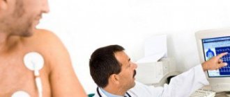
- Electrocardiogram with and without stress.
- X-ray of the chest cavity.
- Electrophysiological study.
- Magnetic resonance imaging.
- ECG or Holter tonometry.
If the heart “sores” and the cause of the pain cannot be determined, they give a referral to a psychiatrist. During hospitalization, the doctor will find out why the pain is on the left side of the sternum, and why the heart constantly aches. Sometimes it can take weeks to find out the cause, but usually the stay in a psychiatric hospital lasts exactly a week.
Electrophysiological study
How to make a diagnosis
If a girl or woman has heart pain, the list of priority studies includes:
- Detailed medical history collection and familiarization with the patient's outpatient card . The doctor clarifies the location, nature, duration of pain, other symptoms, and medications. Collect information about past infectious diseases, toxic exposures (occupational and household, radiation).
- Then the patient is examined (skin color, blood pressure, respiratory rate, pulse are measured, heart and lungs are auscultated).
- Laboratory tests (general and biochemical blood tests) that help determine some non-cardiac causes of pain.
Instrumental diagnostic methods:
- ECG is a highly informative and accessible research method that helps quickly determine cardiac pathology. If a heart attack is suspected, a blood test is performed to determine the content of troponin T or I - specific markers of myocardial necrosis;
- echocardiography (helps evaluate the structure and functional capacity of the heart);
- bicycle ergometry (determines coronary heart disease);
- Holter monitoring ECG;
- load tests;
- coronary angiography is the gold standard in assessing the patency of the coronary vessels;
- multislice computed tomography and cardiac MRI.
If organic pathology of the cardiovascular system is excluded, the diagnostic search includes:
- X-ray and MRI of the spine (if osteochondrosis, hernia, disc protrusion are suspected);
- fibrogastroduodenoscopy (helps assess the condition of the esophagus, stomach and duodenum);
- determination of hormone levels - thyroid and sex hormones.
Causes
Coronary heart disease can have many causes, including:
- smoking;
- physical inactivity;
- diabetes;
- high blood pressure;
- excess weight.
The main factor in the occurrence of coronary heart disease is the high concentration of cholesterol in the blood, which occurs due to the abuse of fried foods or foods containing large amounts of cholesterol.
Until recently, doctors believed that coronary heart disease most often occurs in men. However, recent research has shown that this is not the case. Coronary heart disease affects both men and women equally; it just occurs in women at a later age.
Methods of treatment and observation of women
The tactics for treating the symptom of heart pain in a woman directly depends on its etiology.
For diseases of the organ, therapy with cardiac drugs is used to normalize blood pressure, dilate blood vessels, reduce cholesterol, and prevent the formation of blood clots. In addition, metabolic therapy medications are prescribed: Quercetin, Trimetazidine, L-arginine.
If a woman has an imbalance in the autonomic nervous system, the doctor considers herbal remedies as an option. A good choice are herbal remedies based on hawthorn, valerian, motherwort, passionflower, mint, yarrow, St. John's wort and calendula. Of course, herbal medicine does not always have an effective effect, and it is replaced or supplemented with pharmacological drugs (sedatives, hypnotics, beta-blockers).
Effective methods of treating psychosomatic cardialgia are psychotherapy, normalization of the daily routine, good nutrition, and sports. In case of hormonal imbalance, appropriate replacement therapy is carried out. In osteochondrosis and other inflammatory diseases of the musculoskeletal system, pain is relieved with NSAIDs, glucocorticosteroids, and painkillers.
Differential diagnosis of pain in the heart area.
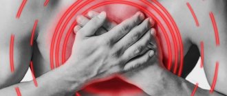
The nurse should regard every attack of acute pain in the heart area as an attack of angina pectoris . The pain manifests itself mainly behind the sternum, less often in the region of the heart, and occurs as a result of acute myocardial ischemia . Measures aimed at eliminating the attack must begin immediately! 1. The patient must be reassured.2. Place the patient reclining in bed or sit him in a chair, on a chair.3. Free the chest from clothing that is constricting it.4. Provide fresh air access to the room.5. If a painful attack occurs for the first time or the patient claims that the pain previously disappeared after taking validol , then give him a validol tablet under the tongue (or 4-5 drops per piece of sugar).6. If the attack does not disappear 2-3 minutes after using validol, then give the patient nitroglycerin or sustak .
If a patient experiences side effects (headache, noise in the head) after taking nitroglycerin, reassure the patient and explain to him that this is not a contraindication to further administration of nitroglycerin and that such side effects go away without a trace. When a patient takes nitroglycerin or sustak for the first time, give him 1/4 of the tablet first and monitor its effect.7. Apply 1-2 mustard plaster to the patient's heart area.8. Apply a warm heating pad to your left shoulder blade.9. If the pain radiates to the left hand with numbness in the fingers, take a hot local bath for the left hand. The most common causes of heart failure are angina, myocardial infarction, coronary thrombosis, pleurisy, rib fractures, cholecystitis, biliary colic, cholelithiasis, myositis (inflammation of the intercostal muscles, intercostal neurolia, osteochondrosis, herpes zoster.
Of great prognostic and diagnostic importance is the identification of attacks of thoracic goiter in a patient, which occurs as a result of myocardial ischemia, against the background of spasm or atherosclerosis of the coronary arteries. This attack is accompanied by nerve receptors producing impaired metabolism.
In typical cases of angina, retrosternal pain of a pressing nature (or squeezing) pain radiates to the left half of the chest, left shoulder, left arm, left shoulder blade and is accompanied by several minutes and disappears after taking nitroglycerin. A distinction is made between angina at rest and after overexertion.
Myocardial infarction is an extremely dangerous disease. In which foci of ischemic necrosis form in the heart muscle. The problem is that the previous angina pain turns out to be more intense and prolonged and drags on for several hours. Accompanied by fear of death and general severe weakness.
1 pain relief
2 drugs that reduce blood clotting
3 drugs that normalize blood pressure, heart rate, respiratory rate
After 5-7 days, they bring you to the card department from the intensive care unit. There the patient is strictly on bed rest. Prevention is a must. Special care for seriously ill patients, rehabilitation therapy
Insufficient blood circulation is the inability of the cardiovascular system to provide the organs and tissues of the body with the required amount of blood.
Heart failure – heart defects, coronary artery disease, hypertension.
Heart failure can be acute or chronic.
Attacks of cardiac asthma occur when blood stagnates in the pulmonary circulation and is characterized by the patient developing a feeling of lack of air (suffocation) with severe shortness of breath at rest and subsequent cyanosis. If you do not provide help, an asthma attack will turn into pulmonary edema; signs are bubbling breathing and the release of a large volume of foamy sputum. If help is not provided, the patient will die.
Help - semi-sitting position, air flow, mustard plaster on the lower limbs, foot baths or a tourniquet on the upper thigh. Suction off foamy vomit and give humidified oxygen
Ticket number 33
Preparing the patient for x-ray examination of the esophagus and stomach.
Preparing the patient for an x-ray examination of the stomach and small intestine The functions of the gastrointestinal tract, as well as its anatomical changes, are studied using the x-ray method. For the study, a contrast agent is used - a suspension of barium sulfate (100 g in 150 g of boiled water; to study the esophagus, prepare a thicker suspension, the consistency of sour cream). During the study, the patient drinks this suspension in sips.
The examination consists of exposure to X-rays (fluoroscopy), and if necessary, photographs can be taken ( radiography ). The nurse must be able to prepare the patient for an x-ray examination of the stomach. The purpose of preparation is to maximally free the stomach and intestines from their contents: food debris, liquid and especially gases. Preparing the patient for an X-ray examination of the stomach and small intestine begins 3 days before the examination. For 3 days, the patient should receive food that is easily digestible and contains as few carbohydrates as possible. If the patient is worried about bloating, then to combat increased gas formation, carbohydrates in food are limited, and during the same 3 days he is given a warm infusion of chamomile (you need to take one tablespoon per glass of water, boil, infuse and strain) - one glass per day or carbolene - 2-3 tablets 3-4 times a day. The night before and early in the morning, 2-3 hours before the test, the intestines are washed with an enema of 5-6 glasses of warm chamomile infusion. In the evening, the patient should be warned that 7-8 hours before the examination he should not eat, drink, take medications or smoke. X-ray examination of the stomach is best done in the morning on an empty stomach, but if this is not possible, then 5-8 hours before the examination the patient is allowed to eat a light breakfast consisting of a glass of sweet tea, semolina porridge or jelly. If it is known in advance that the patient has impaired gastric emptying, then before the study you need to insert a thick probe into him and suck out the contents of the stomach with a rubber balloon.
**** ) Preparation for X-ray examinations 1. X-ray examinations of the chest cavity, heart with contrasted esophagus, larynx, paranasal sinuses, skull bones, limbs, cervical and thoracic spine are carried out without special preparation of the patient. 2. To examine the esophagus, stomach and duodenum, the patient must come for the examination on an empty stomach (do not drink, do not smoke, do not eat). The patient does not require additional preparation. In the absence of contraindications, an endoscopic examination of the stomach is performed first. 3. When examining the lumbosacral spine, pelvic bones, bladder, uterus, and when conducting survey urography, a cleansing enema is necessary the night before. Patients arrive at the radiology department after a light breakfast. 4. Intravenous urography is performed on an empty stomach
Performing venipuncture.
Venipuncture is the insertion of a needle into a vein through the skin to draw blood or infuse medicinal solutions, blood, or blood substitutes. Intravenous infusion is usually performed into a vein in the elbow. The intended injection site must be thoroughly treated with alcohol. Above the elbow, a tourniquet or any rubber tube is applied to the middle third of the shoulder so as to cause swelling of the veins, but it is important not to compress the arteries. Apply a tourniquet so that it can be easily released. To enhance venous stagnation, the patient is asked to clench and unclench his fist several times or lower his hand before applying a tourniquet.
During the procedure, the patient sits or lies. His hand should lie on the table or bed in the position of maximum extension at the elbow joint; To do this, place a flat pillow or cushion under your arm, covered with a sterile napkin or clean towel.
you need to have a syringe with a capacity of 10-20 ml with a well-selected needle, carefully sterilized. All rules of asepsis must be observed. It is necessary to administer the medicine intravenously and collect blood for research only while wearing rubber gloves.