An ECG can diagnose most heart pathologies. The reasons for their appearance are due to concomitant diseases and lifestyle characteristics of the patient.
What does this mean if changes in the myocardium are detected on the ECG? In most cases, the patient requires conservative treatment and lifestyle modification.
Description of the procedure
An electrocardiogram (ECG) is one of the most informative, simple and accessible cardiological studies. It analyzes the characteristics of the electrical charge that causes the heart muscle to contract.
Dynamic recording of charge characteristics is carried out in several areas of the muscle. The electrocardiograph reads information from electrodes placed on the ankles, wrists and chest skin in the area where the heart is projected, and converts them into graphs.
Each of these graphs reflects the work of a specific part of the heart muscle. Find out about the standards for deciphering the ECG of the heart from a separate article.
Types of disease
- Acute form of ischemia. This includes myocardial infarction and sudden death, also called coronary death.
- Coronary form of ischemia. This is heart failure, all types of arrhythmia and angina pectoris. Symptoms may appear all at once or just one.
There is also a transient form that can occur even in a completely healthy person. This may be a kind of reaction to cold, exercise or stress due to spasm of an unchanged artery.
Normal and deviations - possible reasons
Normally, the electrical activity of the myocardial areas, which is recorded by the ECG, should be uniform. This means that intracellular biochemical exchange in heart cells occurs without pathologies and allows the heart muscle to produce mechanical energy for contractions.
If the balance in the internal environment of the body is disturbed for various reasons,
the following characteristics are recorded on the ECG :
- diffuse changes in the myocardium;
- focal changes in the myocardium.
The reasons for such changes in the myocardium on the ECG can be either harmless conditions that do not threaten the life and health of the subject, or serious dystrophic pathologies requiring emergency medical care.
One of these serious pathologies is myocarditis, or inflammation of the heart muscle. Regardless of its etiology, areas of inflammation can be located either in the form of foci or diffusely throughout the heart tissue.
Causes of myocarditis:
- rheumatism as a consequence of scarlet fever, tonsillitis, chronic tonsillitis;
- complications of typhus, scarlet fever;
- consequences of viral diseases: influenza, rubella, measles;
- autoimmune diseases: rheumatoid arthritis, systemic lupus erythematosus.
One of the reasons for changes in muscle tissue may be cardiodystrophy - a metabolic disorder in heart cells without damage to the coronary arteries. Lack of cell nutrition leads to changes in their normal functioning and impaired contractility.
Causes of cardiac dystrophy:
- Ingress of toxic metabolic products into the blood due to severe impairment of kidney and liver function;
- Endocrine diseases: hyperthyroidism, diabetes mellitus, adrenal tumor, and, as a result, excess hormones or metabolic disorders;
- Constant psycho-emotional stress, stress, chronic fatigue, starvation, unbalanced diet with nutritional deficiencies;
- In children, a combination of increased stress with a sedentary lifestyle, vegetative-vascular dystonia;
- Lack of hemoglobin (anemia) and its consequences - oxygen starvation of myocardial cells;
- Severe infectious diseases in acute and chronic form: influenza, tuberculosis, malaria;
- Dehydration of the body;
- Avitaminosis;
- Alcohol intoxication, occupational hazards.
What are moderate changes in the myocardium?
Moderate changes in the myocardium are most often detected by ECG examination, ultrasound diagnostics or echocardiography.
Changes in the myocardium are caused by inflammatory processes, diseases, hormonal imbalances and some external factors. Also, in most cases, age may be the cause.
In old age, as a result of the aging of the body, and in adolescence and childhood, these are still incomplete processes of heart development.
Changes in the myocardium
As a result of cardiac activity, the human body receives the oxygen and nutrients it needs through the bloodstream. Therefore, if any disorder is diagnosed, it can affect the functional characteristics of other organs and lead to various troubles.
Moderate changes come in several types:
- Diffuse. In itself, such a disorder cannot be a separate disease, but is only a syndrome, the cause of which must be identified during examination by a doctor. This phenomenon can be explained as follows. Some of the cells, which are directly involved in biochemical processes, begin to work incorrectly and shrink. As a result, the electrical activity will be non-uniform. In simple terms, diffuse changes in the myocardium mean a part of the changed cells through which the conduction of electrical impulses is impaired.
- Focal. In this case, large or small scars form in the myocardium. The scars themselves are made of connective tissue, which is not inert and cannot conduct electrical impulses.
To find out what causes such changes, it is necessary to undergo a thorough examination. For a more detailed understanding of the etiology, a certain type of changes in the myocardium is identified.
Types of changes and reasons
As we have already understood, the etiology of such a disorder includes both harmless causes and quite serious diseases. Pathological changes are caused by various processes, for example, inflammatory changes.
Here the root cause is myocarditis (inflammation of the heart muscle) of an infectious or aseptic nature.
In the area of the left and right ventricles, as well as the left and right atria, myocarditis can occur in the following diseases:
- rheumatism;
- infectious diseases (rubella, measles, scarlet fever, typhus, diphtheria, etc.);
- systemic autoimmune diseases.
Such altered areas are most often located in a diffuse manner, but focal lesions still occur.
Depending on the causes and specifics of the manifestations, the following changes are distinguished:
1. Diffuse. Damage to the heart muscle occurs uniformly in all parts - this includes the ventricles and atria. It appears more often as a result of inflammatory phenomena (described above), or when the water-salt balance is disturbed or after taking certain groups of medications.
2. Metabolic. It occurs as a result of metabolic disorders and the heart not receiving enough nutrients for this reason. This problem is reversible and can occur as a result of the following factors:
- being overweight;
- hypothermia of the body;
- nervous and physical stress;
- presence of chronic diseases.
Moderate changes may also be associated with impaired sensitivity to stimuli and stress. They can also occur as a result of constant disruptions in the body.
3. Dystrophic. Here we can talk about the discrepancy between energy expenditure and its entry into the heart muscle. Most often, such moderate changes “do not make themselves felt,” but they can also manifest themselves in the form of shortness of breath and increased fatigue.
Changes in the myocardium can spread throughout the heart or be localized in a specific area.
Changes in the left ventricular myocardium
Diffuse changes in the myocardium of the left ventricle occupy a special place among cardiovascular disorders. This pathology of the left ventricle can cause conditions that threaten the life and health of the patient.
The likely chances that such a disorder will appear in a person directly depend on the functioning of the heart, the patient’s age, gender and the presence of hypertension. Therefore, in such a situation, we can safely say that any changes in the muscular layer of the left ventricle most often occur in older people with a history of hypertension.
In children, due to the metabolism not yet being fully formed, changes in the myocardium of the left ventricle can be considered a completely acceptable norm. Adults with this problem should definitely be examined to identify the cause. It is possible that the tissue change was caused by some serious disorder.
Nonspecific changes
Nonspecific changes in the myocardium appear as a result of those provoking factors that are not associated with cardiac activity. In this case, transformations of the muscle layer of the right or left ventricle, right or left atrium may be a consequence of the following phenomena:
- hormonal imbalance in the human body;
- history of previous illnesses;
- improper diet;
- smoking and alcohol abuse;
- metabolic disorders, etc.
In itself, such a disorder does not require any specific treatment, since it does not pose a danger. Moderate changes are considered reversible and in this case it is necessary to lead a healthy lifestyle, as well as treat concomitant diseases.
If a person continues to eat poorly, subject the body to physical stress and does not pay due attention to health, then in frequent cases this can “develop” into a serious pathology, for example, angina pectoris, myocardial infarction and heart failure.
Is treatment necessary?
Symptoms such as:
- the appearance of pain and discomfort in the heart area;
- heart rhythm disturbance;
- dyspnea;
- interruptions in the functioning of the heart muscle.
The doctor, having collected an anamnesis and listened to the patient’s complaints, will send for examination to understand the overall picture of the disorder. Next, treatment will be prescribed depending on the type and degree of the disorder:
- proper balanced nutrition;
- maintaining a sleep schedule;
- getting rid of bad habits;
- prescription of medications.
You should always listen to the work of your heart. Sometimes even minor changes in the myocardium become the reason for the development of serious diseases. As soon as you notice unpleasant symptoms, you must immediately undergo an examination to identify the cause of their occurrence.
(5 4,40 of 5) Loading...
Source: https://SosudInfo.com/heart/umerennye-izmeneniya-v-miokarde.html
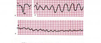
Determination by cardiogram
With diffuse lesions of the heart, deviations from the normal pattern are observed in all leads. They look like numerous areas with impaired conduction of electrical impulses.
This is expressed on the cardiogram as a decrease in T waves, which are responsible for ventricular repolarization. With focal lesions, such deviations are recorded in one or two leads. These deviations are expressed on the graph as negative T waves in the leads.
If focal changes are represented, for example, by scars remaining in the connective tissue after a heart attack, they appear on the cardiogram as electrically inert areas.
Additionally, the ECG will be able to show signs of enlargement of the heart (hypertrophy of the right or left ventricle, right or left atrium), rhythm disturbances, and conduction.
Signs of illness
Symptoms of the disease depend on its form. However, it is possible to identify general signs that indicate that there is a malfunction in the functioning of the heart.
The first thing worth mentioning is any painful sensations. Of course, these are subjective signs, however, the sooner you pay attention to them, the less consequences there will be due to timely treatment. In addition, pain may occur in the chest area during various types of stress, emotional or physical.
Cardiac ischemia sometimes takes decades to develop. During progression, the forms of the disease and clinical manifestations may change. Symptoms can appear individually or in different combinations. Complications such as heart failure, intracardiac conduction, and cardiac arrhythmias may also occur. Let's clarify the symptoms that are characteristic of different forms of coronary disease.
Angina pectoris manifests itself in the form of attacks behind the sternum. They are periodic in nature and appear during periods of emotional or physical stress. In addition, discomfort and burning may be felt. The attack stops as soon as the load disappears or after taking nitroglycerin.
The pain may radiate to the left shoulder blade or arm. Stable exertional angina can be diagnosed as a result of ECG changes or persistent manifestations of the disease. If effective treatment is not prescribed, this stage will develop into a progressive stage, the symptoms of which are more frequent and severe, and can also appear even at rest.
The main symptom of an acute form of ischemia, that is, myocardial infarction, is pain behind the sternum. You may feel discomfort, pain in the left shoulder blade, arm, or abdomen. The pain may last from 15 minutes to an hour. Symptoms of heart failure may appear, such as arrhythmia, profuse sweating and cough. A heart attack at the initial stage can be confused with angina pectoris. However, the subsequent course of the disease, the ineffectiveness of nitroglycerin, the inability to stop the attack in the first hours, arrhythmias, elevated blood pressure and temperature indicate that this is not angina pectoris at all, but a myocardial infarction.
An ECG can diagnose most heart pathologies. The reasons for their appearance are due to concomitant diseases and lifestyle characteristics of the patient.
What does this mean if changes in the myocardium are detected on the ECG? In most cases, the patient requires conservative treatment and lifestyle modification.
An electrocardiogram (ECG) is one of the most informative, simple and accessible
cardiological research. It analyzes the characteristics of the electrical charge that causes the heart muscle to contract.
Dynamic recording of charge characteristics is carried out in several areas of the muscle. The electrocardiograph reads information from electrodes placed on the ankles, wrists and chest skin in the area where the heart is projected, and converts them into graphs.
Diagnostics
Decoding electrocardiogram data takes 5-15 minutes . Its data can reveal:
- Size and depth of ischemic lesion;
- Localization of myocardial infarction, how long ago it occurred in the patient;
- Electrolyte metabolism disorders;
- Enlarged heart cavities;
- Thickening of the walls of the heart muscle;
- Intracardiac conduction disorders;
- Heart rhythm disturbances;
- Toxic damage to the myocardium.
Features of diagnosis for various myocardial pathologies:
- myocarditis – the cardiogram data clearly shows a decrease in the waves in all leads, a violation of the heart rhythm, the result of a general blood test shows the presence of an inflammatory process in the body;
- myocardial dystrophy - ECG indicators are identical to the data obtained for myocarditis; this diagnosis can only be differentiated using laboratory test data (blood biochemistry);
- myocardial ischemia - ECG data show changes in the amplitude, polarity and shape of the T wave in those leads that are associated with the ischemic zone;
- acute myocardial infarction – horizontal displacement of the ST segment upward from the isoline, trough-shaped displacement of this segment;
- necrosis of the heart muscle - irreversible death of myocardial cells is reflected on the ECG graph as a pathological Q wave;
- transmural necrosis - this is an irreversible damage to the entire thickness of the heart muscle wall, expressed in the cardiogram data as the disappearance of the R wave and the acquisition of the QS type by the ventricular complex.
In the case of a hypertensive crisis, decompensated heart failure, electrolyte disturbances or suspicion of acute myocardial infarction, a coronary T scar appears on the ECG graph.
When making a diagnosis, you should additionally pay attention to the symptoms of concomitant diseases . This may include pain in the heart with myocardial ischemia, swelling of the legs and arms with cardiosclerotic changes, signs of heart failure as a result of a heart attack suffered on the legs, hand tremors, sudden weight loss and exophthalmos with hyperthyroidism, weakness and dizziness with anemia.
The combination of such symptoms with diffuse changes detected on the ECG requires an in-depth examination .
Features, treatment and consequences of infarction of the posterior wall of the heart
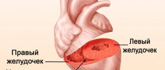
First, let's try to figure out what the posterior wall of the left ventricle is. The heart is a hollow muscular organ that circulates blood throughout the body. It consists of 4 chambers: 2 ventricles and 2 atria. The main component of the muscle pump is the left ventricle, which supplies oxygenated blood to all tissues of the body.
The thickness of the left ventricular myocardium is approximately 2-3 times greater than other parts of the organ and averages from 11 to 14 mm.
Therefore, due to its large size, this part of the heart requires a larger volume of blood, which it receives through the right coronary artery and its circumflex branch.
Any damage to the vessels bringing fresh oxygen quickly affects functional activity and can lead to the death of cardiomyocytes.
Due to the features described above, myocardial infarction in 99.9% of cases affects exclusively the left ventricle.
About 10-15% of vascular accidents occur in the posterior wall, which, for the convenience of doctors, is divided into two main sections:
- diaphragmatic;
- basal.
Recent scientific research works of cardiac surgeons, as well as my personal experience, have made this problem more urgent.
If myocardial infarction of the posterior wall of the left ventricle develops, it is practically invisible on the ECG and is often hidden under the mask of angina pectoris. As a result, the patient does not receive the necessary range of treatment measures.
Organ cells continue to die, and many adverse consequences are observed in the future.
Fortunately, in 60-70% of cases, infarction of the posterior wall of the heart is combined with necrosis of neighboring areas (posteroinferior, posteroseptal, posterolateral), which is clearly reflected in the electrocardiogram curve.
Causes
In fact, there is a huge list of factors leading to damage to the coronary arteries, but the most significant are:
- Atherosclerosis. Occurs in most people over 60 years of age due to lipid metabolism disorders (increased total cholesterol, LDL and TAG, decreased HDL). As a result of the formation of pathological deposits on the walls of blood vessels, their obstruction occurs. The condition is further aggravated by the sedimentation of thrombotic masses. I have not met cardiac patients without signs of this disease.
- Migration of blood clots from distant sites. This phenomenon is most typical for people suffering from varicose veins of the lower extremities, and is much less common against the background of prolonged physical inactivity (severe somatic diseases) in the absence of antiplatelet therapy. As a rule, middle-aged and elderly people generally do not pay attention to changes in the venous bed in the legs. However, young girls who worry about their attractiveness care much more about it.
- Vasospasm. May occur against the background of disorders of the central nervous system (neuroses, systematic stress).
Factors that predispose to the development of myocardial infarction include:
- arterial hypertension;
- obesity (increased BMI more than 30 kg/m2);
- physical inactivity (WHO recommends walking at least 8,000 steps daily);
- lipid profile disorders;
- presence of bad habits (smoking, systematic use of alcoholic beverages and drugs);
- male gender;
- age from 45 years.
You can assess the presence of risk factors yourself. If there are at least 3 of the above, then the likelihood of a fatal complication from the cardiovascular system increases by 2.5 times. It's not too late to change everything and secure a healthy future for yourself.
Clinical manifestations
It is quite possible to suspect an impending posterior myocardial infarction and other vascular complications (for example, a stroke or hemorrhage in the eyeball) at home.
As a rule, they are preceded by conditions such as:
- hypertensive crisis;
- attack of unstable angina (with a history of coronary artery disease);
- episodes of arrhythmias;
- change in general condition and behavior (sudden sharp headaches, increased sweating, weakness, chills).
Pain
Pain and discomfort behind the sternum are the only things that unite all people with developed myocardial infarction.
Pain has specific characteristics:
- duration over 15 minutes;
- localization behind the sternum;
- lack of effect from nitroglycerin and other nitrates;
- possibility of irradiation to the left scapula, shoulder, forearm and little finger.
It is extremely rare that a “silent picture” is revealed, when pain is completely absent, and only weakness and increased sweating are observed.
An important sign is the duration of pain. Stable angina pectoris never lasts so long. If you have discomfort in the chest for more than 15 minutes, urgently call a team of doctors, since the heart cells are already experiencing acute hypoxia, which may soon enter an irreversible stage (necrosis).
Violation of the functional activity of the heart
Important pathways do not pass through the posterior wall of the left ventricle, therefore, rhythm disturbances are not typical, but sometimes occur (in my memory, such situations have never been observed).
By shutting down significant masses of the myocardium from work, stagnation phenomena can be detected from the small (shortness of breath, cough with streaks of blood) and large (swelling in the legs and body cavities, enlarged liver, pallor of the skin with a bluish tint in the distal parts) circles blood circulation
Diagnostics
The fundamental method of diagnosis is electrocardiography.
Acute basal isolated myocardial infarction cannot be detected under any circumstances. Damage to the diaphragmatic part of the posterior wall can be recognized by indirect signs. There are no changes in the ECG characteristic of the stages of vascular pathology (acute, acute, subacute, scarring).
So, the doctor will suspect a heart attack based on the following criteria:
- an increase in the amplitude of the R wave in V1 and V2;
- decrease in the depth of the S waves in the 1st and 2nd chest leads;
- the voltage of the S and R waves in the first two leads is the same;
- bifurcation of the R wave (often diagnosed as right bundle branch block);
- T wave elevation in V1-V
The national guidelines for physicians describe variants of small-focal diaphragmatic infarction with the appearance of a characteristic pathological Q wave and ST segment elevation. However, in personal practice it has never been possible to record such changes on a cardiogram, although the clinic was present.
Instrumental diagnostics
Echo-CG is used to establish cardiac wall dysfunction. Ultrasound waves accurately identify areas of myocardial hypo- or akinesia, allowing one to suspect necrotic or cicatricial transformations in them.
Coronography is widely used to locate the source of coronary artery obstruction. After the administration of a contrast agent, a series of x-rays are taken, in which the areas of narrowing are clearly visible.
Laboratory diagnostics
To confirm the diagnosis, the following may be used:
- Complete blood count (increased number of leukocytes and ESR);
- Troponin test - increases in the presence of necrosis of the heart or any skeletal muscle. Damage to the posterior wall is always minor, which is why the troponin level may not increase, leading to diagnostic errors.
Both methods allow you to confirm myocardial infarction only after 6-7 hours. And the golden window in which the cause of occlusion can be eliminated and “barely alive” cardiomyocytes can be restored is only 3 hours. An extremely difficult choice, isn't it? Echo-CG and other highly informative methods (MRI) are not available at all medical institutions.
Emergency help
If you happen to meet a person with a myocardial infarction, the procedure will be as follows:
- Call an ambulance immediately.
- Place the patient on the bed, raising the head end of the body.
- Provide fresh air flow (open windows).
- Make breathing easier (remove tight outer clothing).
- Every 5 minutes, give any nitro drug (“Nitroglycerin”) under the tongue, simultaneously measuring blood pressure and heart rate before a new dose. When the heart rate increases more than 100 beats per minute or blood pressure decreases below 100/60 mm. rt. Art. therapy is stopped.
- Suggest taking Acetylsalicylic acid (0.3 g) orally.
It is impossible to attempt to eliminate coronary pain with conventional analgesics. Is a painkiller capable of preventing necrosis of heart cells? In addition, the clinical picture may be erased, which will complicate diagnosis.
Treatment
Immediately after diagnosis, emergency treatment with the following drugs is carried out:
Name of medication Dose
| Aspirin (if not given previously) | 0,3 |
| Metoprolol | 0,0250 |
| Morphine 1% | 1 ml |
| Heparin | Up to 4000 units |
| Clopidogrel | 0,3 |
| Oxygen therapy (40% O2) | Until signs of heart failure are eliminated |
The patient is urgently hospitalized in the cardiac intensive care unit. Systemic or local thrombolysis is performed (if less than 6 hours have passed since the onset of the disease). In the long term, stenting or coronary artery bypass grafting is indicated.
The main directions of therapy are as follows:
- Prevention of rhythm disturbances. Beta blockers (Metoprolol, Carvedilol, Bisoprolol) and calcium channel antagonists (Amlodipine, Verapamil, Bepridil) are used.
- Antiplatelet and anticoagulant therapy (Clopidogrel, Xarelto, Pradaxa).
- Relieving pain symptoms.
- Statin therapy (Rosuvastatin, Atorvastatin, Simvastatin).
Complications
The consequences of a heart attack can be significant. Typically, several life support systems are affected at once.
Heart failure
Dead heart cells are no longer able to pump blood in full. The fluid begins to actively move from the vascular bed into the surrounding tissues with the development of multiple edema.
Organs suffer from hypoxia, against the background of which foci of dystrophic changes form. As experience shows, the brain is the first to take the hit (a decrease in all functions is observed: attention, memory, thinking, etc.).
Episodes of loss of consciousness, dizziness, and staggering when walking appear.
The most dangerous is pulmonary edema. It can be acute (occurs instantly) or chronic (increases over several days or months). Exudate begins to leak into the lower parts of the paired organ, as a result of which a large number of alveoli cease to perform the respiratory function.
Progression of IHD
As you know, our body has a wide ability to adapt.
Efficient heart tissue undergoes hypertrophy (gain of muscle mass), which significantly increases the volume of required oxygen, but the functionality of the remaining segments of the coronary bed is not unlimited.
The frequency of angina attacks increases, they become more pronounced and prolonged. The risk of recurrent myocardial infarction increases 3-5 times.
Myocardial remodeling
Against the background of inadequate load and hypertrophy of the myocardium, after a few years, dilatation is observed - thinning of the walls with the formation of bulges - aneurysms. The consequence is always the same - tissue rupture with cardiac tamponade (outflow of blood into the pericardial cavity). This complication is fatal in 8 out of 10 patients.
Forecast
The prognosis for infarction of the posterior wall of the heart in the absence of emergency assistance in the first hours after development is conditionally unfavorable.
There will be a gradual increase in dysfunction of the heart muscle, which will ultimately lead to the death of a person.
To avoid any undesirable consequences, you should try your best to prevent a heart attack, especially if you are at risk.
The process of aneurysm formation
In conclusion, I would like to cite an interesting case from personal experience, which proves the complexity of recognizing ischemic damage to the posterior wall of the left ventricle.
Patient D., 66 years old. He was repeatedly admitted to our cardiology department by ambulance with a diagnosis of Acute Coronary Syndrome. For reference, I would like to say that this term refers to two pathologies. This is a myocardial infarction and an episode of unstable angina. Only after an examination (ECG, troponin test) is the exact nosology determined.
The patient complained of pain in the chest, lasting 35-50 minutes. Each time an examination was carried out (ECG, CBC, troponin detection test), which did not reveal signs of necrosis. Nitroglycerin in the form of a 1% solution and Aspirin were used.
Unfortunately, a few days ago the patient died in a car accident. At autopsy, it was discovered that the patient had suffered 3 small-focal myocardial infarctions during his life, caused by damage to the circumflex branch of the posterior coronary artery. More than 2 years have passed since the last one.
Thus, infarction of the posterior wall of the left ventricle represents a huge problem for modern cardiology due to the almost complete lack of timely diagnosis.
Although such a vascular complication is extremely rare, the likelihood of its development cannot be ignored.
Prevention is always based on a healthy lifestyle and adequate treatment of any diseases (especially those related to the cardiovascular system).
Source: https://cardiograf.com/ibs/nekroz/infarkt-zadnej-stenki-serdca.html
What diseases do they accompany?
Pathological changes in the myocardium detected on the ECG may be accompanied by impaired blood supply to the heart muscle, reprolarization processes, inflammatory processes and other metabolic changes.
A patient with diffuse changes may exhibit the following symptoms:
- dyspnea,
- chest pain,
- increased fatigue,
- cyanosis (blanching) of the skin,
- rapid heartbeat (tachycardia).
Such manifestations most often become the reason for an electrocardiogram.
In medical practice, there are many examples when myocardial pathologies did not cause noticeable changes in the well-being of patients and were discovered during preventive examinations. Diseases accompanied by changes in the heart muscle:
- Myocardial dystrophy is a violation of biochemical metabolic processes occurring in the heart;
- Allergic, toxic, infectious myocarditis - inflammation of the myocardium of various etiologies;
- Myocardiosclerosis – replacement of cardiac muscle cells with connective tissue as a consequence of inflammation or metabolic diseases;
- Disturbances of water-salt metabolism ;
- Hypertrophy of parts of the heart muscle.
Additional examinations are needed to differentiate them.
Symptoms
In my practice, I constantly encounter the fact that patients with coronary heart disease do not seek help immediately. This happens because the first symptoms of coronary artery disease increase gradually.
Pain
The most common complaint of a patient with angina pectoris or a heart attack is pain in the chest area - for most it is accompanied by a feeling of fear and panic. Irradiation is usually observed under the shoulder blade, into the arm, and part of the lower jaw on the maiden side. Some complain of numbness of the upper limb, aching in the wrist joint. With a lack of oxygen in the area of the posterior basal region of the heart, pain spreads to the stomach area. And very rarely it is noted in the right hand.
When I ask to characterize the type of pain, the person indicates that it is:
- baking;
- pressing;
- compressive.
The appearance of pain is associated with physical activity - when a person ran, walked quickly, or climbed stairs. Sometimes an attack was observed after stress, strong emotional stress, or going outside in cold weather. A sharp flow of blood to the heart is also observed when standing up from a lying position. All these factors lead to increased blood pressure, increased heart rate and increased myocardial oxygen demand.
The pain of angina is short-lived and does not last more than 5-15 minutes. As soon as a person stops, sits down, and calms down, it subsides, as the causes of acute ischemia are eliminated. Experienced patients always carry nitroglycerin with them, which quickly relieves symptoms. If the intensity of the unpleasant sensation does not subside after taking the drug, then this most often indicates a non-cardiac pathology, or indicates the development of a heart attack.
With vasospastic or spontaneous angina, pain and other signs of myocardial ischemia develop without connection with physical activity; it is noted in the morning, often provoked by exposure to cold. They can be easily removed only with calcium antagonists.
Other common manifestations
Other signs of coronary heart disease do not always appear; in some patients during an attack, I observed the following symptoms:
- nausea, vomiting;
- severe fatigue;
- severe shortness of breath with difficulty breathing;
- sweating;
- pale skin;
- increase or decrease in blood pressure;
- increased (less often decreased) heart rate, arrhythmia.
In some cases, instead of a classic attack, indicating the presence of coronary artery disease in the heart, you can see its equivalents: shortness of breath, fatigue after minimal exertion.
Additional diagnostic tests
These cardiograms, despite their information content, cannot be the basis for making an accurate diagnosis. In order to fully assess the degree of changes in the myocardium, the cardiologist prescribes additional diagnostic measures:
- General clinical blood test - assesses the level of hemoglobin and such indicators of the inflammatory process as the level of leukocytes in the blood and ESR (erythrocyte sedimentation rate);
- Analysis of blood biochemistry - indicators of protein, cholesterol, glucose levels are assessed to analyze the functioning of the kidneys and liver;
- General clinical urine analysis - kidney function indicators are assessed;
- Ultrasound for suspected pathology of internal organs - according to indications;
- Daily monitoring of ECG indicators;
- Carrying out an ECG with stress ;
- Ultrasound of the heart (echocardiography) – the condition of the parts of the heart is assessed to determine the cause of myocardial pathology: expansion (dilatation), hypertrophy of the heart muscle, signs of decreased contractility of the myocardium, disruption of its motor activity.
After analyzing the medical history and laboratory and instrumental examination data, the cardiologist determines the method of treating the changes.
Myocardial ischemia - Main symptoms and methods of treatment of ischemia, advice from doctors
Diseases of the cardiovascular system are the leading cause of death in the world today.
More than 50% of people die due to vascular problems and heart failure.
The question of what is cardiac ischemia is asked by more than one person every day. The phrase “myocardial ischemia” means insufficient oxygen supply to the heart muscle.
Ischemia on the ECG is manifested by a characteristic set of symptoms and pathological changes that cannot be confused with another disease.
Etiology and pathogenesis of myocardial ischemia
The main cause of the disease is a decrease in the supply of arterial blood rich in oxygen to the myocardium.
Pathological processes causing myocardial ischemia:
- atherosclerotic narrowing of the heart arteries due to the formation of plaques in the stroma of the vessel. Each plaque consists of low- and very low-density lipoproteins, as well as platelets. When a plaque ruptures, acute ischemic injury of the myocardium is formed, otherwise known as a heart attack;
- inflammatory changes in the vascular wall: vasculitis, arteritis, systemic connective tissue diseases lead to impaired vascular permeability;
- viral and bacterial agents that multiply in the intima of blood vessels;
- toxic damage to the coronary vessels, causing their spasm. Can be formed due to excessive consumption of alcohol, nicotine, poisoning with heavy metals and mercury salts;
- a stressful situation that causes a massive release of adrenaline into the blood leads to a narrowing of the vascular lumen.
- hormone-dependent tumors of various organs and tissues.
As a result of malnutrition of the heart muscle, vasospasm develops, impaired oxidation of free fatty acids, accumulation of under-oxidized protein breakdown products, and lipid peroxidation is activated.
These changes lead to disruption of the nutrition of mitochondria, the main sources of energy production in cells.
Due to mitochondrial dysfunction, ion channels in the cardiac tissue do not function, and an imbalance of ions develops, which leads to impaired contractile function of the heart.
Types of myocardial ischemia
Depending on the location of the area of the heart that does not receive blood, ischemia of the entire myocardium is divided into subepicardial and subendocardial forms.
Subepicardial myocardial ischemia is localized directly under the outer layer of the heart muscle - under the epicardium. The damage zone is of sufficient size and extent from the epicardium to the endocardium, affecting all layers of the anterior and posterior walls.
With subendocardial ischemia, the inner part of the heart muscle, the endocardium, is damaged. The endocardium contains a large number of small capillaries and blood vessels that provide nutrition to the heart, so damage to it is particularly difficult.
Clinical picture of the disease
Typical ischemia of the entire myocardium consists of the following manifestations: pressing, burning pain behind the sternum with irradiation to the jaw, scapula or shoulder; feeling of lack of air and shortness of breath, weakness, cold sweat and pale skin.
The pain is accompanied by fear of death and goes away within 15-20 minutes after taking nitroglycerin.
In the abdominal form, the disease begins with nausea and vomiting, possibly diarrhea and abdominal pain.
After a few minutes, chest pain, rapid breathing and palpitations are added to these unpleasant sensations. Symptoms disappear within a few minutes after taking nitro-containing medications.
If the disease manifests itself in the form of an asthmatic attack, there is a high probability of confusing myocardial ischemia with an attack of suffocation. In the asthmatic form, a sharp lack of air initially develops, accompanied by chest pain, impaired sweating and pale skin.
After some time, the discomfort in the chest goes away, and only breathing problems remain. The most severe form for diagnosis is non-painful myocardial ischemia. It is practically asymptomatic for the patient, leaving necrotic changes in the affected vessels.
For the painless form, the most typical occurrence is the appearance of myocardial infarction at the end of the disease.
The cerebrovascular form occurs as an acute cerebrovascular accident and is characterized by loss of consciousness, impaired spinal reflexes, and loss of the ability to read and write for a short period of time.
After the attack ends, the patient may experience the following symptoms: dizziness, headache, nausea and weakness.
The peripheral form of ischemia begins with pain in the arms or legs, gradually moving to the heart area.
The limbs become cold and pale, wet to the touch. The pulse quickens, the frequency of respiratory movements gradually increases. Symptoms of damage to the cardiovascular system appear after a few hours.
Diagnostic measures
To diagnose this disease, many reliable laboratory and instrumental techniques are used. Most hospitals use a primary medical examination and patient interview, which allows one to suspect ischemic changes in the heart and identify specific symptoms and symptomatic complexes.
When listening to the patient's chest, rhythm and conduction disturbances can be detected, indicating a pathological process occurring in the body.
To confirm the diagnosis, the following laboratory methods are used:
- a general analysis of urine and blood with determination of the number of platelets allows us to judge disorders in the hematopoietic system and bone marrow pathology;
- biochemical analyzes provide information about the main markers of myocardial necrosis: troponins, myoglobin, creatine phosphokinase fraction. You also need to measure the main indicators of the liver and observe them over time;
- A coagulogram and hemostasiogram are used to determine blood clotting time and diagnose disorders in the blood coagulation system.
Of the instrumental methods, electrocardiographic examination of the heart is most widely used.
An ECG is taken at the time of an attack, with the participation of stress tests (pharmacological tests, cold and heat tests, stress test, through esophageal stimulation of the ventricles, bicycle ergometry), and 24-hour ECG monitoring, otherwise called Holter, is also very informative.
Myocardial ischemia on the ECG is manifested by ST segment elevation above the isoline.
X-ray examination of the chest in both projections reveals an increase in the size of the ventricles, which indicates a functional overload of certain parts of the heart. You can also see changes in the aorta: this vessel is most susceptible to changes.
On phonocardiography, you can see changes in heart sounds, the appearance of pathological noises, indicating rhythm and conduction disturbances, as well as indicating heart defects.
An echocardiogram of the heart or ultrasound allows you to see the size of the heart cavities, the thickness of the walls, determine the location and nature of the location of necrosis, and pathological changes in the myocardium.
It is a highly sensitive research method that minimizes the risk of making an incorrect diagnosis with these initial data.
Treatment of myocardial ischemia
Coronary heart disease is a severe multifactorial disease that requires an integrated approach to treatment and prevention of further complications. The first and most important step should be changing your lifestyle and giving up bad habits.
It is necessary to strictly observe the work and rest regime, avoid unnecessary stressful situations, and also regularly apply dosed physical activity in order to improve the health of the body as a whole.
Walking at a brisk pace up to several kilometers a day, exercising in a gym or on a playground, visiting a swimming pool or therapeutic exercises reduce the risk of a recurrence of an ischemic episode by almost half.
Changing your eating habits is an important step on the path to a healthy heart.
First of all, you should avoid fatty and fried foods with a lot of salt and spices, avoid fast food, carbonated and alcoholic drinks, packaged juices and preservatives. You should add vegetables and fruits, fresh berries and herbs to your daily diet, and eat more cereals.
Butter should be replaced with vegetable oil, mayonnaise with low-fat sour cream, and honey should be used instead of sugar. Proper nutrition will reduce the risk of developing cardiovascular pathology and atherosclerosis.
In addition to changing your lifestyle, you should not forget about traditional medicine.
The most commonly used drugs are the following:
- Statins. They help reduce cholesterol levels in the body, strengthen the vascular wall and reduce the amount of “bad” fats, while simultaneously increasing the level of “good” ones.
- Anticoagulants and antiplatelet agents prevent thrombus formation in the body, thin the blood, preventing it from forming clots that can cause ischemic damage to the myocardium.
- Nitro-containing drugs relax the vascular wall, reduce the tone of vascular smooth muscles, causing a drop in blood pressure and eliminating coronary spasm.
Treatment of myocardial ischemia is a long and labor-intensive process that must be supervised by a qualified specialist.
If you follow all the recommendations for sports training, nutrition, drug treatment, and giving up bad habits, you can achieve truly stunning results: cardiac ischemia will never bother you and your loved ones again, and in this case you can be calm and not worry about further consequences.
Loading…
Source: https://KardioBit.ru/bolezni-serdtsa/ishemicheskaya-bolezn/ishemiya-miokarda
Treatment for focal and diffuse disorders
In the treatment of myocardial pathologies, various groups of drugs are used:
- Corticosteroid hormones – as an antiallergic agent;
- Cardiac glycosides - for the treatment of diffuse changes in the myocardium, manifestations of heart failure (ATP, Cocarboxylase);
- Diuretics - to prevent edema;
- Means for improving metabolism (Panangin, Magnerot, Asparkam);
- Antioxidants (Mexidol, Actovegin) - to eliminate the negative effects of lipid oxidation products;
- Antibiotics – for anti-inflammatory therapy;
- Drugs for the treatment of concomitant diseases;
- Vitamin preparations.
If conservative treatment does not lead to significant improvements in the condition of a patient with myocardial diseases, he undergoes surgery to implant a myocardial pacemaker.
In addition to medications, the patient is recommended to change his lifestyle and establish a balanced diet. For a patient with such pathological manifestations, physical activity, drinking alcohol and smoking are unacceptable. He is prescribed physical therapy and feasible labor.
Basic principles of dietary nutrition:
- The consumption of salt and excess liquid is limited to a minimum;
- Spicy and fatty foods are not recommended;
- The menu should include vegetables, fruits, lean fish and meat, and dairy products.
Myocardial changes detected on the ECG require additional laboratory and instrumental examination . If necessary, the cardiologist will prescribe treatment in a hospital or on an outpatient basis. Timely measures taken will help avoid serious complications.
Treatment
Treatment of changes in the myocardium of the ventricles of the heart is based on complex therapy. The initial stage is based on changing the usual lifestyle and creating a diet taking into account the specific nature of the disease.
Diet
With diffuse changes of a non-inflammatory nature in the initial stages, a properly selected diet, good sleep and adequate rest are of utmost importance.
To fully nourish the heart muscle with the necessary energy resources, you should follow a diet that excludes foods with high cholesterol levels, flour and sweets, alcohol and coffee, spices that irritate the walls of the stomach.
Foods and vitamins to strengthen your heart
It is recommended to divide the meal into six approaches. Must be used:
- Lean meat and chicken.
- Sea fish.
- A sufficient amount of vegetables and fruits rich in iron and calcium (apricots, bananas, carrots, potatoes, spinach, nuts).
- Whole milk products.
- Whole grain cereals are rich in complex carbohydrates.
Drugs
Drug therapy consists of prescribing drugs that improve cellular metabolism.
Typically, when there is a change in the myocardium, drugs such as:
- Panangin, asparkam avexima, magnesium orotate dihydrate, magne B6 - are responsible for the conduction of nerve impulses, serve as the prevention of cardiac arrhythmia, and stimulate metabolic processes in the myocardium.
- Actovegin and Mexidol are antioxidants that help activate energy processes in the cell and affect its functional metabolism.
- Retinol, ascorbic acid, tocopherol, thiamine and pyrodoxine.
If the cause of diffuse changes is a cardiac disease, then therapy is aimed at treating it. For example, the prescription of drugs that replenish iron deficiency in case of disturbances in hemoglobin levels, hormonal therapy for disorders of the endocrine system, drugs aimed at stabilizing blood pressure in case of essential hypertension.
Do not be upset if the ECG results reveal diffuse changes in the myocardium. The presence of minor changes in the absence of symptoms of serious cardiovascular diseases can occur with metabolic disorders. But you shouldn’t put off visiting a doctor. If necessary, only a cardiologist can prescribe examination and treatment.