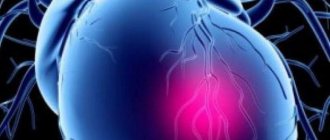
While left ventricular failure has been the subject of intense discussion for decades, right ventricular heart failure has generally received minimal attention. Indeed, the right half of the heart has long been considered a relatively passive channel for blood flow between the systemic and pulmonary circulations; accordingly, its disorders were considered relatively mild.
Right ventricular heart failure (RHF) is a heart disease in which the functioning of the right ventricle (RV) is impaired. Today, left ventricular failure is more often diagnosed, although it is believed that isolated PVCF has a more unfavorable prognosis.
According to the course, right ventricular heart failure is divided into acute and chronic, and in each case there are characteristic features in terms of clinical presentation, diagnosis and treatment.
The PVC is also known as the “pulmonary heart” because the right ventricle pumps blood through the pulmonary circulation through the lungs. The more impaired the activity of the pancreas, the more pronounced the clinical picture of the disease and the higher the risk of pulmonary edema, which can subsequently result in the death of the patient. Therefore, it is extremely important to begin treatment of the disease on time and carefully prevent a recurrent attack of pancreatic failure.
Video Right Sided Heart Failure – Explained in 2 Minutes (Right Ventricle Failure)
Causes
There are two broad groups of factors contributing to the development of right ventricular heart failure.
- Primary right ventricular failure in the absence of pulmonary hypertension. Myocardial infarction
- Pulmonary embolism
More recently, a high mortality rate has been identified among patients with isolated RV myocardial infarction, which has forced a reassessment of the risk of the disease.
Various congenital heart defects are often associated with RV failure, usually due to increased workload, volume overload, or both. Septal defects are commonly associated with PVC; usually because blood is shunted from the left side of the heart to the right. Tetralogy of Fallot is another birth defect in which RV hypertrophy and eventual RV failure occurs due to obstruction of the pulmonary artery that receives blood from the RV. In an adult with repaired tetralogy of Fallot, RV failure may still occur due to pulmonary regurgitation, especially when the tricuspid valve has been used to repair the structure of the affected vessel.
Symptoms
Manifestations of right ventricular heart failure begin with the appearance of signs of stagnation of the pulmonary circulation and are unchanged.
- tachycardia;
- decrease in pressure;
- shortness of breath, feeling of lack of air;
- chest pain perceived as pressure;
- decrease in skin temperature;
- cyanosis of the limbs and nasolabial triangle;
- the jugular veins become noticeable and their pulsation is observed;
- possible fainting;
- Pulmonary edema may occur.
The mechanism of development of deficiency involves increased symptoms. The clinic of blood stagnation in the systemic circle, which is usually accompanied by left ventricular failure, is connected:
- pain appears in the right hypochondrium due to liver enlargement;
- the legs swell, and in more severe cases, the abdominal wall;
- ascites and hydrothorax develop;
- hemodynamics in the kidneys are disrupted, which can cause serious complications;
- the amount of urine produced decreases.
Moderate shortness of breath. Severe cyanosis of the face, neck, and limbs. Swelling and pulsation of the neck veins during inhalation and exhalation. Increased epigastric pulsation. Swelling in the legs, often ascites and hydrothorax, enlarged and painful liver. Pulse is rapid, low filling. The ECG shows signs of overload on the right side of the heart.
Acute right ventricular heart failure causes pulmonary hypertension, which gradually reduces blood output from the left ventricle.
The patient in such conditions suffers from various symptoms:
- heart rate increases;
- pressure in the arteries decreases;
- worried about the problem of lack of air;
- capillary permeability increases, which leads to pulmonary edema;
- the jugular veins swell and pulsate;
- blue skin due to insufficient blood supply to small vessels. For the same reason, skin temperature decreases.
The flow of blood in the systemic circulation is gradually disrupted, due to which:
- the liver increases in size and its capsule becomes stretched, which is accompanied by pain on the right under the ribs;
- the lower limbs swell, and later the anterior abdominal wall;
- fluid accumulates in the chest cavity and abdomen;
- blood flow to the kidneys is disrupted;
- urine production decreases.
Risk factors
Risk factors for right heart failure may include:
- Age: Men between 50 and 70 years of age often experience right-sided heart failure, especially if they have previously had a heart attack.
- Chronic diseases: Lung pathologies such as COPD or pulmonary fibrosis, diabetes, HIV, hyperthyroidism, hypothyroidism or iron/protein accumulation can lead to right-sided heart failure.
- Irregular heartbeats (arrhythmias): Abnormal heart rhythms, especially if they are very fast and fast, can weaken the heart muscle.
- Lung conditions: Blood clots or high blood pressure in the lungs may increase the risk of right-sided heart failure.
- Inflammation of the pericardium: the disease provokes a decrease in the size of the pericardial sac, which compresses and tightens the heart muscle.
- Racial Predisposition: African-American men are at higher risk than others for developing right-sided heart failure.
- Certain chemotherapy and diabetes drugs: Certain medications have been found to increase the risk of PHF.
- Valvular heart disease: Damage or defect in one of the four heart valves may prevent effective hemodynamics.
- Viral infection: Some viral diseases can damage the heart muscle, including the right ventricle.
Pathogenesis
The main stages of the formation of PZHSN:
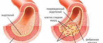
- Dilatation of the RV chamber leads to enlargement of the tricuspid annulus, which causes regurgitation through the tricuspid valve, further exacerbating ventricular dilatation.
- Over time, pancreatic hypertrophy occurs as a natural response to increased stress.
- As the RV enlarges, the crescentic shape of the ventricular cavity is disrupted.
- The interventricular septum protrudes into the LV cavity. This occurs because the pericardium limits the space available for cardiac expansion, and therefore an increase in RV size must be accompanied by a decrease in LV volume.
- Shift of the septum impairs LV filling and therefore impairs its function. This phenomenon is called ventricular dependence.
- As LV dysfunction develops, systemic blood pressure and coronary artery supply decrease, further impairing RV function.
With the unfavorable development of PZHS, high venous pressure is determined in combination with a decrease in systemic arterial pressure, which impairs the perfusion of major organs. Such disturbances can lead to persistent circulatory failure, which ultimately causes failure of all organs and death.
Clinic
At the beginning of the development of right ventricular heart failure, signs are mild. With the addition of LV dysfunction, the clinical picture becomes more pronounced.
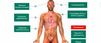
Signs and symptoms of RV heart failure include the following:
- The appearance of shortness of breath, first during physical activity, and then at rest
- Orthopnea (semi-sitting position)
- Chest pain
- Low blood pressure
- Rapid heartbeat
- Tachycardia
- Fatigue and weakness
- Lack of urination or little urine output
- Anorexia, weight loss, nausea
- Exophthalmos and/or visible eye pulsation
- Wheezing in the chest, often distantly audible
- Central or peripheral cyanosis, pallor
In severe cases, there is ascites, an increase in the size of the liver, or even anasarca.
Video Heart failure - symptoms and treatment
Acute heart failure – Insufficientia cordis acuta
Emergency care algorithm for acute heart failure
Acute heart failure is a polyetiological symptom complex that arises as a result of impaired myocardial contractility, leading to a decrease in blood supply to organs (output insufficiency) and relative stagnation of blood in the venous system and in the pulmonary circulation (inflow insufficiency).
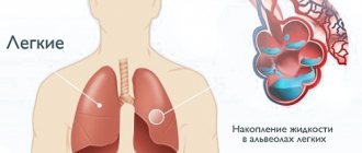
Pulmonary edema is the accumulation of fluid in the interstitial tissue and/or alveoli of the lungs as a result of extravasation of plasma from the vessels of the pulmonary circulation.
ETIOLOGY AND PATHOGENESIS
The contractility of the myocardium decreases as a result of: ■ a decrease in the functioning mass of the myocardium, ■ hemodynamic overload of the left or right heart, ■ a decrease in the compliance of the chamber walls.
Causes of acute heart failure
■ Violation of diastolic and/or systolic myocardial function with: □ myocardial infarction (the most common cause); □ inflammatory or dystrophic diseases of the myocardium; □ tachycardia, tachy- and bradyarrhythmias.
■ Sudden myocardial overload during: □ hypertensive crisis; □ heart defects; □ severe anemia; □ hyperthyroidism; hypervolemia.
■ Acute disturbances of intracardiac hemodynamics with: □ rupture of the interventricular septum; □ septal myocardial infarction; □ infarction or separation of the papillary muscle; □ bacterial endocarditis with perforation of valve leaflets; □ rupture of chords; □ injury.
■ Increased load on the decompensated myocardium in patients with severe chronic congestive heart failure with: □ physical activity, □ psycho-emotional stress, □ fever, □ increased blood volume (for example, when consuming too much fluid or massive infusions); □ increasing inflow in a horizontal position, etc.
■ Drug overdose.
For left ventricular acute heart failure:
■ pressure increases in the pulmonary circulation, the pulmonary artery system; ■ pulmonary arterioles constrict in response to increased pressure in the left atrium; ■ external respiration and blood oxygenation deteriorate; ■ interstitial edema develops (cardiac asthma syndrome), and then alveolar edema (pulmonary edema syndrome).
For right ventricular acute heart failure:
■ the ability of the heart to pump blood into the pulmonary circulation decreases or is lost; ■ venous congestion occurs in the systemic circulation; ■ acute respiratory failure develops.
CLASSIFICATION
Based on the type of hemodynamics, the following types of acute heart failure are distinguished:
■ Congestive type: left ventricular acute heart failure (cardiac asthma, pulmonary edema) and right ventricular acute heart failure (venous congestion in the systemic circulation); ■ Hypokinetic type: cardiogenic shock.
In myocardial infarction, there are 4 classes of acute heart failure.
Classification of acute heart failure during myocardial infarction
| Class | Clinical signs | Frequency, % | Mortality, % | Treatment |
| There are no wheezes or third sounds in the lungs | 33 | 8 | Not required | |
| II | Wheezing in the lungs no more than 50% of their surface or III tone | 38 | 30 | Reducing preload with diuretics |
| III | Wheezing in the lungs over more than 50% of their surface, often pulmonary edema | 10 | 44 | Reduce preload with diuretics; if ineffective, increase cardiac output with non-glycoside inotropic agents |
| IV | Cardiogenic shock | 19 | 80-100 | Analgesic, infusion, inotropic therapy |
CLINICAL PICTURE
Left ventricular acute heart failure is characterized by the appearance of several of the following symptoms:
■ increasing shortness of breath of varying severity (up to suffocation); ■ orthopnea position; ■ sometimes Cheyne-Stokes breathing (alternating short periods of hyperventilation with respiratory arrests);
■ cough (first dry, and then with sputum), later - foamy sputum, often colored pink;
■ feeling of fear, anxiety, fear of death; ■ pallor; ■ acrocyanosis; ■ heavy sweat; ■ tachycardia (up to 120-150 per minute); ■ normal or reduced blood pressure;
■ moist rales may not be heard at first, or a meager amount of fine bubbling rales is detected over the lower parts of the lungs; swelling of the mucous membrane of the small bronchi can manifest itself as a moderate picture of bronchial obstruction with prolongation of exhalation, dry wheezing and signs of pulmonary emphysema; ■ with alveolar edema, ringing moist rales of different sizes are detected over all the lungs, which can be heard at a distance (bubbly breathing).
Right ventricular acute heart failure:
■ shortness of breath; ■ swelling of the neck veins; ■ congestion in the veins of the upper half of the body; ■ Kussmaul's sign (swelling of the jugular veins on inspiration); ■ enlarged liver; ■ intense pain in the right hypochondrium, aggravated by palpation; ■ swelling in the lower parts of the body (in a horizontal position - on the back or side), ascites; ■ in some cases, dyspepsia (stagnant gastritis); ■ more pronounced cyanosis; ■ tachycardia; ■ possible development of arterial hypotension up to a picture of shock.
In global acute heart failure, a combination of the above symptoms is observed.
DIFFERENTIAL DIAGNOSTICS
It is carried out with non-cardiogenic pulmonary edema, which develops as a result of increased permeability of the alveolar membranes (with pneumonia, sepsis, aspiration, pancreatitis, poisoning with irritating and toxic gases, etc.).
) and is called adult respiratory distress syndrome. Features of therapy include avoiding the use of nitrates and cardiac glycosides.
The feasibility of prescribing glucocorticoids to reduce membrane permeability and stimulate the formation of pulmonary surfactant should be assessed.
ADVICE FOR THE CALLER
■ Help the patient assume a sitting position with his legs down.
■ Provide warmth and comfort.
■ For chest pain, give the patient nitroglycerin under the tongue (1-2 tablets or spray 1-2 doses), if necessary, repeat the dose after 5 minutes.
■ If an angina attack lasts more than 15 minutes, let the patient chew 160-325 mg of acetylsalicylic acid.
■ Find the medications the patient is taking, ECGs taken previously, and show them to the emergency medical personnel.
■ Do not give anything to drink or eat.
■ Do not leave the patient unattended.
Attention! In case of hypotension (cardiogenic shock), a position with a raised foot end, nitroglycerin is contraindicated!
ACTIONS ON CALL
Diagnostics
REQUIRED QUESTIONS
■ How long does shortness of breath bother you?
■ Was the onset sudden or did the shortness of breath increase gradually?
■ What are the conditions under which shortness of breath occurs (at rest, during physical activity, etc.)?
■ What symptoms preceded the present condition (chest pain,
palpitations, hypertensive crisis, etc.)?
■ What medications did the patient take independently and their effectiveness?
■ Has the patient recently suffered a myocardial infarction or an episode of congestive heart failure?
■ Does the patient suffer from diabetes?
INSPECTION AND PHYSICAL EXAMINATION
■ Assessment of general condition and vital functions: consciousness, breathing, blood circulation.
■ Patient position: presence of orthopnea.
■ Visual assessment: skin (pale, high humidity), the presence of acrocyanosis, swelling of the neck veins and veins of the upper half of the body, peripheral edema (lower extremities, ascites).
■ Calculation of respiratory rate: tachypnea.
■ Pulse examination: correct or incorrect.
■ Heart rate calculation: tachycardia or rarely bradycardia.
■ Measuring blood pressure: the presence of hypotension (with severe myocardial damage) or hypertension (with the body’s stress response); decrease in SBP 110 mm Hg. repeat after 10 minutes.
■ Furosemide (Lasix) 1% solution in ampoules of 2 ml (10 mg/ml).
□ Children: the initial single dose in children is 2 mg/kg, the maximum is 6 mg/kg.
□ Adults: 20-80 mg IV over 1-2 minutes.
■ Morphine (morphine hydrochloride*) 1% solution in ampoules of 1 ml (10 mg/ml). □ Children: under 2 years of age are more sensitive to the inhibitory effect of morphine on the respiratory center.
□ Adults: dilute 1 ml in 20 ml of 0.9% sodium chloride solution and administer intravenously in fractional amounts of 4-10 ml every 5-15 minutes until pain and shortness of breath are eliminated or until side effects appear (arterial hypotension, depression breathing, vomiting).
■ Dopamine, 4% solution in ampoules of 5 ml (40 mg/ml).
□ Children: IV at a dose of 4-6 (maximum 10) mcg/kg/min). Use with caution.
□ Adults: IV at a dose of 2-10 mcg/kg/min).
■ Dobutamine, 50 ml ampoules (5 mg/ml). □ Children: IV at a dose of 5-20 mcg/kg/min). The minimum effective dose for children is often higher than for adults, while the maximum dose for children is lower than for adults.
□ Adults: IV at a dose of 2.5-10 mcg/kg/min).
“,”author”:””,”date_published”:”2020-02-25T20:20:00.000Z”,”lead_image_url”:”//1.bp.blogspot.com/-5ziaRbKcYwY/TiLx6oP-sgI/AAAAAAAAABfw/ xaCj4DZKp7s/w1200-h630-pk-no-nu/%25D1%2581%25D0%25B5%25D1%2580%25D0%25B4%25D0%25B5%25D1%2587%25D0%25BD%25D0%25B0%25D1%258F%2B %25D0%25BD%25D0%25B5%25D0%25B4%25D0%25BE%25D1%2581%25D1%2582%25D0%25B0%25D1%2582%25D0%25BE%25D1%2587%25D0%25BD%25D0%25BE%25D1 %2581%25D1%2582%25D1%258C.bmp”,”dek”:NULL,”next_page_url”:NULL,”url”:”//ambulance-russia.blogspot.com/2011/09/blog-post_3905.html ”,”domain”:”ambulance-russia.blogspot.com”,”excerpt”:”бДог о Ñ Ñ‚Ð°Ð½Ð´Ð°Ñ€Ñ‚Ð°Ñ… Ñ ÐºÐ¾Ñ€Ð¾Ð¹ мед RESULTS омощи,”,”word_count”:2001,” direction”:”ltr”,”total_pages”:1,”rendered_pages”:1}
Source: //ambulance-russia.blogspot.com/2011/09/blog-post_3905.html
Diagnostics
At an appointment with a cardiologist, a physical examination of the patient is required, during which it can be determined:
- peripheral edema;
- pulsation of the jugular veins;
- hepatomegaly;
- systolic murmur of tricuspid regurgitation;
- second tone accent.
After analyzing the general condition of the patient, instrumental research methods are necessarily prescribed. Due to the unusual anatomy of the pancreas, assessing its function is challenging. However, technological advances, especially in echocardiography and cardiac MRI, are helping to determine right ventricular function and volumes, as well as measuring pulmonary artery pressure.
Instrumental research methods for pancreatic heart failure:
- Echocardiography
- Electrocardiography
- Magnetic resonance imaging
- Chest X-ray
- Invasive diagnostic methods
- Maximum load testing
- Pulse oximetry (determining the amount of oxygen in arterial blood)
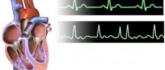
Electrocardiography
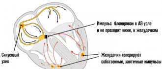
The ECG is assessed within normal limits in most cases. When right axis deviation is determined, pulmonary embolism is suspected.
Chest X-ray
This research method has limited capabilities and does not provide specific identification of right ventricular heart failure. However, dilation of the base of the pulmonary artery and the veins approaching the heart, as well as oligemia of the cardiac chambers (Westermarck sign) all indicate pulmonary embolism and PVC.
Invasive monitoring
Most often, pulmonary artery catheterization is performed, which makes it possible to measure pressure in the RV, PA, determine PA occlusion and cardiac output. When each of these indicators is obtained, primary PVC heart failure can be distinguished from secondary heart failure, combined with an increased load on the heart. The transpulmonary gradient, the mean arterial pressure in the pulmonary artery, is especially important. The study also determines pulmonary vascular resistance, calculated by dividing the transpulmonary gradient by cardiac output. Important limitations to consider when using a PA catheter are the unreliable measurement of cardiac output thermodilution with severe tricupidal regurgitation and the risk of flotation balloon inflation in patients with severe pulmonary hypertension.
Echocardiography
Today, transesophageal echocardiography (TE echocardiography) is preferred for assessing PZHF. In experienced hands, this technique can immediately identify a dilated, hypertrophic, or poorly contracting RV and associated disorders such as tricuspid regurgitation and ventricular septal shift.
The following tests may be useful in the initial evaluation of suspected right ventricular heart failure:
- Complete blood count (CBC)
- Determination of the amount of iron
- Analysis of urine
- Electrolyte level
- Kidney and liver function tests
- Fasting blood glucose level
- Lipid profile
- Thyroid stimulating hormone (TSH) level
- B-type and pro-B-type natriuretic peptide levels
Treatment
When determining right ventricular heart failure, various treatment strategies are used. The main ones are used first, and then the additional ones.
Basic therapy for PZHF includes:
- Prevention of recurrence of an attack.
- Normalization of blood volume.
- Reducing the load on the heart.
- Prevention of hypercapnia and hypoxia.
Blood volume correction
The increased blood volume is normalized by reducing the amount of fluid and salt consumed. If this is not enough, diuretics and renal replacement therapy are used.
Artificial ventilation
Helps reduce pressure in the capillary network in the lungs, which helps improve the general condition of the patient.
Inotropes and vasopressors
- The most commonly used drugs are beta-agonists, calcium sensitizers, and phosphodiesterase inhibitors. With their help, the load on the heart is reduced, contractility and oxygen consumption by the myocardium are increased.
- Levosimendan is indicated in cases where it is necessary to reduce pulmonary arterial pressure and improve pancreatic function.
- Norepinephrine, phenylephrine and vasopressin improve ventricular contractility and thus have a positive effect on coronary artery perfusion, but if not used properly, increase myocardial oxygen consumption.
- Milrinone and amrinone increase cardiac contractility through a non-beta adrenergic mechanism.
Load reduction
- Sildenafil is an oral drug that contains a phosphodiesterase enzyme.
- Milrinone (PDE-III inhibitor): reduces pulmonary pressure, can be administered via nebulizer.
- Systemic vasodilators - hydralazine and others, which help normalize coronary perfusion.
- Recombinant BNP (brain natriuretic peptide) is a neseritide that reduces the preliminary and subsequent load on the heart, thereby improving myocardial function without inotropic effect.
Surgical treatment to support the pancreas
If medications are not effective in treating right heart failure or symptoms are very severe, a ventricular pacing device or heart transplant may be needed.
- Ventricular assist device (VAD) surgery : Once implanted, a special device helps the weak heart muscle contract more efficiently.
- Heart transplantation: surgery is performed when all other treatments for heart failure have failed. The damaged heart is surgically removed and replaced with a healthy organ from a deceased donor.
Emergency care for acute right ventricular failure
Therapy for this disease will be successful only if, in addition to symptomatic treatment, the underlying disease is treated.
Before the emergency team arrives, the patient must be seated in a room with an open window. If the patient is unconscious, you can put him in a supine position with his head raised high or place a couple of pillows under his head. If the patient is conscious and can answer what he was sick with before, he needs to take the medications that he receives constantly. For example, you should use inhalers during an attack of bronchial asthma (salbutamol, berodual, etc.).
After the team arrives, oxygen therapy begins (oxygen supply through a mask), for asthma - prednisolone and aminophylline are administered intravenously, for a heart attack - narcotic analgesics intravenously, nitroglycerin sublingually, for thromboembolism - heparin and fibrinolytics (streptokinase, etc.).
In the intensive care unit, cardiology or pulmonology, treatment continues. Thus, for pneumonia, the administration of antibiotics is indicated, and for pneumo- or hydrothorax, pleural puncture is often performed - a puncture of the skin and intercostal muscles in order to evacuate air or fluid. After stabilization of the patient’s condition, optimal therapy is selected in the future in order to prevent the development of severe stages of chronic failure and prevent the formation of cor pulmonale.
Forecast
Overall, despite significant improvements in medical and device therapy, mortality among patients with heart failure after hospitalization is 10.4% in the first 30 days, 22% at 1 year, and 42.3% at 5 years.
Heart failure associated with acute myocardial infarction has an inpatient mortality rate of 20–40%; the same figure reaches 80% in patients who suffer from hypotension (for example, with cardiogenic shock).
Heart failure associated with systolic dysfunction has a mortality rate of up to 50% after 5 years.
The prognostic conclusion of hospitalized patients is additionally influenced by the following risk factors:
- age (elderly and older);
- presence of concomitant pathology;
- length of sick day.
In the absence of timely and adequate treatment, the risk of death from pulmonary edema or cardiac arrest is very high.
Prevention
Not all risk factors can be corrected, which is why it is important to take steps to help reduce the likelihood of developing PWS.
- If you have diabetes, you need to monitor what you eat, how regularly you eat, and what your blood glucose level is.
- Moderate exercise helps improve blood circulation and reduces stress on the heart muscle.
- The diet should be balanced: It is important to limit the amount of sugar, animal fat, cholesterol and salt. You need to eat more fruits, vegetables, whole grains and low-fat dairy products.
- If you experience new symptoms or have side effects from your medications, you should contact your doctor.
- Maintaining a healthy weight is important: Losing extra pounds and maintaining a healthy weight reduces the strain on your heart.
- When determining sleep apnea, a CPAP/BIPAP device should be used every night.
- Reducing your stress levels can help prevent fast or irregular heartbeats.
- It is worth remembering the prescribed medications that should be taken as prescribed:
Additionally, it is important to reduce your alcohol intake; in some cases, you may need to stop drinking completely. You should also quit smoking, since smoking has an extremely negative effect on the cardiovascular system.
Video Heart failure. What makes the heart weak?
4.83 Aug. rating ( 95 % score) – 6 votes – ratings
Reasons for development
Acute left ventricular failure can occur in people of any age. The chances of this increase in the presence of other pathologies of the cardiovascular system and after 50 years.
Most often, this diagnosis is made to elderly men suffering from coronary artery disease.
Cardiological causes of left ventricular failure are considered diseases in the form of:
- Myocardial infarction and subsequent tissue necrosis. The development of the disease occurs with extensive transmural infarction. The patient in this case is in serious condition and consequences can be provoked depending on the degree of damage.
- Myocarditis. These are inflammatory processes in the tissues of the heart.
- Congenital and acquired defects that disrupt the architecture of the heart.
- Atrial fibrillation, ventricular tachyarrhythmias.
- Hypertensive crisis, in which the pressure in the arteries reaches high levels.
Some pathologies of other organs and systems also contribute to the development of left ventricular failure. The disease may occur due to:
- Thromboembolism. In this case, the pulmonary artery becomes blocked by a blood clot when there is significant damage to the vessel.
- Pneumonia.
- Acute intoxication with various substances.
- Anemia.
- Electric shock.
- Asphyxia.
- Severe chest injuries.
- Pathologies of the kidneys and liver in the terminal stage.
There are also certain factors that increase the likelihood of developing left ventricular failure. The disease occurs if a person has heart pathologies, and he is exposed to excessive physical and emotional overload, visits baths and saunas, abuses alcohol, and smokes.
Therefore, it is important to monitor the condition of your heart and try to avoid the influence of such factors, since acute left ventricular failure develops under their influence.