How does hemosiderin deposition occur?
A disease such as hemosiderosis mainly develops as a result of other diseases that appeared earlier, but at the same time it can also be independent in nature. Basically, the disease occurs as a result of the following reasons:
- Disruption of the process of destruction of red blood cells, as well as an increased amount of iron absorbed by the body.
- As a result of blood transfusion.
It is no secret that for the normal functioning of the body, it requires a certain amount of a microelement such as iron every day. As a rule, the daily dose is 15 mg. If, for even a short period of time, the body receives a large amount of this element, this threatens with an excess amount of iron-containing pigment accumulated in the tissues, which in turn leads to the development of various diseases.
There are two main forms of development of such a disease as hemosiderosis:
General hemosiderosis. Excess hemosiderin in general hemosiderosis is found in the liver, spleen, bone marrow, and other internal organs. It is also worth noting that due to the absence of the property of destroying parenchymal cells, the functions of the organs in which hemosiderin has accumulated are not impaired.
Local hemosiderosis. Occurs as a result of intravascular destruction of red blood cells. When hemoglobin disappears from red blood cells, they turn into pale bodies. After this, the released hemoglobin, as well as part of the red blood cells, are covered by other cells and are generated into hemosiderin. Deposition of hemosiderin in the brain, as well as other organs, is often observed. The disease hemosiderosis can occur both in a specific area of tissue and in the whole organ.
Complications
There are no critical complications of skin hemosiderosis. The most common consequences may include:
- cosmetic defect;
- scratch infection.
The following conditions may also occur:
- iron deficiency anemia – a decrease in the number of red blood cells and hemoglobin due to a lack of iron in the blood;
- arthralgia – joint pain;
- heart rhythm disturbances. Most often this is sinus tachycardia - an acceleration of the heartbeat while maintaining the correct rhythm;
- arterial hypotension is a regular, persistent decrease in blood pressure.
But these conditions are direct complications not of skin hemosiderosis, but of a general disorder of iron metabolism, the manifestation of which is the described disease.
How does excess hemosiderin accumulation manifest itself?
The clinical picture primarily depends on which organ has an excess amount of accumulated iron-containing pigment. At the same time, the symptoms may be very similar, or manifest themselves to a similar extent. It is also noteworthy that hemosiderosis can develop differently in everyone, and therefore the symptoms manifest themselves individually for each patient. The most common manifestations of this disease include the following:
- Cough with bloody discharge. This manifests itself as a result of the accumulation of hemosiderin in the sputum.
- Problem breathing.
- Change in skin color.
- Pain in the heart and other organs.
- Change in liver size.
Differential diagnosis
Differential (distinctive) diagnosis of skin hemosiderosis must be carried out with such diseases and pathological conditions as:
- atypical forms of lichen - a non-inflammatory skin lesion that manifests itself as papules (flat plaques);
- atypical forms of dermatoses;
- Kaposi's sarcoma - multiple malignant skin lesions in the form of spots that turn into nodes;
- some forms of nevi - pigment spots of various colors (including those close to red).
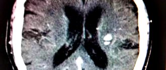
Accumulation of hemosiderin in the brain and other internal organs
A disease such as hemochromatosis occurs as a result of excessive accumulation of hemosiderin in tissues, as well as internal organs of the body. As a result of the fact that iron tends to accumulate in almost any organ, it causes the development of various diseases, in particular diabetes mellitus, liver cirrhosis, heart or kidney failure, etc. The clinical picture is mainly accompanied by general malaise, fatigue, joint pain, and heart failure. When hemosiderin accumulates in the body in large quantities, the color of the affected organs begins to change, and they acquire a yellowish or brown tint.
In order to diagnose the disease, it may require the participation of several doctors at once, in particular a dermatologist, pulmonologist and others. Taking into account the form of the disease, various tests may be prescribed, which will help specialists establish an accurate diagnosis and prescribe appropriate treatment. For example, if hemosiderin is detected in the urine (normally it is absent), then this may indicate damage to the body from poisons, infections, and the like. Depending on this, the necessary treatment is prescribed.
How to remove hemosiderin if it has accumulated in excess? Basically, experts prescribe glucocorticosteroids to patients, but they are effective only in 40-50% of cases. If such drugs do not bring the desired effect, immunosuppressants and other medications are used. What are macrophages with hemosiderin?
Macrophages are special cells that are designed to capture various bacteria and dead cells and are one of the lines of defense of the immune system, which helps protect the body from various pathogens. They originate in the bone marrow, where a cell such as the monobullast is initially synthesized. Considering the fact that the accumulation of hemosiderin can be observed in various organs, thereby disrupting their functionality, it is not difficult to assume that hemosiderosis causes a disruption in the development of macrophages. In addition, without proper treatment of the pathological accumulation of iron-containing pigment, other serious diseases may occur, which not only negatively affect the patient’s quality of life, but can also lead to death.
Skin hemosiderosis
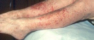
Hemosiderosis of the skin is a dermatological disease in which age spots and various rashes appear on the skin of patients. The appearance of areas of pigmentation and petechiae is caused by the accumulation of hemosiderin in the dermis and damage to the capillaries of the papillary layer.
cutaneous hemosiderosis
The spots on the skin vary in color and size. Fresh rashes are usually bright red, while older ones are brownish, brown or yellow. Spots up to three centimeters in size are localized on the lower extremities, hands and forearms. Petechiae, nodules, papules, and plaques often appear on the affected skin. Patients complain of slight burning and itching.
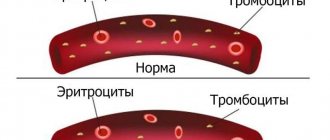
In the patient's dermis, the structure of the endothelium of the capillaries changes, and the hydrostatic pressure in them increases. Plasma leaves the vascular bed, and red blood cells are excreted along with it. The capillaries dilate, clumps of hemosiderin are deposited in the endothelium, histocytes and endothelial cells are affected, and perivascular infiltration develops. This is how the process of hemosiderin deposition occurs in the skin. In patients, clinical blood tests usually reveal thrombocytopenia and impaired iron metabolism.
Hemosiderosis of the skin can occur in various clinical forms, among which the most common are: Majocchi disease, Gougerot-Blum disease, orthostatic, eczematid-like and pruritic purpura.
Schamberg's disease
Schamberg's disease deserves special attention. This is a fairly common chronic autoimmune pathology, characterized by the appearance of red dots on the skin, similar to the mark of a regular injection. Circulating immune complexes are deposited in the vascular wall, autoimmune inflammation of the endothelium develops, and intradermal petechial hemorrhages appear. Hemosiderin accumulates in large quantities in the papillary layer of the dermis, which is clinically manifested by the appearance of symmetrical brown spots on the skin. They come together and form plaques or entire areas that are yellow or brown. Bright red rashes appear along the edges of such plaques. In patients, petechiae and hemorrhages the size of a pea are also present on the skin, which merge with each other and form large lesions. Over time, the plaques in the center atrophy. The general health of the patients remains satisfactory. The prognosis of the pathology is favorable.
Cutaneous hemosiderosis, unlike pulmonary and general hemosiderosis, can be easily corrected. Patients feel well and recover quickly.
Causes
In adults, iron accumulation in the body does not exceed 5 grams. If the human body experiences an excess of iron, then hemosiderin accumulates in the tissues of the brain and other organs. This abnormal accumulation contributes to the development of a number of other diseases.
Other reasons for the rapid accumulation of hemosiderin in the brain are:
- Acute infectious diseases (brucellosis, malaria);
- Chemical and toxic poisoning;
- Abnormal effects of various medications due to their excessive use (sorbifer, maltofer);
- Systematic blood transfusion;
- Genetic diseases transmitted by inheritance (enzymopathy, hemoglobinopathy);
- Systematic blood transfusions;
- Circulatory disorders;
- Autoimmune diseases.
Etiology and pathogenesis
Post-traumatic hemosiderosis
Its appearance is preceded by trauma - bruise, injection, skin incision, etc. As a result, blood enters the soft tissues, where oxidation of hemoglobin and ferritin occurs with subsequent accumulation of hemosiderin. Other causes and predisposing factors are:
- swelling of the lower extremities;
- diabetes;
- diseases of the cardiovascular system;
- high blood pressure;
- venous hypertension;
- chronic venous insufficiency;
- lipodermatosclerosis.
Pigmented progressive dermatosis of Schamberg
First described in 1901 by English dermatologist Jay Frank Schamberg. The main cause of dermatosis is considered to be increased permeability of small vessels and capillaries located near the surface of the skin, through which red blood cells can pass. Once in the tissues, they are destroyed, resulting in the release of hemoglobin from the red blood cells and subsequently iron. It gradually accumulates in a certain area, forming a specific skin tone.
Annular telangiectatic purpura of Majocchi
Described by the Italian dermatologist Dominico Maiocchi at the end of the 19th century. The etiology of the disease is associated with several factors: increased blood pressure, poor circulation in the legs, chronic spasm of small vessels, diapedesis (leakage) of blood cells into the tissue.
Lichenoid purpuric pigmentary dermatitis Gougerot-Blum
Described by dermatologists Henry Gougereau and Paul Bloom in 1925. The exact cause of the disease is unknown; venous hypertension, systemic diseases, infections, blood thinning medications, increased capillary fragility, etc. play a role.
Eczematid-like purpura of Doukas-Kapetanakis
Etiology unknown. Possible participation in the pathogenesis of increased permeability of small vessels with subsequent diapedesis of blood cells.
Classification
Hemosiderosis has 2 forms of development:
This pathology is characterized by extravascular destruction of red blood cells or so-called extravascular hemolysis in places where hemorrhage occurred. In this case, red blood cells are deprived of hemoglobin and transform into pale round bodies.
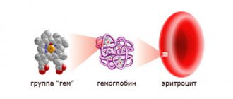
Once released, hemoglobin and the remainder of the red blood cells are taken up by other cells (leukocytes, endothelium and epithelium) and are initially generated in the cytoplasm into hemosiderin.
In areas of excessive blood accumulation, hemosiderin is deposited in the brain and other tissues. In areas where there is no oxygen, hematoidin crystals begin to settle. Depending on the conditions of occurrence, hemosiderosis occurs in a specific area of tissue or an entire organ (hemosiderosis of the lungs, liver, brain).
Has a characteristic excess of iron due to intravascular hemolysis or abnormal absorption of iron from food. Hemosiderin is deposited, as a rule, in the bone marrow, liver and other organs. In this case, the pigment does not have the property of destroying parenchymal cells, as a result of which the functions of the organs are not damaged.
Occurs in diseases such as anemia, leukemia, brucellosis, malaria and others. If an excess of iron occurs, in which tissue begins to be damaged, resulting in a decrease in the functionality of organs, this leads to the disease hemochromatosis.
There are also 3 types of hemoteriosis as independent diseases:
This disease is hereditary. It is characterized by pathology of the lung tissue, resulting in frequent hemorrhage into this tissue, as well as the development of pulmonary sclerosis. When diagnosed, hemosiderin has a distinct brown color. The disease is characterized by extensive pathological symptoms of the lungs, such as a persistent cough with pain and frequent bleeding, shortness of breath, nausea and malaise.
This disease is also hereditary and is manifested by a disorder of iron metabolism with abnormal accumulation in the tissues and organs of the patient. Iron tends to accumulate in almost all human organs, as a result of which it provokes diseases such as heart failure, diabetes, arthritis, and cirrhosis of the liver. Symptoms are pronounced and manifest themselves in the form of weakness, fatigue, low blood pressure, joint diseases and heart failure.
This pathological disease is characterized by the deposition of hemosiderin in the dermis. Hemosiderin in the brain and tissues of other organs, due to hemorrhages, begins to be deposited in the skin, as a result of which characteristic pigment spots with a diameter of 0.5 - 3 cm, yellow or brownish, begin to appear on the skin. Symptoms manifest themselves in the form of external skin rashes, while the internal organs of a person are in perfect order.