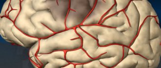
What is cerebral hemangioma
Hemangioma formed in the brain is a benign formation. It is formed from vessels and capillaries.
Most often, the tumor is diagnosed in the right or left hemisphere of the brain, but it can also occur in other parts of the organ, for example, affecting the cerebellum.
In appearance, the formation resembles a tangle, which consists of blood vessels. They are closely intertwined with each other. The tumor develops inside the head.
On this topic
- central nervous system
How does a headache with a brain tumor hurt?
- Natalya Gennadievna Butsyk
- December 3, 2020
Hemangioma is quite common in newborns. At the same time, it resembles a red spot and has a different size.
In children, hemangioma of the skin is not dangerous and often completely disappears by 5-7 years. In contrast, brain hemangioma is a dangerous disease, as it negatively affects the functioning of the organ. As a result, serious consequences and complications arise.
The pathology does not manifest itself for a long time and gradually develops over many years.
Symptoms of skin angioma
Skin angioma, the symptoms and treatment of which depend on its type, has its own characteristics.
You can determine the type of angioma by its shape, color and contours:
- Flat angiomas or port-wine stains - in most cases they are located on the head and can have any shade of red: from purple to dark cherry. With age, they often enlarge and change shade. When you press on their surface, they turn pale. They are a dense interweaving of capillaries covered with endothelial cells into one ball. Found in 80-90% of all angiomas.
Skin angioma. Photos, symptoms and treatment depend on the type - Swollen - venous hemangiomas. They have a bluish tint.
- Protruding above the skin are cavernous hemangiomas. The skin in the pathological area is warmer to the touch and turns pale when pressed. It has a characteristic appearance: in the form of various shapes of uneven protruding nodes of a bluish color. When the body bends, the angioma tends to increase, as there is an influx of blood into its vessels.
- Colorless angiomas are lymphangiomas that are found on the skin near the mucous membranes. They protrude slightly above the surface of the skin, are identical in color, and are painless.
- Combined - combine undeveloped capillaries and cavities filled with blood. The color of the angioma is bright red, without clear contours. It protrudes slightly above the skin with an uneven, bumpy surface.
Classification
In medicine, there are several different types and types of cerebral hemangiomas.
They are distinguished depending on the type of vessels from which the formation is formed. The most common diagnosis is an arteriovenous tumor. It consists of veins and arteries.
Arterial and venous formations are also distinguished.
Vascular tumors are divided into subtypes:
- Cavernous . Leads to frequent hemorrhages.
- Branched . The formation consists of many tortuous arterial cords.
- Bone . Located on the bone tissue of the skull.
Also, all brain hemangiomas are divided into the following types:
- Venous . In appearance it resembles a tangle consisting of vessels or an enlarged vein.
- Capillary . The formed node consists of capillaries in which blood circulation is impaired.
- Cavernous . This is a formation delimited by septa and having vascular cavities. It is considered the most dangerous, since as a result of impaired blood circulation it leads to frequent hemorrhages in the brain.
Depending on the time of onset of symptoms and the nature of the course, hemangioma is divided into sluggish and hemorrhagic, which is prone to bleeding.
Types of skin angioma
The following types of skin angioma are recognized:
- Simple (capillary) angioma – formations of capillaries and vessels. May occur on the surface of the skin or mucous membranes. When you press them, they can change color to a lighter color.
- Cavernous angioma is a formation that protrudes above the surface of the skin and does not have clear edges. Inside the angioma there are cavities filled with blood clots.
- Branched angioma - consists of different types of vessels.
- Granuloma is a formation on the mucous membranes.
Regarding vascular damage, angiomas are divided into the following types:
- Hemangiomas are lesions of blood vessels: arteries, veins, capillaries.
- Lymphangiomas are lesions of the lymphatic vessels.
Also recognizes:
- Monomorphic - formation occurs from structures of one type.
- Polymorphic - originate from different structures.
Reasons for the development of hemangioma
The exact reasons for the formation of a tumor in the brain have not been established. It is believed that the main reason for its appearance is a violation of embryonic development.
As a result of the process change, more vascular tissue is laid down than necessary. Therefore, the disease is also called vascular hyperplasia.
Experts have identified a number of factors that can influence and increase the risk of developing a tumor in the brain:
- Infectious lesions and viral pathologies during pregnancy.
- Unfavorable environmental conditions.
- Negative effects of various substances such as nicotine, alcohol , drugs, toxins, poisons and chemicals.
- Heredity.
Also, one of the causes of the disease is considered to be the pathological course of pregnancy.
Clinical picture
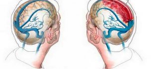
In adults, cerebral hemangioma may not show symptoms for a long time. But in cases where the tumor reaches a significant size and begins to put pressure on neighboring tissues, a number of signs appear.
First of all, patients complain of constant headaches. They vary in duration, but there are no apparent reasons for their appearance.
One of the signs of a tumor in the brain is impaired coordination of movement. There are also problems with the sensation of one’s own body in space.
On this topic
- central nervous system
What are the dangers of a porencephalic cyst of the brain?
- Olga Vladimirovna Khazova
- May 27, 2020
Patients experience disturbances in vision, hearing, smell, and taste. Dizziness and nausea may occur. In certain cases, loss of consciousness is observed.
Most patients experience epileptic seizures and convulsions. There is a disturbance in the thought process.
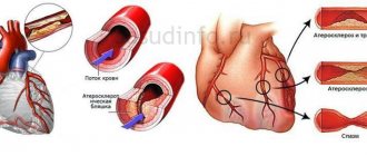
The danger of the disease is the thinning of the walls of blood vessels and their stretching. As a result of such changes, frequent and extensive hemorrhages occur. In the absence of medical attention, they lead to death.
Photo
Examples of angiomas in different parts of the body in the photo:
For example, the simple form is a flat or slightly raised tumor consisting of tortuous and dilated vessels. They often appear on the face and can reach the size of a palm.
When a cavernous or cavernous angioma , you can sometimes see it pulsating; such a tumor has a dark purple hue. They can form in the liver, especially in older people, and even in the bones.
How is the disease diagnosed?
Thanks to modern diagnostic methods, a vascular tumor in the brain can be detected even in the early stages of its development. But it is almost impossible to detect brain hemangioma without special equipment, which is due to its long asymptomatic course.
If the presence of a formation is suspected, a number of instrumental methods are carried out, which allow not only to confirm or refute the diagnosis, but also to establish the characteristics of the course of the pathology.
Contrast angiography
When diagnosing various formations, contrast angiography was the most popular and informative method.
First of all, the patient is injected with a special contrast agent intravenously. It is not dangerous to the body and allows you to determine the condition of the blood vessels in the image.
After a few minutes, an X-ray examination is performed. But today the technique is practically not used, since the radiation from the device is quite strong and can harm the body.
Superselective angiography
The technique is considered completely harmless and is prescribed if abnormalities in the development of the brain and blood vessels of the organ are suspected.
In this case, the contrast agent is injected directly into a certain part of the organ in which, according to assumptions, the formation is located.
CT or MRI
Computed tomography or magnetic resonance imaging is a non-invasive diagnostic method. They are also considered the safest, since the integrity of the soft tissues is not compromised during the procedure.
Based on the results of the study, the doctor can determine the location of the tumor, its size, type, shape and other features.
Diagnostic methods
It is impossible to determine a hematoma without examination. To make an accurate diagnosis, doctors need to know the signs of the disease. When there is a suspicion of an accumulation of blood vessels in the head, the following procedures are performed:
- Angiography involves the use of a contrast agent in the vessels.
- MRI or CT. Such techniques are among the most accurate and harmless to people.
- Superselective angiography. Contrast liquid is injected pointwise into the site of formation of a cluster of blood vessels.
With the help of modern diagnostic methods, the size, structural components and localization of the tumor change. Normal therapeutic procedures can be performed without difficulty.
Treatment methods
The only treatment is to remove the tumor. The method of resection when identifying a cerebral hemangioma depends on the presence of symptoms, concomitant diseases, complications, localization of the formation and other features.
The choice of method is made by the attending physician based on research results, the patient’s condition and the presence of contraindications.
Surgical intervention
The method is used to remove large tumors located close to the surface of the meninges. During the procedure, the tumor is completely removed using a scalpel.
But surgical intervention can lead to injury to neighboring tissues, which will lead to irreversible consequences.
In cases where the tumor is not large and is located deep in the brain tissue, the classical method of removal using a scalpel is not used.
Endovascular embolization
A modern method in which the location of the hemangioma is blocked artificially. The indication for the procedure is the location of the tumor deep in the brain tissue.
On this topic
- central nervous system
Everything you need to know about pituitary adenoma
- Olga Vladimirovna Khazova
- February 28, 2020
The method is minimally invasive. A catheter is inserted into the artery, with the help of which the drug is delivered to the required place. It is this that contributes to the blockage of the tumor.
The procedure is carried out under the control of an x-ray machine, which allows the specialist to insert the catheter into the desired location. But endovascular embolization cannot always guarantee a successful cure.
Radiosurgical method
In modern medicine, treatment using radio waves is widely used. The technique is safe and effective. Radiation that affects a specific area of the brain where the tumor is located has no effect on the patient’s body.
Thus, with the help of radiotherapy, specialists are able to remove small brain hemangiomas.
Possible complications
Hemangioma is a benign neoplasm, but despite this, it causes severe consequences. First of all, it leads to cerebral hemorrhage, which causes various complications. When enough blood accumulates, convulsive and epileptic seizures occur.
Bleeding caused by a hemangioma in the brain can be fatal in severe cases.
In case of timely diagnosis, the formation does not pose a danger to the patient and is successfully treated. That is why experts recommend regular preventive examinations and, if unpleasant symptoms occur, consult a doctor.
What is the danger?
Despite the fact that hemangioma is a benign tumor and in 3% of cases harmless, there are cases where the tumor is complicated by rupture and bleeding. The accumulation of blood causes focal or local seizures. Head injuries, a sharp increase in blood pressure, stress, sudden movement can provoke a rupture of the vascular wall. When an angioma ruptures, bleeding leads to a stroke and reduced blood flow to other parts of the brain. A fatal outcome is possible in case of hemorrhage near the brain stem, which is responsible for vital functions.
Prognosis and prevention
The prognosis is favorable with timely treatment. Thanks to modern medicine, it is possible to remove the tumor and avoid the development of complications. In severe cases, when the tumor reaches a significant size, death occurs in more than half of the cases due to complications.
Since the exact reasons for the development of pathology have not been established, experts recommend following general rules of prevention. First of all, you need to lead a healthy lifestyle and eat right.
You also need to give up bad habits such as smoking and drinking alcohol, especially during pregnancy. For the purpose of timely diagnosis, you should regularly visit your doctor for preventive examinations.