Radiofrequency ablation or RFA is a minimally invasive cardiac surgery performed under the control of an X-ray machine. The procedure is carried out to treat arrhythmia, tachycardia, extrasystole and other disorders of the heart. The therapeutic effect is achieved through the targeted impact of high-frequency current on damaged areas of the heart. The source of currents is the endovascular catheter. The result of such an intervention is the blocking of pathological impulses that are sent to areas of the heart with impaired conduction, as well as the normalization of sinus rhythm.
Radiofrequency ablation is an operation that is a low-risk procedure. At the same time, the effectiveness of the procedure is quite high; some heart rhythm disturbances after the use of RFA can be eliminated 100%.
The main goal pursued by cardiac surgeons is targeted destruction of those areas of the heart that reproduce pathological impulses. Their ablation makes it possible to maximally neutralize these pathogenic arrhythmogenic foci of excitation, resulting in recovery.
Radiofrequency ablation is a method of treating life-threatening cardiac arrhythmias. Moreover, ablation in some cases is more preferable to a full-fledged cardiac operation and is more effective than drug treatment. RFA is performed in cardiology hospitals equipped with the necessary equipment to monitor cardiac activity. Venue: X-ray operating room.
Indications for radiofrequency ablation
The indications for radiofrequency ablation should be determined by a cardiac arrhythmologist who is well acquainted with the patient’s medical history. Of great importance is the frequency of attacks, the presence of episodes of loss of consciousness, ECG data, 24-hour ECG, echocardiography, transesophageal electrical stimulation.
All patients with heart defects and coronary heart disease must undergo cardiac MRI, coronography and ventriculography before undergoing RFA.
Direct indications for radiofrequency ablation of the heart are:
- The presence of atrial fibrillation and flutter in atrial forms of rhythm disturbance.
- There is a risk of developing heart failure due to cardiac arrhythmia.
- Supraventricular arrhythmias. In this case, success after RFA can reach 98%.
- Ventricular, supraventricular and atrial malignant extrasystoles.
- Atrial fibrillation, suggesting subsequent installation of a pacemaker.
- Cardiomegaly.
- Wolff-Parkinson-White syndrome.
- High risk of sudden cardiac arrest.
- Lack of effect from drug correction, or prolonged patient avoidance of therapeutic treatment against the background of malignant cardiac arrhythmia.
- The impossibility of drug treatment for cardiac arrhythmias in the patient.
Sometimes cardiac ablation is performed not only for planned reasons, but also for emergency indications.
Extrasystoles after RFA - is this normal?
Extrasystole is a fairly common phenomenon, during which an unauthorized strong contraction of the entire heart as a whole or its components occurs (extrasystoles).
The patient feels a strong shock in the sternum, accompanied by a sharp lack of oxygen, a rush of blood to the face and neck, weakness and a feeling of increasing anxiety.
As a result, there is a decrease in the force of blood ejection by the heart, which disrupts coronary and cerebral circulation.
If extrasystoles occur too often, it can lead to deterioration of brain function, aggravation of other heart diseases, atrial fibrillation and angina pectoris, even death. One of the methods of treating extrasystoles is radiofrequency ablation, which is designed to eliminate this disease or reduce the frequency of its manifestation to a minimum.
Extrasystoles are extraordinary contractions of the heart. The contractile process is explained by the formation of ectopic highly active formations in the atrium, right and left ventricles, as well as in the atrioventricular node, while normally such formations should be located only within the boundaries of the sinus node.
In these formations, electrical impulses arise that cause the heart to contract during diastole (complete relaxation of the heart muscles), which causes an extraordinary release of blood.
But due to the fact that the volume of blood ejected during extrasystole is lower than normal, the total volume of blood circulation within one minute decreases noticeably.
Isolated cases of extrasystole can be observed among absolutely healthy people. Statistics show that most people of mature age (over 45 years) experience frequent isolated cases of extrasystoles.
There is a certain classification of extrasystoles that have different clinical significance and the degree of negative impact on the development of heart disease:
- According to the place of formation of foci of excitation of the contraction impulse, ventricular (more than 60% of cases), atrial (about 25% of cases), atrioventricular extrasystoles (more than 10%), impulse of the atrioventricular connection (no more than 3%) and the most rare extrasystoles caused by the main driver of the heart rhythm is the sinus node (less than 1%).
- Based on the frequency of the contraction rhythm, the following are distinguished: parasystoles - the formation of two rhythms (extrasystolic and sinus) at the same time; paired extrasystoles - impulses occurring in a row one after another; volley - multiple extrasystoles, heart contractions occur one after another with virtually no pauses.
- Allorhythmias - regular alternation of extrasystoles and normal systoles in a different order: bigeminy - alternate manifestation of normal contraction of the heart muscles (systole) and abnormal contraction - extrasystole; trigeminy - two normal systole and one extrasystole, occurring one after another; quadrigymenia - extrasystole following every third normal systole.
- According to the time of manifestation, electrosystoles are divided into early, middle and late. With an electrocardiogram, you can see an early extrasystole simultaneously with a T-shaped wave (+- 0.1 seconds), a middle one after half a second and a late one before normal heart systole.
- Based on how often abnormal heart contractions occur, rare extrasystoles (less than 5 per minute), medium (from 5 to 15) and frequent (more than 15 contractions per minute) are distinguished.
- According to the number of places of formation of active foci of extrasystoles, there are monotopic (one focus) and polytopic (2 or more foci of excitation).
- The etiology of extrasystoles can have a functional genesis, as well as organic and toxic.
For extrasystoles of various etiologies, separate reasons for their formation are identified.
Reasons for the manifestation of functional extrasystoles:
- disruption of the nervous and mental activity of the body due to the use of alcohol and drugs, poor-quality food or poisoning with chemicals of various types;
- osteochondrosis;
- vegetative-vascular dystonia;
- regular physical activity;
- menstruation (in women);
- stress;
- frequent consumption of strong coffee.
Functional extrasystoles can occur in absolutely healthy people in a single instance and at any age.
Reasons for the manifestation of organic extrasystoles due to myocardial pathology:
- cardiac ischemia;
- pericarditis (inflammation of the pericardial sac);
- development of cardiosclerosis;
- advanced myocarditis (inflammation of the myocardium);
- extensive myocardial infarction;
- cor pulmonale and other diseases of the bronchopulmonary system;
- cardiomyopathy;
- acute heart failure;
- congenital and acquired heart defects;
- surgical intervention.
Toxic extrasystoles develop against the background of excessive use of various medications, aerobic manifestations of allergies, or poisoning with various chemical gases.
Contraindications to radiofrequency ablation
Naturally, there are contraindications to radiofrequency ablation in which the procedure is strictly prohibited, including:
- Oncological diseases in stages 3 and 4.
- Severe cardiopathologies are acute myocardial infarction, acute cardiomyopathy, unstable angina, circulatory failure, pulmonary hypertension, polymorphic arrhythmias of the ventricles of the heart.
- Aneurysms and vascular thrombosis.
- Acute infectious diseases.
- Endocarditis.
- Thrombosis of the heart.
- Allergy to radiopaque contrast agent.
- Iodine intolerance.
- Occlusion of the veins of the lower extremities.
Causes of extrasystole
For extrasystoles of various etiologies, separate reasons for their formation are identified.
Reasons for the manifestation of functional extrasystoles:
- disruption of the nervous and mental activity of the body due to the use of alcohol and drugs, poor-quality food or poisoning with chemicals of various types;
- osteochondrosis;
- vegetative-vascular dystonia;
- regular physical activity;
- menstruation (in women);
- stress;
- frequent consumption of strong coffee.
Functional extrasystoles can occur in absolutely healthy people in a single instance and at any age.
Reasons for the manifestation of organic extrasystoles due to myocardial pathology:
- cardiac ischemia;
- pericarditis (inflammation of the pericardial sac);
- development of cardiosclerosis;
- advanced myocarditis (inflammation of the myocardium);
- extensive myocardial infarction;
- cor pulmonale and other diseases of the bronchopulmonary system;
- cardiomyopathy;
- acute heart failure;
- congenital and acquired heart defects;
- surgical intervention.
How is radiofrequency ablation performed?
To perform radiofrequency ablation, the patient is given a combined local or intravenous anesthesia. Surgery is performed in the operating room, where resuscitation equipment must be available. Each stage of RFA is strictly controlled by X-ray equipment.
Electrodes are passed through the arteries to pathological areas of excitation in the heart using a flexible conductor. The subclavian and femoral veins are used during ablation of the right heart. If the operation is performed in the left chambers, then the catheter is inserted into the femoral artery, or access is achieved by puncturing the interatrial septum (transseptal insertion method).
The arrhythmogenic area of the heart is determined using electrode sensors, which record the cardiogram both during the patient’s complete rest and during special tests that cause arrhythmia.
Once the pathological focus is detected, a radiofrequency pulse is applied to the heart. In this case, the thinnest tip of the electrode is heated to high temperatures (up to 70 degrees) and precisely cauterizes the arrhythmic area. As a result, it becomes unable to reproduce pathogenic impulses.
To monitor the effectiveness of the procedure, the patient undergoes a repeat electrocardiogram at rest and after performing tests that provoke arrhythmia. It is important that a stable heart rhythm is recorded, which is not disrupted by either medication or electrical impulse tests. If successful, the probes are removed from the vessels, and the catheter installation site is compressed with a pressure bandage.
The timing of radiofrequency ablation varies and depends on the complexity of the operation. The average duration of the procedure is from 90 minutes to 3 hours. Sometimes after ablation, patients are given a cardiac pacemaker or defibrillator.
Rehabilitation period after radiofrequency ablation
Immediately after the operation, the person will experience some discomfort in the heart, which is characterized by a feeling of pressure in this area. As a rule, after 30 minutes the discomfort goes away.
The long-term rehabilitation period after radiofrequency ablation involves taking antiarrhythmic drugs. On the first day, bed rest should be observed, which will stabilize the patient’s condition.
You will also need to adhere to the following recommendations:
- Stop drinking alcohol;
- Limit salt intake;
- Maintain adequate physical activity;
- Stop smoking.
Radiofrequency ablation of the heart (RFA) – review
I had RFA of my heart done in December 2020, i.e. a month ago.
In Krasnodar, at the Center for Thoracic Surgery. This is my friend’s, three punctures, I had a 2cm bruise.
It all started when I had the flu a year ago! It was somehow difficult right away: a furious cough, pain in the chest and it immediately became difficult to breathe... Well, I thought it was bronchitis and because of this it was difficult to breathe, with a doctor we were treated for both the flu and ARVI, and even in the end we began to take Bronchomunal, because of the cough didn't go through at all.
In general, it was then that the ECG showed that my heart rhythm was out of whack. I’m actually 35 years old, to say that I was stunned is an understatement...
The cardiologist from the clinic prescribed beta blockers, there was Biol, there was Betaloc. But none of them helped.
Therefore, I took 24-hour Holter monitoring at a paid clinic and made an appointment with a paid cardiologist! Holter showed that I have 62,000 extrasystoles!!! So you understand: the cardiologist said that above 6000-8000 they already recommend RFA, but here it’s 62000...
Meanwhile, the cardiologist from the clinic was just as calm and treated with the same pills... And she said that she would give a referral to the Thoracic Surgery Center for a consultation, but because... There is a huge queue, you have to wait 4 months.
And I waited, endured incomprehensible somersaults of my heart, worried that it would rise in the middle of the night, the waiting itself was scary.
I went for a consultation in October, had an ECG, an ultrasound, and a surgeon’s consultation there at the Thoracic Surgery Center, everything was done at once and on the same day. They scheduled the operation for December, gave me a list of tests and I left.
In principle, there is not much to take, I worked and took tests at the same time, I managed to do everything. And at the appointed time I arrived at the hospital at 9 am with my things. I was admitted to the Heart Rhythm Disorders department on the 3rd floor.
They took blood again and said that the operation would be the next day. You can’t eat or drink in the morning; the night before, they post a list of who is following whom on the bill.
At 8 am the first one comes, after 1.5-2 hours the second one, at 12 o’clock the third one, etc.
And now the most interesting and scary part). A nurse arrived with a gurney and said: undress completely, lie down, cover yourself with a sheet, remove all jewelry. She lay down.
It’s too cold, it’s uncomfortable under one sheet, they’re being taken through the entire hospital on a gurney, your nerves are going through the roof….
The main thing to understand is that it will be like in a movie, doctors washing their hands up to their elbows, an operating room with lamps in their faces, the smells of alcohol and medicine. If you prepare for this mentally, then you have overcome 80%)
Take them to the operating room, it’s very cold there, they said there’s air conditioning, because the doctors work in X-ray capes or something, and so they can feel comfortable, the air conditioning is turned on...
Okay, tolerable, they pour alcohol where the femoral artery is (the whole body is covered with sheets, except for the artery), inject with a thin needle with an anesthetic, then inject a thick needle and the electrode that will cauterize your heart will go through it.
The head of the department himself operated on me, and a young doctor, about 25 years old, helped him.
His work is also very important, because he is the one who must correctly pierce the femoral artery!! If your femoral artery is pierced, then lying down without bending your legs for only 6 hours is not difficult.
But if a vein is cursed, you have to lie down for 24 hours, and even with a tight bandage on your leg, which is bandaged very tightly, periodically slips (if you bandaged it haphazardly), the leg goes numb, in short, not very...
In general, a young doctor pierced my artery the first time, everything was neat, the manager immediately came and silently began to poke around with an electrode inside) When he just moves it, looking for the cauterization site, it doesn’t hurt at all, as if something was crawling inside, a strange feeling. But when to cauterize...
This lasts 10-15 seconds, you just need to be patient! The young doctor then said that usually everything is felt less (according to patients’ reviews) ... But it hurt me, as if there was a very, very strong burning and bursting of the chest from the inside, but knowing that it was only for 15 seconds, it was possible to be patient. There were total cauterizations...
well, about 10-20 times, to be honest, I lost count.
In the end, I want to say that it is almost impossible to tune in to this, because a person does not know in the smallest detail what and how it will happen. This is why I shared it with you. The girl (whom I became friends with at the hospital) said that my story helped her a lot. She was ready, she knew exactly what would follow. What time, etc.
But she got another surgeon... He made 3 punctures in her, both in the artery and in the vein. As soon as I pushed the catheter in, fortunately everything was injected and there was no pain, only a feeling of strong pressure, as if it would not go away. And then there was a terrible hematoma on her leg. Plus a loincloth, a swollen leg, limping for almost a week. In general, they didn’t do it well for her.
It all depends on the doctor...
I was told that I needed to take care for a month, not to get sick, without sports and strong emotions.
The result of the operation will be visible after 3 months. That the scars on the heart will heal and it will be clear whether all the areas have been cauterized. So far (ugh ugh ugh) I feel almost nothing and am breathing deeply, which has not happened for a whole year))) I rarely feel a heaviness in my heart, but it seems like it happens after an operation, we’ll see...
I'll update my review later in March.
Don't be afraid, read, remember, be prepared and everything will go well. It’s definitely better to suffer for one day than to live on pills for the rest of your life, good health to everyone!!
Source: //irecommend.ru/content/rcha-serdtsa-podrobneishii-otzyv-rekomendatsii-kak-vse-bylo
Complications of radiofrequency ablation
Complications from radiofrequency ablation are uncommon. Sometimes local hematomas are observed at the site of catheter insertion. Deep vein thrombosis, arterial perforation, and pneumothorax cannot be excluded.
From the side of the heart, damage to its valves, thrombosis of the coronary arteries, and microembolism is possible. The very impact of high-frequency currents can lead to perforation of myocardial tissue or sinus node, to spasm of the coronary arteries, and to the development of cerebrovascular disease.
Once again, it should be clarified that these complications are quite rare and the expected benefit from the procedure most often prevails over them. The risks increase if the patient is over 75 years old or has diabetes or bleeding disorders.
Judging by your description, episodes of cardiac dysfunction are not a manifestation of relapse of AF after RFA.
Extrasystole is an independent pathology, subject to treatment in case of severe symptoms. Invasive treatment is indicated in case of ineffectiveness of drug therapy.
Apparently, you should listen to the advice of your treating cardiologist.
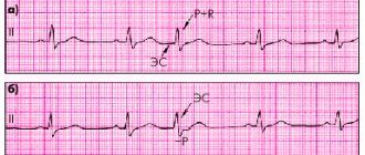
It’s just that recently group ES, lasting 2-4 seconds, have become more frequent. And they, as far as I know, are usually harbingers of atrial fibrillation. . According to pre-operative experience, it all started in approximately the same way; there was only an increase in frequency and prolongation of such cases of ES
This, of course, may be so. But problems are usually solved as they arise. It would be nice to see these extrasystoles on the ECG.
To treat the pathology, it is necessary to specifically determine what exactly is bothering you (ventricular arrhythmia and group supraventricular electrical activity).
Still, if a decision is made on drug treatment of rhythm disturbances, then the effectiveness of therapy can be monitored using daily ECG monitoring over time (in addition to subjective sensations).
CONCLUSION - repeat CM ECG. If you make sure that there are no other arrhythmias (atrial fibrillation or flutter, ventricular tachycardia, etc.), then treatment with propafenone is quite possible. And if they register something else, then . “problems must be solved as they arise” (c)
Performing repeated manipulations increases the effectiveness of the procedure in terms of sinus rhythm control by up to 90%. While the effectiveness of drug therapy is much lower.
I think it is appropriate to clarify: 1. The figure of 90% comes only from the lips of those who perform these operations. 2. The period for evaluating effectiveness is the same - 1 year. It would be interesting to see five years of observation by outside observers.
Regarding the effectiveness of medications: 1. “The odds ratio for maintaining sinus rhythm while taking amiodarone compared with any control drug was 6.8.” McNamara RL, Tamariz LJ, Segal JB, Bass EB Role of Pharmacologic Therapy, Electrical Cardioversion, and Echocardiography . Ann Intern Med., 2003; 139:1018–1033. 2. “The median time to a recurrence of atrial fibrillation was 569 days with amiodarone therapy,” i.e. almost 2 years. N Engl J Med. 2005 May 5;352(18):1861-72. Amiodarone versus sotalol for atrial fibrillation. Singh BN, Singh SN, Reda DJ, Tang XC, Lopez B, Harris CL, Fletcher RD, Sharma SC, Atwood JE, Jacobson AK, Lewis HD Jr, Raisch DW, Ezekowitz MD; Sotalol Amiodarone Atrial Fibrillation Efficacy Trial (SAFE-T) Investigators.
A one-time isolation of the pulmonary veins is rather an exception to the rule; repeated manipulations increase the effectiveness of the procedure in terms of sinus rhythm control by up to 90%. While the effectiveness of drug therapy is much lower.
I think it is appropriate to clarify: 1. The figure of 90% comes only from the lips of those who perform these operations. 2. The period for evaluating effectiveness is the same - 1 year. It would be interesting to see five years of observation by outside observers.
To clarify, it is worth adding that we are talking mainly about symptomatic paroxysms. Now let's wait for the results of research with revils. It will probably be interesting and not very rosy (at least not 90%). :rolleyes:
. the question is how effective repeat RFA is at a later stage. Or will it be at the primary level? Very good question. I understand that you rated the effectiveness of primary RFA quite highly.