In medicine, there is such a thing as propaedeutics, which implies primary diagnosis. Such diagnostics do not require special procedures. Having knowledge from this scientific field makes it possible to make a diagnosis based on an external examination of the patient or taking into account those characteristics that are easy to establish without the use of special devices. One of the methods of this science is auscultation.
This diagnostic method involves listening to sounds produced in the lungs and larynx. Based on their characteristics, one can assume the presence or absence of pathologies in the patient’s organs of the respiratory system.
This becomes possible only if the specialist has the necessary knowledge and sufficient experience, otherwise it will be difficult to draw the right conclusions. You also need to understand that with the help of auscultation it is not always possible to detect a disease or choose one diagnosis from several possible ones.
In this case, other diagnostic procedures must be used. However, in simple situations this method is sufficient, so that the patient does not have to be exposed to UV rays, for example. That is why auscultation is used at the present stage of development of medicine.
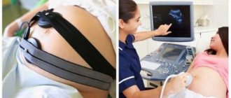
Auscultation of the lungs is especially important for diagnosing respiratory diseases in children. In childhood, many effective diagnostic procedures are harmful to the body, so doctors avoid using them.
As a result, when a child is sick, one has to choose simpler, albeit less accurate, methods of identifying pathologies. It must be said that the methodology for carrying out the procedure in question for children does not differ from that performed for adults. Doctors are guided by the same rules and the same algorithm of actions.
What is it used for?
Auscultation is used to detect a variety of diseases of the lungs, bronchi, heart and circulatory system. To do this, an assessment of the main and secondary breathing noises is carried out. Bronchophony over the entire surface is also assessed. These indicators are then supposed to be compared with normal ones, on the basis of which a conclusion is made about the presence or absence of diseases.
Thanks to auscultation, the following pathological conditions inherent in children and adults can be detected:
pneumonia,- pulmonary infarction,
- presence of a tumor in the lung,
- pulmonary edema,
- tuberculosis,
- pneumothorax,
- accumulation of fluid in the pleural cavity,
- heart failure.
Since the main signs by which such a diagnosis is carried out are noises, it is necessary to find out what kind of noises can be detected during auscultation. This:
- Vesicular respiration. This type of noise is soft and uniform and should be continuous when inhaling. The sound resembles the sound “v” or “f”.
- Bronchial breathing. It is observed in the phases of inhalation and exhalation, similar to the sound “x”. When exhaling, this noise is more sharp than when inhaling.
- Mixed breathing. It can be called intermediate between the first two, since it has the features of both of them.
In addition to the main ones, the doctor may hear additional noises during auscultation, which are signs of pathological phenomena. This:
- Wheezing. Can be dry or wet. They appear as a whistling, buzzing or buzzing sound (dry) or resemble the sound of bursting bubbles (wet).
- Crepitus. This phenomenon is a jerky squeaking sound.
- Pleural friction noise. When this noise is detected, it can be assumed that its source is very close to the surface. It sounds like the crunch of snow or the rustle of paper.
In order for the diagnosis to be correct, the doctor must take into account not only the existing extraneous noises, but also the characteristics of the main noises. In addition, it is necessary to take into account the symptoms that the patient names, his individual characteristics and much more.
Medical Encyclopedia - Auscultation
Auscultation (listening) is one of the main methods of clinical examination of a patient, which consists of listening to sound phenomena that spontaneously arise in the body.
In practice, both direct or direct auscultation is performed (listening directly with the ear attached to the patient’s body) and indirect auscultation using a stethoscope or phonendoscope. With direct auscultation, audibility is much better than when using a stethoscope, since the auscultated noises are not distorted; for example, weak bronchial breathing, see diastolic murmur in aortic insufficiency, is sometimes detected only in this way.
For indirect auscultation, solid stethoscopes made of wood, plastic or flexible binaural stethoscopes, consisting of a funnel and two rubber tubes, a stethophonendoscope, and a phonendoscope are used (Fig. 1-3). The stethoscope is a closed acoustic system; the air in it is the main conductor of sound, especially in a flexible stethoscope; in the case of communication with outside air or the lumen of the tube is closed, auscultation becomes almost impossible. When a solid stethoscope is placed into the auricle, the conduction of sound through the bones of the skull is of some importance. When auscultating with a stethoscope, noises are more or less distorted due to resonance, but better delineation of noises of different origins in a small area is ensured (for example, during auscultation of the heart); Noises are perceived more clearly.
Auscultation is of exceptional importance in examining the lungs, heart and blood vessels, measuring blood pressure according to Korotkoff, and determining bowel sounds.
Auscultation (Latin auscultatio - listening) is one of the main methods of clinical research, which consists of listening to sound phenomena that spontaneously arise in the body.
Auscultation was studied and introduced into medical practice by K. Laennec. The nature of acoustic phenomena detected during auscultation depends on the physical properties (density, tension, mass) of the vibrating bodies, the anatomical structure and intensity of organ function. Vibrations of tissue structures occur during the act of breathing, contraction of the heart, movements of the stomach and intestines; Some of the vibrations reach the surface of the body (skin). Each point on the surface of the body is a source of sound waves propagating in all directions; As you move away from the sound source, the wave energy is distributed over increasingly larger volumes of air, so the amplitude of vibrations quickly decreases, and the sound becomes so quiet that it is not perceived by the ear that is not in contact with the body. The main significance of the stethoscope, as well as the direct application of the ear to the body, is that it prevents the sound from being attenuated by dissipating energy.
All sounds occurring in the body are noises, that is, a mixture of sounds of different frequencies. The ear is most sensitive to sounds around 2000 Hz. As frequency decreases, sensitivity decreases sharply, so that at the same intensity, high-frequency sounds seem louder than low-frequency sounds. The ear can more easily detect changes in frequency or pitch than in sound intensity. A weak sound after a strong one is difficult to perceive; In addition, in a mixture of sounds of different frequencies, strong vibrations of one frequency mask weaker vibrations of other frequencies.
The sounds perceived during auscultation of the lungs and heart do not exceed 1000 Hz in frequency. The ear picks up only about 10% of the vibrations caused by the heart (a normal heart is a source of vibrations with a frequency of 5 to 800 per 1 sec.), since some of the vibrations are too low frequency (below 20), i.e. below the threshold of perception (infrasounds) , and some are of low intensity. Nevertheless, the importance of auscultation is very great. Phonocardiographic studies have confirmed the explanations for almost all sound phenomena of the heart (tones, noises) established by clinical A. (see Phonography).
In practice, both direct, or direct, auscultation is used, as well as indirect with the help of a stethoscope, phonendoscope, etc. With direct auscultation, audibility is much better than when using a stethoscope, since the auscultated noises are perceived directly by the auricle and are not distorted. For example, weak bronchial breathing and diastolic murmur in aortic insufficiency are sometimes detected only with this method. V.P. Obraztsov proposed an original method of direct audio for recognizing an additional tone during a gallop rhythm. A necessary condition for this method of A. is the tight pressing of the ear to the area of the heart, in this case a closed air cavity is formed and the first heart sound and gallop rhythm acquire the character of a ringing sound due to the push of the heart against the chest, since the gallop rhythm is a tone-push.
For indirect auscultation, solid stethoscopes made of wood, plastic or flexible binaural stethoscopes, consisting of a funnel and two rubber tubes, stethophonendoscopes and phonendoscopes are used.
The stethoscope is a closed acoustic system, and the air in it is the main conductor of sound, especially in a flexible stethoscope; in the case of communication with outside air or the lumen of the tube is closed, auscultation becomes almost impossible. When a solid stethoscope is placed on the auricle, the conduction of sound through the bones of the skull is of some importance. With A. using a stethoscope, noises are more or less distorted due to resonance, but better localization and delimitation of noise of different origins in a small area is ensured (for example, with A. of the heart); Noises are perceived more clearly. A necessary condition for successful A. is silence in the ward or office.
Auscultation is an indispensable method of clinical research. It is of exceptional importance in examining the lungs, heart and blood vessels, measuring Korotkoff blood pressure, determining bowel sounds, examining joints, etc.
Auscultation of the heart in children
The auscultation technique in young children and adolescents does not differ from the technique used when listening to the heart in adults. The points and order of auscultation are similar. The only difference is in the interpretation of the results.
For example, in thin children with low body weight, the third and fourth sounds can be auscultated normally. They also have an increase in the sonority of all tones, due to a thin layer of the chest cavity.
Heart rate also differs. If in adults the normal values of heart rate are in the range from 60 to 80 beats per minute, then for a child of the first year of life this will be a severe bradyarrhythmia, since its norm is in the range from 110 to 160 beats.
Types of noises and their significance in diagnosis
All noises, depending on the phase of cardiac contraction, are divided into:
- systolic - heard in case of tricuspid and bicuspid valve insufficiency, stenosis of the pulmonary artery and aorta;
- diastolic - formed due to insufficiency of the valves of the main vessels, stenosis of the atrioventricular orifices.
The nature of the noise has diagnostic significance. Noises of organic origin associated with heart defects have more “musical” properties. Thus, listening to a patient with septic endocarditis reveals a diastolic murmur on the aorta with a howling or whistling tone. This indicates a perforation with separation of the valve leaflet.
For the congenital defect of patent ductus arteriosus, a noise similar to “the rumble of a train in a tunnel” is typical.
To identify the place of greatest sound, palpation is carried out at the same time, the patient is listened to in the interscapular area, above the carotid arteries.
Cardiopulmonary murmurs are rare due to emptying during systole and decreased ventricular size. At the same time, the adjacent section of lung tissue expands and sucks air from the bronchus. The noise is heard at the height of inspiration.
Murmurs of pericardial origin are not audible in a healthy person. A creaking sound accompanies both systole and diastole. Indicates overirritation of the enlarged heart and friction of the pericardial layers.
Heart murmurs
It is customary to compare heart murmurs with the sounds that fluid creates when moving in pipes.
If the inner surface of the heart chambers were perfectly smooth, then there would be no noise. However, the walls are rough to one degree or another, which affects the speed of blood flow. When pumping blood, narrowing will occur in the chambers and adjacent vessels. Also, heart sounds will be louder in the presence of dense obstacles, as well as if they are located near the outlet.
When listening, the noise will have different shades:
- weak,
- hissing,
- whistling,
- howling,
- rude,
- “mosquito squeak.”
The increase in noise is influenced by an increase in the speed of blood pumping with a decrease in its viscosity. Problems with the valve flaps can cause an increase in noise.
For reference. A heart murmur is a sound that appears as a result of disturbances in hemodynamic parameters. The diameter of the valves or vessels, the viscosity of the blood or the speed of its movement changes).
During normal functioning of the cardiovascular system, only tones are auscultated; listening to various noises always indicates the presence of a disease.
Examination using a stethoscope
The device, called a stethoscope, is made in the form of a tube with a funnel-shaped expansion at its ends. Listening occurs by applying one end of this instrument to the listening site, and the other to the doctor’s ear.
A stethoscope allows you to expand the picture of sound perception, but some deviations in sound conductivity and bulky structure make it somewhat inconvenient.
Currently, this type of device is used in gynecological practice; it is convenient to listen to the fetal heartbeat.
How tones are formed, interpretation of deviations from the norm
Be sure to listen to two interconnected beats at each point. These are heart sounds. All healthy individuals have them. It is less common to hear the third and even fourth tone.
The first sound is called systolic; it consists of several components:
- atrial function;
- muscular - caused by vibrations of the tense muscles of the ventricles;
- valvular - considered the main component, formed by the oscillating leaflets of the atrioventricular valves;
- vascular - includes the walls of the aorta and pulmonary artery and their valve apparatus.
Based on the nature of its sound, it can be classified as:
- deaf - with left ventricular hypertrophy, myocarditis, cardiosclerosis, dystrophic changes;
- quiet, “velvet” - with myocardial infarction;
- weak, as if coming from afar - with exudative pleurisy, emphysema, significant thickness of the chest wall;
- loud, clapping - with neurosis, thyrotoxicosis, stenosis of the left atrioventricular orifice, anemia, high fever, extrasystole;
- bifurcated - with bundle branch block, thyrotoxicosis, aneurysm in the apex of the heart, myocardial dystrophy.
The second sound is formed at the beginning of diastole, caused by the slamming of the semilunar valves of the pulmonary artery and aorta. In a healthy person it is concentrated on the aorta. In cases of “pulmonary heart” with hypertension in the pulmonary circle - on the pulmonary artery.
With atherosclerotic lesions of the aorta, vasodilation, the second tone rings and resonates. Bifurcation is observed with aortic aneurysm and mitral stenosis.
You can visually register noises and tones using a phonocardiogram (bottom line), it must be written along with the ECG
The appearance of the third tone creates an auditory picture of a “gallop rhythm”. It is believed that it is formed due to the rapid decrease in the tone of the flabby walls of the ventricles in the diastole phase. In children and adolescents it is heard more often than in adults and indicates functional inferiority of the myocardium, since pathology is not detected.
For persons 30 years of age and older, it is a characteristic sign of hypertension, cor pulmonale, myocarditis, cardiosclerosis, myocardial infarction and aortic aneurysm.