Fetal echocardiography is similar in procedure to ultrasound examination of the heart. The essence of the method is the use of high-frequency ultrasonic waves and the Doppler effect. This allows you to visualize in detail the cardiovascular system of the unborn child, identify possible abnormalities in the development of the heart at an early stage and take the necessary measures.
What is it?
Fetal echocardiography (ECHO CG of the fetus, ultrasound of the fetal heart) is a diagnostic method based on the use of ultrasound waves to detect congenital heart defects in the unborn child. The use of this technique is determined by the simultaneous combination of safety, speed, accessibility, and accuracy.
Regarding the safety of fetal echocardiography, we can say that ultrasound waves are absolutely safe not only for the mother, but also for the child, which is important.
Who should undergo fetal cardiac echocardiography?
There are certain risk groups for pregnant women who may give birth to a child with congenital heart disease:
- alcoholism, drug addiction, smoking while pregnant;
- the presence of congenital heart defects in the mother, father or other close blood relatives;
- diabetes mellitus, whether gestational or not;
- systemic lupus erythematosus or other autoimmune diseases;
- taking medications that are prohibited for pregnant women (antibiotics, antidepressants, antiepileptic drugs, nonsteroidal anti-inflammatory drugs, etc.);
- the mother has so-called torch and other infections (rubella, cytomegalovirus, parvovirus, toxoplasmosis, Lyme disease, etc.);
- hyperfunction of the thyroid gland;
- maternal hypertension;
- diseases affecting connective tissue;
- age over 35 years;
- polyhydramnios.
Echocardiography of the fetal heart is performed even when nothing is detected on a general ultrasound examination. But sometimes, a woman who is not at risk may also give birth to a child with a congenital heart defect.
Some signs on echocardiography of the fetus and not only may indicate defects:
- cardiac arrhythmia in a child;
- the presence of fluid in various organs of the fetus (stomach, lungs, head);
- detection of chromosomal diseases;
- signs of cardiac pathology during ultrasound examination (single umbilical artery, fetal anemia, agenesis of the ductus venosus, etc.);
Sometimes, a sign indicating the presence of a congenital heart defect is located outside the fetal cardiovascular system. Such signs are called extracardiac. For example: atresia of any part of the intestine, congenital kidney pathology.
Carrying out echocardiography
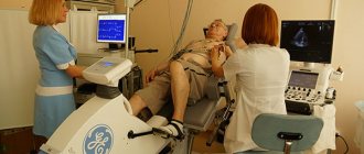
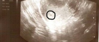
The most reliable results can be obtained at 18-24 weeks of pregnancy. The study uses transabdominal access, however, if necessary, at a gestational age of less than 18 weeks, echocardiography can be performed transvaginally. The fetal echocardiography procedure lasts about 30-40 minutes and the procedure is similar to a regular ultrasound. During the fetal echocardiography procedure, the pregnant woman should be calm and relaxed; She may have to stop taking certain medications a few days before the planned test, on the advice of her doctor.
A standard echographic examination includes assessment of a four-chamber section of the heart (the position of the heart in the chest cavity, the structure of the ventricles and atria, the formation of the interventricular and interatrial septa, the movements of the mitral and tricuspid valves), the main arteries (the relative position and diameter of the aorta and pulmonary trunk), characteristics of cardiac activity ( Heart rate, rhythm of atrioventricular contractions). The obstetrician-gynecologist monitoring the pregnancy must be familiar with the fetal echocardiography protocol, who will comment on the diagnostic results to the woman and explain all the risks.
The quality of ultrasound imaging depends on the level of qualifications and experience of the specialist, the type of equipment used, gestational age, fetal position, low or polyhydramnios, and the thickness of the subcutaneous fat layer on the anterior abdominal wall of the pregnant woman.
Cost of fetal echocardiography in Moscow
The study is carried out in antenatal clinics, perinatal centers and medical clinics by ultrasound diagnostic specialists who have the appropriate certificate. The price of a diagnostic procedure is influenced by the type of echocardiography (transabdominal, transvaginal), the form of ownership of the medical organization, the category of equipment and the qualifications of the diagnostician. If the patient wishes to undergo the study on the day of treatment, the cost may increase. The price increase may also be due to the availability of additional services and the involvement of experts in assessing the results of echocardiography.
Echocardiography is not included in the list of mandatory procedures during pregnancy, so many women do not know what an echocardiogram is. Below in the article you will find information about the indications for this examination, how an echo-kg is done, and what consequences may arise.
What are the types of fetal echocardiography?
With the development of medicine, in particular diagnostics using ultrasound, many types of ultrasound have appeared. There are three main types that are used most often in medical practice.
- Two-dimensional echocardiography is used to study the main anatomical structures, but does not always reveal the cause of cardiac dysfunction.
- M-mode is used to monitor the operation of valves, leaflets and myocardial walls during their functioning.
- Pulsed-wave Doppler echocardiography implies the additional use of fetal Doppler sonography, making it possible to study the functioning of the heart in more detail.
In addition, the use of the third method makes it possible to fully assess blood flows, their volume, direction, both in the heart and in the vessels.
When is ECHO CG screening necessary during pregnancy?
Thanks to fetal echocardiography, the doctor sees and studies the first spark of life - the fetal heart, which is still in the process of formation. This is an invaluable opportunity to detect most cardiac abnormalities well before PDR. During the study, the specialist evaluates in which direction and how quickly the blood moves in the baby’s heart vessels. Moreover, with the help of ECHO CG, it is possible to make a conclusion whether the lumen in the vessels is full and to judge whether a sufficient volume of blood is pumped by the heart, and this is of great importance in the diagnosis of heart diseases.
The list of indications for ECHO CG is quite large:
- there were already children in the family with cardiac anomalies;
- the woman has experienced spontaneous abortion in the past;
- the anamnesis of the parents or their immediate relatives includes a diagnosis of one or another heart disease;
- the patient suffers from diabetes or has hormonal imbalances;
- the expectant mother suffered an infectious disease during pregnancy;
- the doctor has reason to believe that the development of the fetus is in danger due to chromosomal abnormalities;
- the first trimester of pregnancy was associated with the use of antibacterial agents;
- fluid has formed in the baby’s head, lungs or stomach;
- The pregnant woman is approaching 40 years of age;
- the patient is expecting two babies;
- the amount of amniotic fluid in the expectant mother exceeds the norm or, conversely, is reduced.
Why should an ultrasound of the fetal heart be performed?
Echocardiography of the fetal heart allows doctors to detect many different heart conditions early. Based on this, treatment tactics and appropriate measures that can be taken during and after childbirth are selected. However, sometimes it is necessary to resort to termination of pregnancy. This is performed in cases where the heart defect is absolutely incompatible with life, in addition, it may threaten the woman’s life.
However, if fetal echocardiography reveals abnormal heart function, you should not immediately panic. This could be a device error, an artifact during testing, etc. Therefore, after 1-2 weeks it is prescribed again. The obstetrician-gynecologist will indicate a more precise date. When the pathology has already been accurately diagnosed, a mandatory consultation with other specialists will follow. For example, if surgical intervention is necessary for a defect, then it is worth discussing everything with a cardiac surgeon. Another related specialist whose consultation is necessary is a neonatologist.
Not all defects detected on fetal echocardiography require mandatory surgical intervention. Some of them go away on their own, for example, a patent septum between the ventricles or atria may no longer be visible on a subsequent ultrasound examination.
results
The main criterion for normal fetal development is heart rate (heart rate), which changes every week of development:
- at the 5th week the heart begins to beat at a frequency of up to 85 beats;
- in the 6th week the frequency increases to 120;
- from the 7th week, 130-150 beats are considered normal;
- at -11 weeks, heart rate should be 150-180 beats per minute;
- from the 12th week until birth, the heart beats at a frequency of 145-170 beats.
The same amount of time must pass between heartbeats, otherwise the doctor may suspect a congenital malformation or hypoxia. No extraneous noises should be heard (they can also indicate hypoxia or other pathology), the beats should be clear and rhythmic.
At the end of the second trimester, an ultrasound scan should clearly show 2 ventricles (right and left), right and left atria, septa (interventricular and interatrial), and the valve of the foramen ovale. The fetus must have an open oval window, which closes after birth or by the age of 6 months.
It should be noted that some pathologies require treatment after the baby is born. Doctors may also advise you to wait some time after childbirth - in some cases, developmental abnormalities go away with age.
When to do it?
An ultrasound of the child’s heart is carried out from the 18-19th week to the 23-24th pregnancy, because during this period you can get the most reliable information that will help doctors. And of course, if the pathology itself is suspected or detected, further repeated studies will be ordered, the date of which is set by the doctor leading the pregnancy of a particular woman.
How is it carried out?
Before talking about directly conducting echocardiography, an important fact should be pointed out - preparation for an ultrasound examination of the fetal heart is not required.
The duration of the procedure is from 15 to 30 minutes. Echocardiography is performed in the supine position. After a woman lies down on the couch, she should remove clothes from her stomach. Then, a special gel is applied to the area to be examined. It is necessary to improve the sliding of the sensor and eliminate air on the surface, since it also interferes with echocardiography. Finally, the remaining gel is removed from the surface of the skin.
If a defect is detected, then after birth, the child will also undergo an echocardiography.
Principle of preparation for research
In fact, no preliminary preparation is required to carry out this procedure. The only thing is that it is convenient for infant patients to perform ultrasound while they are sleeping. It is also worth noting that infants need to be fed an hour and a half before the examination. Feeding before the procedure is not recommended, since the digestion process is an additional burden on the heart.
Adult patients with a pulse over 90 and high blood pressure over 160 mm Hg. Art., before the study you should consult with your doctor or cardiologist. A specialist will help eliminate all symptoms that have arisen, otherwise the examination results will contain a lot of errors.
Feedback about the study
Fetal echocardiography has extremely positive reviews from both doctors and women. Pregnant women highlight the speed, simplicity and lack of need to prepare for the study. Doctors note the quick and high-quality diagnosis of defects, and, of course, safety.
Echocardiography has helped save more than one child’s life, and moreover, those children who could become disabled due to a defect, thanks to timely diagnosis and high professionalism of doctors, are already living full lives, and their parents, with a calm soul and heart, watch how they grow their children.
At what time can an ultrasound be performed?
During pregnancy, the first fetal echocardiography is performed in the fourth month of pregnancy. In this case, the doctor tries to determine congenital defects and the structure of heart tissue. From the eleventh week, intrauterine ultrasound of the heart is allowed. Due to the fact that at this stage the size of the heart is extremely small, the doctor will be able to identify only a small proportion of possible diseases.
Most often, such diagnostics reveal the following pathologies:
- violation of the structure of the left atrium,
- violation of the structure of the left ventricle,
- atresia of the tricuspid valve,
- blockade of the atrioventricular plexus,
- underdevelopment of the left branch of the pulmonary artery,
- underdevelopment of the pulmonary valve.
Such defects are considered one of the most unsuitable for surgical treatment. Pathologies of the development of great vessels in the early stages are almost impossible to identify, since they are not visible on ultrasound.
If it is possible to detect a violation of the central circulatory system in the first trimesters of pregnancy, then control studies are required at the fifteenth and nineteenth weeks of pregnancy.
In addition, there are many congenital defects of heart structures that cannot be detected in the prenatal period using ultrasound. Such vices include:
- violation of the Bottal duct,
- anomaly of the septa between the atria,
- atresia of several segments of the aorta,
- minor disturbances in the structure of the valves.
Echocardiography is recommended to be performed between the eighteenth and twenty-third weeks of gestation. This is explained by the fact that before the eighteenth week the baby’s heart is too small to detect pathology, and after the twenty-third week the volume of amniotic fluid decreases, which impairs the visibility of the heart.