Dopplerography of blood vessels is the most accurate and safe method for diagnosing circulatory pathologies. The study is based on the use of the Doppler effect. Using it, you can evaluate not only morphological characteristics, but also determine the dynamic performance of the arterial and venous beds.
What is Dopplerography
Duplex scanning (ultrasound with Doppler) is an ultrasound research method aimed at diagnosing vascular pathologies. The examination helps to identify signs of the disease even at the asymptomatic stage.
Subject to study:
- coronary arteries;
- aorta and its branches;
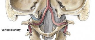
- vessels of the head, neck (BCA, vertebral artery);
- abdominal cavity and aorta;
- genitourinary system (kidneys, uterus, penis);
- vessels of the upper extremities;
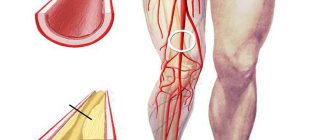
- arteries, veins of the lower extremities.
Doppler ultrasound of the heart arteries is performed if cardiac atherosclerosis is suspected. The disease may not give an obvious clinical picture for a long time. With a significant degree of narrowing, ischemic pain appears like angina pectoris.
Color Doppler mapping of brachiocephalic vessels (CDC BVC) is also aimed at identifying early forms of atherosclerosis. In addition, examination often reveals congenital or acquired abnormalities in the structure of the arterial wall.
Doppleroscopy of the vessels of the extremities is indicated in the presence of a high risk of thrombosis or pulmonary embolism. At the same time, ultrasound scanning of the leg veins reveals wall anomalies, valve incompetence, and the presence of tortuosity and kinks.
To watch a video from a specialist on the topic:
Indications for transcranial Doppler ultrasound
There are many reasons why you may need to undergo Doppler ultrasound. Transcranial Dopplerography of cerebral vessels
may show a large number of deviations from the norm.
Vascular malformation
It is a congenital disease that affects the development of the vascular system. Manifests itself in the form of plexuses of abnormal vessels of various sizes. It is dangerous because it can cause subarachnoid hemorrhage of a non-traumatic nature. Among other things, steal syndrome may develop, and malformations may begin to compress the brain tissue.
Cerebral circulatory disorders
TCD is used quite often to examine cerebral vessels. It may be necessary to identify the cause of problems with cerebral circulation. They may consist of the formation of blood clots, loops and kinks in blood vessels. Often the cause is: the formation of narrowings, aneurysms or embolism.
Atherosclerosis of the arteries of the neck and head
It consists of damage to areas of blood vessels by atherosclerotic plaques, which either significantly narrow the lumen of the vessel or clog it completely. In advanced cases, treatment is performed surgically. TCD is used to examine vessels because it allows one to determine not only the presence of plaques, but also their size and location.
Encephalopathy
Occurs in chronic cerebral vascular diseases that slowly progress. This disease combines cognitive impairment, disruptions in the emotional and motor spheres.
Types of research
Doppler examination is carried out in three modes:
- B-mode evaluates the anatomy of blood vessels and underlying tissues.
- Color Doppler mapping (tissue angioscanning) allows you to obtain a color and volumetric image of structures, and the nature of the blood flow is also determined.
- Spectral Doppler ultrasound (D-mode) - the results are presented in the form of a graph that takes into account changes in blood flow over time.
Dopplerography also varies in the nature of the image.
More common:
- three-dimensional;
- duplex;
- triplex scanning.
Three-dimensional ultrasound is a three-dimensional image that is formed as a result of specific signal transformation. Vessels, organs, tissues can be seen from all sides. The method is especially popular in obstetrics and gynecology. The expectant mother can examine her baby even before his birth. In addition, the examination has proven itself in oncology. It is used to determine the topography, size, and degree of blood supply to the tumor (highly vascularized or avascular formations).
Duplex scanning is a combination of B-mode and Doppler effect. The method allows you to see vessels in real time. It is also possible to assess the condition of soft tissues and their location in relation to other structures. The study is used to diagnose pathologies of the head and neck vessels (sonography, ultrasound of the vertebral arteries).
Triplex Doppler ultrasound combines duplex scanning and color mapping. As a result, the study occurs in three modes simultaneously. This allows you to obtain more accurate data.
This method is used primarily in diagnosing pathologies of the veins of the upper and lower extremities, extracranial and brachiocephalic vessels. The method is also effective for determining the causes of erectile dysfunction (pharmaco-Doppler ultrasonography).
In addition, there are:
- continuous wave Doppler – assesses blood flow in high-speed vessels (ultrasound of the main arteries of the head and neck, examination of the veins of the lower extremities);
- pulse-wave mode - the examination is carried out in a limited area (for pathologies of the heart or blood vessels).
In addition, watch a detailed video from a specialist:
Ultrasound and Doppler Doppler - what is the difference?
The high degree of information content, how ultrasound differs from Doppler ultrasound, allows advanced ultrasound scanning methods to identify not only areas of poor vascular patency and the quality of blood flow, but also the causes of problems with blood vessels. During an ultrasound scan, the sonologist acts blindly, applying the sensor to areas of the body that are usually examined during the procedure, while ultrasound using duplex and triplex scanning opens an overview of areas of blood vessels with critical problems, which is the difference between ultrasound and ultrasound.
Indications
Vascular Doppler is performed if there are strict indications. Their appearance depends on the expected localization of the pathological process.
Indications for the study of cerebral vessels:
- headache;
- dizziness;
- syncope;
- impairment of vision, hearing or smell in the absence of pathologies from the analyzers;
- changes in cognitive function;
- long-term arterial hypertension;
- presence of signs of atherosclerosis;
- history of strokes;
- suffered traumatic brain injuries.
Also at risk for the occurrence of pathologies of cerebral vessels are persons who have hemodynamic disorders (arrhythmia, heart defects). They are recommended to undergo duplex scanning of blood vessels as planned.
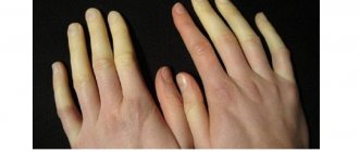
Ultrasound of the vessels of the extremities is performed in the presence of:
- swelling;
- pain in the limb;
- sensitivity disorders;
- changes in skin color;
- signs of varicose veins.
Duplex scanning of renal vessels is carried out:
- with severe and resistant arterial hypertension;
- if thrombosis of the renal vessels is suspected;
- when signs of renal failure appear.
Dopplerography of the abdominal aorta is indicated in cases of severe atherosclerosis or in the presence of signs of an aneurysm (pulsating formation in the abdominal cavity).
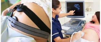
For pregnant women, duplex scanning is performed in the third trimester, before the woman in labor is transferred to the maternity hospital.
Early examinations are carried out for the following symptoms:
- changes in fetal motor activity;
- suspicion of a violation of the feto-placental-uterine blood flow;
- developmental delay;
- signs of fetal hypoxia.
Duplex scanning helps not only to determine the violation, but also to identify its suspected cause.
Examination in children
Ultrasound examination is mandatory for all infants at the age of 1 month. This makes it possible to identify a violation of the blood supply to the brain at the earliest stages and correct it. Without timely diagnosis and treatment, this condition will lead to serious neurological problems and intellectual impairment in the child.
For older children, vascular ultrasound is prescribed for complaints of headaches, fatigue, mental retardation, decreased attention and memory. The study allows you to prescribe appropriate therapy and improve the child’s condition.
How to prepare for Doppler
No special preparation is required for Doppler ultrasound. When duplex scanning of abdominal vessels, it is necessary to adhere to a diet low in carbohydrates and fiber. The day before the procedure, fruits, vegetables, flour, sweets, and carbonated drinks are excluded from the diet. If you are lactose intolerant, you should limit your intake of dairy products.
If duplex scanning of the female reproductive organs is performed, it is necessary to perform an ultrasound on the 5-7th day of the menstrual cycle.
An important point is skin care before the study. Do not apply greasy creams and oils to the skin. This can worsen signal transmission and reduce the information content of Doppler ultrasound.
For the procedure itself, you must take a towel or cotton diaper with you.
How is Doppler done?
Doppler ultrasound is performed in the same way as standard ultrasound. The duration of the procedure does not exceed 15-20 minutes.
First, a special gel is applied to the study area, which improves contact and signal transmission. Then, using a sensor, ultrasound is emitted, reflected and received. In the process, the signal is transformed into a graphic image, which is displayed on the device’s screen.
Duplex scanning is an absolutely safe method. The study has no contraindications. It can be done as often as the clinical situation requires.
The doctor explains and shows how the procedure is performed:
Operating principle of ultrasound scanner
Blood is a liquid moving medium. Ultrasound ultrasound sends waves into the blood, which are reflected depending on how the properties of the fluid change in space. That is, sound waves are reflected from blood particles, which depends on blood flow.
In Doppler studies, two sensors are used: the first to generate ultrasonic waves, the second to record reflected waves. Schematically it looks like this: the first sensor sends an initial signal with the specified parameters. It is reflected from solid moving objects and comes back. Now the original and received signal are compared. The difference between them in the form of information is converted into an image.
Ultrasound scanning uses blood biophysics, which has hydrodynamic properties in the form of two liquid flows:
- Laminar flow. The difference between the central and wall blood flow in the vessel is taken into account. These two layers should not be mixed. Laminar flow is recorded with normal blood flow in the vessel.
- Turbulent flow. The central and peripheral flow mix, forming “chaos”. Turbulent flow is recorded in pathology when there is an obstruction in the vessel or it is excessively narrowed.
Decoding the results
After the procedure is completed, the doctor fills out a research protocol (Dopplerogram), which indicates the anatomical features and identified pathologies. Further interpretation and analysis is carried out by a specialized specialist (neurologist, cardiologist, vascular surgeon).
In duplex mode, both paired and unpaired vessels are scanned. The scope of the study depends on the clinical manifestations of the pathology. If a widespread process is suspected, it is necessary to perform an ultrasound of the entire anatomical area. If the disorders are localized in only one artery, only that one should be studied in detail.
During the duplex scanning process, it is important to evaluate the following parameters:
- nature of blood flow;
- its changes (strengthened or weakened);
- the speed of blood flow in different phases of the cardiac cycle;
- morphological characteristics of vessels;
- dynamic parameters (resistance and pulsation index);
- pathological narrowings (stenosis), the degree of their severity.
The obtained data are compared with the norm. Based on this, a conclusion is made about the state of the system and compensation for violations.
Depending on the area being examined, duplex scanning has its own characteristics and additional parameters.
What the study shows
During the deciphering process, the following vascular pathologies are most often identified:
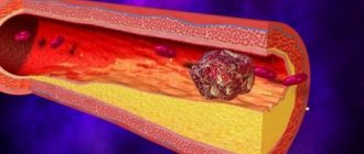
- atherosclerosis;
- abnormalities of the heart valve apparatus;
- aneurysms;
- stenosis;
- occlusion;
- pathological course, tortuosity;
- underdevelopment of the vascular network;
- varicose veins, incompetent venous valves.
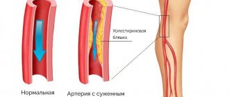
Atherosclerotic changes can be observed in various vessels. More often the process is generalized. In this case, the clinical picture of ischemia occurs only with a significant decrease in the lumen.
Doppler ultrasound reveals hyperechoic plaques that change the nature and speed of blood flow. It slows down, and a turbulent current may occur.
Video of an ultrasound showing signs of atherosclerosis:
Anomalies of heart development can be either congenital or acquired. The cause of congenital defects is a violation of the formation of the cardiovascular system in the embryonic period. Acquired defects are formed as a result of rheumatic damage to the valve apparatus.
With duplex scanning, you can see the disorder itself (valvular insufficiency, expansion of the valve ring, dilatation of the chambers, additional communication pathways) and compensatory hemodynamic reactions to it. First of all, the phase filling of the chambers changes. The pressure in the cavities also increases in diastole.
As the disease progresses, the heart muscle and the anatomical structures located in it undergo changes. On ultrasound you can see myocardial hypertrophy and chamber dilatation.
Aneurysms most often affect the aortic arch or its abdominal part. Duplex scanning shows a sac-like expansion of the vessel cavity and thinning of its walls. Linear blood flow is replaced by turbulent one. His speed decreases.
You can see the aneurysm in the video:
Stenoses are characterized by a ring-shaped narrowing of the lumen and poststenotic dilation of the vessel. Doppler ultrasound reveals a significant slowdown in blood flow and a compensatory change in the vascularization of the blood supply area (development of collaterals).
Unlike stenoses, with occlusion, collaterals do not have time to develop. Clinical manifestations of acute insufficiency of blood supply come first. Duplex examination reveals complete occlusion of the vessel lumen. Moreover, above the site of occlusion, expansion of the artery is observed.
The ultrasound monitor shows mitral stenosis:
The pathological course of the vessel and the underdevelopment of the arterial network are often congenital pathologies. Due to the high degree of compensation, there may be no clinical manifestations. With weak development of collaterals, a decrease in blood supply to organs and ischemic phenomena is determined. At the same time, a large number of thin, convoluted vessels attract attention.
Special mention should be made of poor blood flow in the placenta and umbilical cord. Insufficient blood supply to the fetus leads to hypoxia and death. The examination reveals underdevelopment, torsion, and inflammatory changes in the umbilical cord. Pathologies of the placenta on duplex scanning are represented by chorionic villi hypoplasia, improper attachment of the placenta, and chorioamnionitis.
Varicose veins most often affect the lower extremities. Deformation of the wall and an increase in the lumen of the vessel are observed. In this case, the valve apparatus does not change, which leads to its functional failure.
Features and advantages of Doppler ultrasound of the heart
Modern devices for diagnosing diseases of the heart and blood vessels allow performing ultrasound examinations at a high level. Vascular Dopplerography, in combination with cardiac ultrasound, has a number of advantages over other research methods, such as:
- high quality images;
- visualization of individual elements of organs that are inaccessible for research by other methods;
- the ability to track small details of the research object;
- is a non-invasive and safe diagnostic method with no contraindications;
- the method does not require the introduction of contrast agents into the body for examination;
- diagnostics can be carried out over time repeatedly, as needed;
- the method allows you to observe the work of the heart and blood vessels in real time;
- is an indispensable research tool in obstetric practice.
Thanks to Doppler ultrasound of the heart, it became possible to conduct more widespread preventive examinations among the population. This method contributes to the early detection of diseases of the cardiovascular system and the prevention of complications.