What is Holter monitoring?
| Holter monitoring (hereinafter referred to as HM) is a method of continuous recording of an electrocardiogram (ECG) on a solid-state medium or magnetic tape (practically not used today in modern systems) in several leads in conditions of free activity of the patient, followed by off-line decoding using special decoders. The method is named after the scientist who first proposed it in 1954. You can also hear alternative names for this method: dynamic electrocardiography, 24-hour ECG monitoring, Holter monitoring. |
It is generally assumed that ECG recordings are performed over a 24-hour period, but shorter or longer recording periods may be used as indicated, technical capabilities, or circumstances (e.g., overnight only, period of specific sport activity, multi-day monitoring).
Manufacturers may include various additional options in their devices in addition to ECG assessment (analysis of heart rate variability, automatic options for assessing the QT interval, analysis of late potentials of the atria and ventricles, pneumogram or other parameters). Such monitoring is called multifunctional. It is an extension of the traditional technique, often an independent research method (for example, 24-hour blood pressure monitoring - ABPM). Modern devices also record the patient’s breathing, movement, and body position in space. There may be other parameters that are logged. Such a study requires more time for analysis and additional specialist knowledge.
What devices are used for monitoring
| In addition to conventional devices that record ECG and other parameters continuously throughout the day, there are devices for 2 and 3 day recording. Monitors have also appeared for multi-day monitoring with telemetric data transmission via telephone or Internet channels. This makes it possible to register rare attacks of palpitations and arrhythmia, rare attacks of chest pain or suffocation, fainting and semi-fainting conditions, and also makes it possible to monitor the effectiveness and safety of the prescribed treatment on a daily basis. The patient wears a monitor and gives the recording to the doctor daily for interpretation. The doctor contacts the patient by phone or the Internet and gives him appropriate recommendations on treatment and life activities. At the same time, the patient continues to lead his normal life, including work or study. |
An alternative to such devices are implantable monitors, which are sutured subcutaneously. Monitors are also used that are turned on to record by the patient himself or his relatives in the event of a clinical event (an attack of irregularities, palpitations, fainting or pain).
Features of interpretation
When carrying out differential diagnosis, some difficulties arise with the interpretation of deviations of the ST segment from the normal position. For example, a shift of a segment below the isoline is detected not only with ischemic disease, but also with hypertrophy of the ventricles of the heart, electrolyte disturbances, autonomic disorders, and hormonal imbalance. Changes in the ECG can be caused by pathological changes in the cardiovascular system, including arterial hypertension, bradycardia, cardiomyopathy.
Clinically significant ST fragment depression was detected in half of the subjects without a history of cardiac pathology. The dynamics of the ST segment of an actually healthy person is influenced by a large number of factors. The most significant reasons include: psycho-emotional state, smoking, changes in body position, parasympathicotonia.
Taking certain medications can affect the level of the ST segment on the ECG diagram: adrenergic blockers, antiarrhythmic drugs, some cytostatics, antidepressants.
A dependence of the occurrence of ischemic episodes on the time of day was revealed. Most often, attacks are recorded in the early morning hours (4–6 am) and late evening (10–12 pm). ST segment deviations are associated with circadian rhythms - natural daily fluctuations in the activity of various processes within the body. Thus, in the daytime and in the morning, due to a natural increase in the sympathetic effect on the heart, the ST segment may shift downward. In the late evening and at night, segment elevation is recorded.
Analysis of T wave changes
The T wave reflects the cycle of repolarization (restoration) of the ventricles of the heart. Its analysis as an indicator of coronary disease does not have as much weight as the study of the ST segment. Cicatricial fluctuations often occur due to positional changes in the heart. During the day, the T wave can decrease, thicken, and transform into a negative wave. Most researchers agree that changes in the wave cannot be considered a manifestation of coronary artery disease.
However, some researchers insist: if inversion is observed or a huge wave lasting more than a minute occurs, the presence of ischemia of myocardial tissue can be assumed.
QT interval
The QT interval (ventricular electrical systole) corresponds to the cycle of excitation of all parts of the ventricles of the heart with their subsequent repolarization. It is one of the most significant parameters of the electrocardiogram.
Since coronary artery disease reduces the contractility of the heart muscle, the length of the QT interval should be taken into account when diagnosing. The lengthening of this segment on the cardiogram indicates a decrease in the speed of electrical impulse transmission through the atrioventricular junction. Deviation from the norm of the ST segment and T wave on the cardiogram is considered a sign of ischemic disease, provided that the QT interval increases by more than 1.1 times.
Who needs Holter monitoring?
1. This study is necessary for those who are being examined for:
- heart attacks,
- interruptions,
- fainting and semi-fainting states,
- chest pain that occurs both at rest and during physical activity;
- as well as those who have no complaints, but rhythm and conduction disturbances were detected on an ECG or during examination;
- for those who, based on changes in the ECG, are suspected of having ischemic heart disease or another disease associated with ischemic changes or arrhythmia.
2. Monitoring is also mandatory for patients with an already established diagnosis, such as:
- ventricular rhythm disturbances (ventricular extrasystole, ventricular tachycardia);
- atrial extrasystole and atrial tachycardia;
- atrial fibrillation (atrial fibrillation), both constant and paroxysmal;
- sinoauricular blockades (SA blockades) and atrioventricular blockades (AV blockades) of varying severity;
- arrest or autonomic dysfunction of the sinus node;
- sick sinus syndrome;
- WPW syndrome.
In these cases, chemotherapy is carried out at least once every 6-12 months
according to the doctor’s decision in order to monitor the number and arrhythmias, to evaluate the treatment being carried out, to decide whether it is necessary to apply surgical treatment methods.
3. Monitoring of patients with coronary heart disease (CHD), especially those who have had a myocardial infarction or heart surgery, is mandatory. The study allows you to assess whether there is myocardial ischemia, which is often “silent”, that is, not felt by the patient. Also, such patients often have rhythm and conduction disturbances, sometimes not felt by them.
4. Patients with heart defects, including those who have undergone heart surgery, with various cardiomyopathies (hypertrophic, dilated, arrhythmogenic right ventricular dysplasia and other cardiomyopathies), channelopathies (such as congenital long or short QT syndrome, Brugada syndrome, etc.) d.)
5. Patients with heart failure.
6. Patients on hemodialysis.
7. Patients with diabetic neuropathy.
With all of these diseases, cardiac arrhythmias are common, including life-threatening ones. Therefore, it is necessary to undergo regular monitoring to select the correct treatment. Including high-tech.
8. CM is mandatory for patients with implanted pacemakers (pacers), cardioverters-defibrillators, since during the study it is possible to evaluate both the operation of the implanted device and the presence of arrhythmias, assess their danger and select treatment accordingly.
A blood pressure monitor is necessary to monitor ongoing antihypertensive therapy, as well as to assess blood pressure at night, during physical activity, during stressful situations, and when elevated blood pressure is detected only in the doctor’s office (“white coat hypertension”). Modern devices can detect obstructive sleep apnea syndrome (OSA), a breathing disorder during sleep, usually accompanied by snoring. The severity of this syndrome can be assessed. This is important, as it can cause arrhythmias and increased blood pressure at night. With severe OSA, the patient is referred for further examination and selection of treatment to a somnologist.
CHILDREN, ADOLESCENTS and PREGNANT WOMEN are monitored for the same reasons as adults.
Chemotherapy for athletes is carried out according to the same indications as for people not involved in sports. This method also allows you to evaluate changes in heart rate and the appearance of ischemia under conditions of a specific sports load, which is not always possible to do with other methods (for example, in parachutists).
Causes of tachycardia
Non-cardiac causes of tachycardia include:
- Patient detraining (low level of physical fitness)
- febrile conditions
- pathology of the thyroid gland (hyperthyroidism)
- anemia (decreased hemoglobin level in the blood)
- hypoxia (decreased oxygen levels in the blood)
- lower blood sugar levels
- arterial hypotension
- shock
- pheochromocytoma
- chronic bronchopulmonary diseases accompanied by the development of respiratory failure
- significant weight loss
- decreased potassium levels in the blood
- intoxication with cardiac glycosides
- hyperkinetic syndrome and anxiety states (increased activity of the sympathetic nervous system - for example, with vegetative-vascular dystonia and in women during menopause)
- excessive consumption of alcohol, coffee, energy drinks
- taking sympathomimetics (inhaled beta-adrenergic agonists for bronchial asthma)
- use of atropine derivatives (for example, ipratropium bromide for chronic obstructive pulmonary disease)
- taking certain psychotropic, hormonal and antihypertensive drugs
- work of toxic substances
- diseases of other organs: pathologies of the gastrointestinal tract, skull injuries
Cardiac causes of tachycardia include:
- ischemic disease, including acute myocardial infarction and cardiosclerosis that developed as a result of a heart attack
- cardiomyopathy
- cardiac malformations of various etiologies
- hypertension
- mitral valve prolapse
- hereditary predisposition: congenital abnormalities in the development of the conduction system and the presence of a pathological additional conduction pathway (APP)
How is HM carried out?
As a rule, the patient comes to the clinic to install the device. But if necessary, an ECG monitor (without blood pressure) can be installed and removed by a nurse at home. The patient's skin is treated with alcohol or another antiseptic, and it is recommended to rub (scrub) it to improve skin contact with the electrode.
IMPORTANT!
If there is hair on the skin, it must be shaved. Otherwise, the recording quality will be extremely poor.
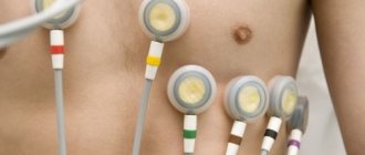
| After treatment, electrodes in an amount of 5 to 10 pieces are glued to the skin. This depends on how many ECG leads the device will record. For examination of rhythm and conduction disorders, 3 or 6 leads can be used. To diagnose ischemic changes, 12 leads are needed. Wires are attached to the electrodes and go to a wearable recording device. The device itself is small in size and fits into a handbag, which can be worn over the shoulder or around the belt, under or over clothing at the patient’s request. There are very small devices, they do not require a handbag, they hang on your neck like a small telephone. |
IMPORTANT!
If you are scheduled for blood pressure monitoring, your clothing should have wide sleeves to fit the blood pressure cuff underneath.
IMPORTANT!
Be sure to wear a thick cotton T-shirt (or tank top) on your body. This will allow the electrodes to be pressed more tightly to the skin, reducing the amount of interference and improving recording quality. The nurse will instruct you on how to behave during monitoring and how to fill out the diary correctly. After this, you will leave the clinic and come back to take the monitor in a day (after a different period of time if multi-day monitoring is carried out).
It doesn't really matter whether the monitoring lasted exactly 24 hours, the miles were a little shorter or longer.
In some cases, the patient can remove the monitor at home and ask relatives (friends) to bring it to the clinic at the appointed time.
How to behave during research?
It is best if you are as active as possible on this day. That is, it is better to choose a working day rather than a weekend. It is also advisable to perform the maximum physical activity possible for you at a given time, unless the doctor prohibits you from doing it. Then the research will be the most informative.
In addition to water procedures, you can perform all your usual activities, including sports, sex, smoking, and drinking alcohol. While you sleep, you can place the monitor next to you or under your pillow, or leave it in your purse slung over your shoulder.
IMPORTANT!
Before installing the monitor, you must take a shower. While you are wearing the monitor, you will not be allowed to swim, shower or take a bath!
IMPORTANT!
Don't forget to take with you reading glasses, a wristwatch (not a mobile phone), reports from previous monitors and ECGs, a certificate with the diagnosis and doctor's prescriptions.
IMPORTANT!
Patients who have a pacemaker or cardioverter-defibrillator must have its passport and a certificate of the latest reprogramming.
the PATIENT DIARY correctly
. Then, when deciphering the monitor, the doctor will be able to compare your complaints, discomfort and your actions (for example, smoking, stress at work, driving) with changes in the ECG and blood pressure numbers. The nurse will instruct you on how to fill it out correctly.
Patient diary
The diary must include:
1. The time of the beginning and end of night sleep and its quality (did you wake up at night; if so, at what time, in connection with what, were there any complaints from the heart).
2. Climbing stairs.
During ECG and blood pressure monitoring, it is recommended to perform three physical activities in the form of climbing stairs, of course, if you do not have a restriction on the motor mode, which is established by the attending doctor. Climbing the stairs must be done three times, distributing them throughout the day, for example, in the afternoon, in the evening and in the morning immediately after climbing. You should climb at a normal pace, without stopping, until any unpleasant sensations appear in the heart area (shortness of breath, palpitations, pain, interruptions, etc.) or physical fatigue. Stop as you always do - don't try to do more work than usual! It is important for us to assess exactly your daily exercise tolerance. If there are no unpleasant sensations, the ascent continues to the end of the stairs and ends there. No need to go up and down several times in a row!
The load should be performed according to the following method:
- Before going up, on the first floor, you should stand and rest for 3 minutes to restore your heart rate (pulse);
- Press the button on the ECG monitor or the “sensation” button on the blood pressure monitor, and then continue climbing at your usual pace;
- At the end of climbing the stairs, press the same button again.
In the diary you should note:
- the number of steps passed (not flights),
- start time of ascent
- its duration (or the end of the rise)
- unpleasant sensations that forced me to stop exercising.
3.
Positional changes on the ECG
The diary indicates 4 positions, in each of which you need to lie down for 1 - 2 minutes - on your back, on your stomach, on your right side, on your left side. This should be done at any convenient time (for example, before going to bed), in a row in all positions noted in the diary. Opposite each position, it is necessary to note the execution time (from_ to_).
4. Actions and sensations during the day
During daily monitoring, the patient should keep a daily diary, which should indicate the actions performed during the examination period. In the column “Other actions during the day,” the actions performed, the time of their implementation and the sensations to which you want to draw the doctor’s attention are noted in detail. It is important to record the state of rest or physical activity (movement on the street and indoors), as well as emotional experiences and stressful situations during the day, the time of meals and medications. If the diary does not record the stress, medication and your sensations, the doctor will not be able to assess their effect on the heart and the study may be useless.
Some features of tachycardia in adults
Tachycardia in women
The above mechanisms for the development of tachycardia are similar in men and women. But, if we talk about the characteristics of the female body, it is worth noting that periods such as menopause, pregnancy, and premenstrual syndrome bring their own characteristics. During these periods, women may experience sinus tachycardia, sometimes quite pronounced, which requires correction of the underlying cause and the prescription of additional medications.
This occurs as a result of an increase in the tone of the sympathetic nervous system, as well as under the direct influence of hormonal levels. During exercise, heart rate increases sharply. In addition, if the patient experiences heavy menstruation, this often leads to the development of chronic anemia, which may be accompanied by sinus tachycardia.