Thrombus in the left atrial appendage
Content
hide
1 Left atrial appendage closure as an alternative to anticoagulants
2 Occlusion of the left atrial appendage
3 Occlusion of the left atrial appendage
4 Prevalence of AF:
5 How the left atrial appendage is associated with stroke in patients with AF
6 How occlusion of the left atrial appendage is performed
7 Preparing for the study
8 Pain relief
9 Monitoring your heart function
10 What is the Amplatzer Cardiac Plug?
11 Who should not have the Amplatzer Cardiac Plug implanted?
12 What happens after the occluder installation procedure 12.1 Will an MRI interfere with or disrupt the operation of the occluder?
12.2 Is the procedure possible for pregnant women and nursing mothers?
13 Possible complications associated with the Amplatzer Cardiac Plug installation procedure
14 How to know which treatment option is right
Left atrial appendage closure as an alternative to anticoagulants
1 Left atrial appendage closure as an alternative to anticoagulants
2 – one of the most important causes of stroke and death
3 Epidemiology of atrial fibrillation Framingham Study, Wolf, 1991%
4 Ok, atrial fibrillation Gon is more frequent in the elderly but is this also a frequent cause of stroke in the elderly?
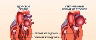
5 Thromboembolism AFibrillation ARRHYTHMIA decreased activity of atrial contraction THROMBUS FORMATION: Stagnation of blood in the atria Formation of blood clots in the appendages of the atria Separation of a thrombus or part of it Thrombus entering the left ventricle Thrombus entering the vascular beds ARTERY EMBOLISM OCCUPATION OF BLOOD FLOW Embolism of a cerebral artery Embolism of a cardiac artery Embolism of the arteries of the lower extremities IN SULT INFARCTION GANGRENE
6 Factors that increase the risk of thromboembolism in patients with atrial fibrillation Age > 65 years Condition after stroke Arterial hypertension Diabetes mellitus Coronary heart disease Poor cardiac function
7 Prevention of thromboembolism Use of anticoagulants drugs to reduce blood clotting: Coumadin and its derivatives Warfarin Markumar
8 Prevention of thromboembolism Anticoagulants reliably counteract thrombus formation, protecting against stroke and heart attack, BUT are often contraindicated and not convenient in everyday life
9 Prevention of thromboembolism Contraindications to the use of anticoagulants Any situation in which the risk of bleeding is higher than the benefit of taking an anticoagulant Pregnancy Tendency to bleeding Bleeding disorders Conditions after or planned operations on the nervous system Conditions after or planned operations on the eyes Cerebral aneurysms Aortic dissection Pericarditis Endocarditis Eclampsia Senile disorders Liver dysfunction Alcoholism Psychosis Gastrointestinal bleeding Spinal cord puncture Bleeding from the genitourinary tract Therapeutic or diagnostic Bleeding from the respiratory tract Cerebrovascular bleeding Procedures with a potential risk of bleeding Poorly controlled hypertension
10 Prevention of thromboembolism Blood thinning is a matter of balance Bleeding Stroke protection
11 Closure of the left atrial appendage 90% of all cardiac thrombi form in the left atrial appendage
12 Closure of the left atrial appendage 90% of all cardiac thrombi form in the left atrial appendage A. Thrombus (T) in the left atrial appendage B. Thrombus is squeezed into the left atrium C. Thrombus in the left atrium D. The thrombus is no longer observed Where is it now? In the brain? In hand? In the intestines?
13 Closure of the left atrial appendage 90% of all cardiac blood clots form in the left atrial appendage and cause blockage of blood vessels in the brain (stroke) A. Computed tomography B. Blockage of blood vessels in the brain (white arrow) C. Computed tomography of the heart with data on the presence of a blood clot in left atrial appendage
14 Closing the left atrial appendage If you close the left atrial appendage, blood clots will have nowhere to accumulate and there will be no need to take coagulants
15 Closure of the left atrial appendage Special devices isolate the appendage cavity from the rest of the atrium and prevent thrombosis
16 Closure of the left atrial appendage Special devices isolate the cavity of the appendage from the rest of the atrium and prevent thrombosis. Duration of the procedure is 40 minutes. Reliability of the procedure 99.5% Duration of hospital stay 2 days
Blood clot in the heart: causes, consequences, treatment and prognosis
To start the process of thrombus formation, several conditions are necessary:
- damaged vessel wall;
- decreased blood flow speed;
- disorders of the rheological properties of blood.
These factors are the trigger for a number of biochemical reactions involved in the formation of a blood clot.
There are three main stages of the process:
- Release of the enzyme thromboplastin from destroyed platelets.
- Thromboplastin, with the help of Ca2+ ions, accelerates the conversion of inactive plasma protein prothrombin into thrombin.
- Under the influence of thrombin, insoluble fibrin is formed from fibrinogen. The threads of the latter form a mesh in which blood cells are retained. The resulting structure tightly covers the damaged area, stopping bleeding. Normally this process takes 5-10 minutes.
After healing of the affected area, the resorption of the formed blood clot is ensured by the fibrinolysis system. An imbalance between the interaction of these two systems determines the risk of the occurrence and development of thrombosis.
Why does a clot form?
Normally, thrombus formation is a physiological process that does not lead to the development of pathologies. And only under the influence of certain factors, the resulting clots do not dissolve, but attach to the vessels, clogging their lumen and disrupting blood flow.
Risk factors include the following diseases of the cardiovascular system:
- cardiac aneurysm;
- atrial fibrillation;
- myocardial infarction;
- cardiac ischemia;
- congenital and acquired valve defects;
- dilated cardiomyopathy;
- chronic heart failure (CHF).
The risk of developing thrombosis increases significantly if the patient has several of the above diseases.
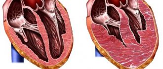
The resulting thrombus in the heart is classified into right- or left-sided, atrial and ventricular (parietal). A special type of clot (spherical) occurs with mitral stenosis.
Complications and their consequences
The most dangerous complication of cardiac thrombosis is separation of the floating part and blockage of blood vessels. When a blood clot is located in the veins of the systemic circulation, right atrium or ventricle, the greatest danger is pulmonary embolism. The severity of the condition depends on the size of the blocked vessel.
When large ones are obstructed, a pulmonary infarction occurs. In this case, patients may feel pain in the chest, difficulty breathing, fever and severe weakness. There may be a drop in blood pressure and an increase in heart rate. The prognosis is unfavorable—in most cases, immediate death occurs.
Blood clots enter the systemic circulation from the left sections, from where they can move in two directions - up and down. If a blood clot breaks off in the heart and moves upward, it eventually ends up in the blood vessels of the brain. As a result, symptoms of ischemic stroke develop.
Thromboembolism of the arteries of the lower extremities, damage to the renal and mesenteric vessels occurs when the thrombus moves downwards. The most difficult course of thrombosis of the mesenteric arteries is the development of clinical peritonitis with subsequent necrosis of the mesentery. Obstruction in the lower extremities has a more favorable outcome due to the developed collateral blood flow in them.
Severance of a blood clot from the left side of the heart can lead to the following consequences:
- thrombosis of the arteries of the brain with the clinical picture of ischemic stroke;
- obstruction of the jugular vein, which is characterized by severe headache, dizziness, palpitations and visual disturbances;
- clinic of acute myocardial infarction (MI) when an embolus enters the coronary arteries;
- renal artery thrombosis is accompanied by severe pain in the lumbar region and difficulty urinating;
- blockage of mesenteric vessels is manifested by peritonitis followed by intestinal necrosis;
- the presence of a blood clot in the arteries of the extremities is accompanied by paleness and blueness of the skin, the disappearance of pulsation in them, and in the absence of timely assistance, gangrene can form.
Each of these complications requires specially selected therapy, the main goal of which is to remove the detached clot and avoid the appearance of new ones. In addition, it is important to remember that detachment of a blood clot, regardless of its primary location, is the most common cause of heart attacks.
Prevention of intracardiac thrombosis
Prevention of the onset and progression of this disease consists of proper nutrition, regular exercise and maintaining normal blood viscosity. Also important in preventing the development of thrombosis is timely and adequate treatment of diseases that contribute to it.
There are special scales that can be used to classify the degree of risk of developing venous or arterial thromboembolism. The latter include:
- patient age over 65 years;
- the presence of malignant neoplasms;
- increased blood clotting;
- pregnancy;
- prolonged bed rest after injury;
- obesity;
- taking hormonal medications (oral contraceptives, steroid therapy for rheumatological pathologies);
- major abdominal operations;
- the presence of concomitant vascular pathologies (atherosclerosis, thrombophlebitis, varicose veins).
In addition, the patient’s general condition is assessed, the presence of signs of heart failure (total or for an individual ventricle) and symptoms from other organs and systems.
Difficulties in diagnosing intracardiac thrombosis arise due to the fact that immobile blood clots do not manifest themselves in any way, which only enhance the characteristic signs of the underlying disease.
Diagnosis and treatment of the patient
After identifying a patient from a high-risk group, it is necessary to conduct a series of studies. The standard electrocardiography (ECG) procedure in this case is not informative. Laboratory markers of increased blood clotting and inhibition of fibrinolysis are not specific, as they are characteristic of many diseases of the cardiovascular system.
To verify the diagnosis you will need:
- Doppler ultrasound – displays the speed and direction of blood flow in the heart;
- scintigraphy - determines the localization of disorders in the coronary vessels and the degree of blood supply to the myocardium;
- MRI – displays the condition of cardiac tissues;
- X-ray of the heart - allows you to diagnose aneurysm, myocardial hypertrophy, dilated cardiomyopathy, as well as the presence of thrombotic plaques;
- X-ray kymogram - allows you to diagnose the location of the thrombus.
Diagnosis of thrombosis requires initiation of treatment. Drugs of choice for long-term drug therapy:
- antiplatelet agents that reduce the degree of platelet aggregation and adhesion. These include Acetylsalicylic acid, Dipyridamole, Clopidogrel;
- anticoagulants, the mechanism of action of which is aimed at inhibiting the processes of activation of blood coagulation factors. The most commonly used are Dabigatran, Rivaroxaban, and Heparin.
For patients with pulmonary embolism, myocardial infarction and ischemic stroke, thrombolytic therapy (Alteplase, Urokinase, Tenectoplase) is indicated, and subsequently antiplatelet and anticoagulant agents are added.
The thrombolysis procedure is contraindicated in the presence of an aortic aneurysm, intestinal bleeding, stroke, or a history of severe skull trauma. Retinal diseases, pregnancy and lactation, high or low blood pressure are relative contraindications.
In addition to side effects, thrombolytic therapy may be accompanied by the following complications:
- reperfusion arrhythmias;
- phenomenon of “stunned myocardium”;
- re-occlusion;
- bleeding;
- arterial hypotension;
- allergic reactions.
It is recommended to stop thrombolytic therapy if its use poses a greater threat to the patient’s life than the disease itself.
Removal of intracardiac clots by surgical method is possible only in specialized departments. The essence of the operation lies in the extraction of thrombotic masses using an endoscope passed into the heart cavity.
Also effective in the case of coronary artery thrombosis will be coronary artery bypass grafting and stenting under X-ray control (photos are constantly displayed on the screen in real time). The essence of the first operation is to bypass the affected area using vascular prostheses, and the second is to install a special frame in the lumen of the vessel to expand it.
It is important to remember that surgical intervention does not eliminate the pathological process itself, but is carried out to restore blood flow or to avoid possible complications in the event of a clot rupture.
The choice of treatment method and recommendations for rehabilitation are individual in each specific situation. It is necessary to take into account all possible risks and contraindications to obtain the most positive result.
conclusions
Today, the prevention and treatment of cardiac thrombosis is an advanced area of cardiology.
The process of thrombus formation itself has two sides: on the one hand, protecting the body from large blood losses, on the other, the occurrence of serious diseases with the risk of death.
Therefore, it is necessary to know what diseases lead to the pathological formation of blood clots, symptoms and possible complications of cardiac thrombosis in order to receive medical help in time and have a chance for a full recovery.
Source: https://cardiograf.com/bolezni/neotlozhnye/tromb-v-serdce.html
Left atrial appendage occlusion
Left atrial appendage occlusion using Amplatzer Cardiac Plug (USA)
The left atrial appendage is a muscular bursa connected by a lumen to the left atrium and is part of the normal anatomy of the heart. However, in most cases, it is the left atrial appendage that is the main source of blood clots and thrombotic complications in patients with atrial fibrillation.
Atrial fibrillation (AF) is a major risk factor for the formation of blood clots (thrombi), which can block blood flow to the brain and lead to cerebral infarction (stroke).
of AF : – in patients over 60 years of age, AF occurs in 4%; – in patients over 80 years of age, AF occurs in 9%.
In more than 90% of cases, thrombi are located in the left atrial appendage and, therefore, occlusion of the left atrial appendage is effective in preventing and developing thrombotic complications such as thromboembolism.
How the left atrial appendage is associated with stroke in patients with AF
With AF, disruptions occur in the conduction system of the heart and irregular electrical impulses occur in the upper parts of the heart (atria), which leads to their trembling and irregular contraction. Irregular heartbeats lead to decreased blood flow, rapid heart rate, difficulty breathing, and shortness of breath. These irregular heartbeats lead to an increased risk of developing blood clots. The left atrial appendage has a long, tubular shape and connects to the left atrium. During AF, blood can pool in the atrial appendage and lead to the formation of blood clots. When the heart rhythm returns to normal, these blood clots can fly out of the appendage into the left atrium and then travel through the blood throughout the body, causing blockage of arteries in the brain and leading to the development of a stroke.
How is left atrial appendage occlusion performed?
The procedure is performed using an endovascular, minimally invasive technique in a cath lab. By puncturing the femoral vein (usually on the right), the x-ray surgeon inserts a thin, flexible and long tube (catheter) into the right side of the heart. Next, puncture of the interatrial septum is performed and special instruments are carried out to the mouth of the left atrial appendage. During the entire procedure, X-ray images, as well as transesophageal echocardiography (TEE-CG) data, are used to monitor the implementation of instruments and their correct placement in the cavities of the heart.
Preparing for the study
Before the procedure, a consultation with a cardiologist and/or a neurologist is necessary, who will tell you in detail about all stages of the study, possible results and complications. A detailed allergy history is also collected to determine if there is an allergy to medications and/or contrast used during the procedure.
Your doctor will tell you which medications you are taking to stop taking on the day of your procedure. The patient should not independently decide to stop taking medications and can do this only after agreement with the cardiologist. It is advisable to avoid taking liquids and food several hours before the procedure.
Anesthesia
The examination will be performed under local anesthesia, but you will be under intravenous anesthesia during the entire procedure. This is due to the need to conduct TEE throughout the entire procedure to fully control the placement of the occluder in the cavity of the left atrial appendage. During the operation, a thin, flexible and long tube (catheter) is inserted into the heart cavity.
Monitoring your heart function
During the entire study, an ECG will be recorded and recorded on the computer hard drive. Electrodes (small metal circles) will be attached to your arms and legs. The electrodes are connected to a computer and record every heartbeat.
What is the Amplatzer Cardiac Plug
The Amplatzer Cardiac Plug is a device specifically designed for non-surgical, low-impact closure of the left atrial appendage.
The device is folded in a thin catheter (
4 mm in diameter) and is delivered folded to the orifice of the left atrial appendage. Next, the occluder is released from the catheter and takes on the shape as shown in the figure.
The occluder is securely fixed to the delivery cable and, if necessary, the x-ray surgeon can repeatedly remove the occluder again into the lumen of the catheter until he is sure that the occluder is securely fixed in the cavity of the ear. Only after this is the occluder disconnected from the delivery device.
Amplatzer Cardiac Plug is manufactured at the plant in Minnesota (USA) from a special alloy Nitinol (nickel-titanium alloy). Nitinol is absolutely not susceptible to corrosion, its strength exceeds both titanium and steel, and also has a special property of “shape memory”, when, when straightened, it acquires its original shape as shown in the figure.
Who should not have the Amplatzer Cardiac Plug
the Amplatzer Cardiac Plug into the atrial appendage if the following conditions exist
– blood clots in the cavities of the heart; – active infectious process; – placement of the occluder will interfere with the functioning of other structures or devices in the heart (for example, a pacemaker lead).
What happens after the occluder installation procedure
Because the occluder procedure is minimally invasive, recovery is likely to be quick and easy. Many patients are discharged from the hospital within the next 48 hours, with subsequent medication recommendations to continue treatment and recovery in an outpatient setting. It is necessary to conduct a control TEE-CG 3 and 6 months after installation of the occluder to monitor the process of endothelialization of the installed device. Endothelialization is the growth of the occluder with connective tissue and, in fact, its ingrowth into the wall of the heart. This is a normal and desirable process. In 99% of cases, complete endothelialization of the occluder occurs within several months. The patient returns to his normal lifestyle within the first month.
Is it possible to travel with an implanted device ? Will there be problems going through the metal detector at airport security?
The metal parts of the Amplatzer Cardiac Plug are very small and will not normally trigger an alarm in an airport metal detector frame. However, for your comfort and peace of mind, you will be given a special card confirming the fact that the occluder has been installed.
Will an MRI interfere with or disrupt the occluder?
Most modern devices do not in any way affect the operation of the occluder, and the presence of an occluder does not affect the operation of the devices. However, it is best to alert staff to the presence of implanted devices before undergoing any medical procedure. Magnetic resonance imaging (MRI) is acceptable and the Amplatzer Cardiac Plug will not affect the performance of an MRI in any way, even at 3 Tesla. It is necessary to inform the staff of the MRI department about the presence of an implant.
Is the procedure possible for pregnant women and nursing mothers?
The risk of exposure of a child to X-ray radiation and the benefits of treatment must be weighed, and the correct and most effective tactics must be adopted. If it is necessary to implant a device during pregnancy, all possible measures will be taken to minimize radiation exposure to the fetus and mother.
There is no evidence of the effect of installing an occluder on the lactation process in nursing mothers.
Amplatzer Cardiac Plug installation procedure
There are some potential risks associated with the placement of an occluder, as well as additional risks associated with the vein puncture procedure itself. It is necessary to consult with a radiologist about the possible risks of implanting the device.
Potential risks include, but are not limited to the following:
– air embolism (an air bubble that can move through the vessels and block the work of some of them); – allergic reactions to contrast; – allergic reaction to anesthetic drugs; – cardiac arrhythmias (the occurrence of irregular heart rhythms); – bleeding; – cardiac arrest; – cardiac tamponade (rupture of the heart muscle); - death; – fever; – hypertensive or hypotensive reactions of the body; – infections; – multiple organ failure; – myocardial infarction (heart attack); – perforation of the heart cavity or vessel; – pericarditis (excess fluid in the pericardial sac); – renal failure/renal dysfunction; – cerebrovascular accidents (temporary or permanent); – arterial thrombosis; – valvular regurgitation or insufficiency.
How to Know Which Treatment Option Is Right
Every person is unique. Your attending physician will help you learn about the possible treatment options available to you and choose the optimal and most effective option, taking into account the entire clinical picture of the disease.
Clinical example of occluder implantation in the left atrial appendage
Causes of formation and separation of a blood clot in the heart
Thrombosis is a concern for many, even relatively healthy people. How often do we hear about the sudden death of a person due to a blood clot in his heart. To better understand the danger of such a disease, let’s look at its signs and the necessary preventive measures.
Irina, 32 years old: “How I got rid of spider veins in 2 weeks + PHOTO”
Read completely…
Mechanism of clot formation
Normally, blood constantly circulating in the body should not clot during a person’s life if the vessel has not been damaged. As a result of changes in the composition of the blood due to various reasons, a violation of its coagulation occurs.
A clot, whose main function is to prevent blood loss, is formed by platelets and fibrin. But when it comes to thrombosis, clots pose a serious danger, clogging blood vessels.
Thrombi are divided into parietal or obturating - those that completely block the lumen of the vessel. The latter threaten the patient's life.
Thrombosis is a concern for many, even relatively healthy people.
There are also so-called emboli - clots that have broken off from the vascular wall and circulate in the bloodstream. They are dangerous because they can cause blockage of any artery. A thrombus in the left ventricle of the heart is especially dangerous. It is this section of the organ that serves as the beginning of blood circulation.
Etiology
This disease is constantly being studied by medical practitioners. Today, the main goal of medicine is to prevent thrombosis.
But for this it is necessary to determine the exact reasons for its occurrence.
It was found that a heart clot is formed due to vascular atherosclerosis or increased blood clotting, and the causes of these pathologies are different. Risk factors include:
- heart valve defects;
- atrial fibrillation;
- myocardial infarction;
- hypertension;
- endocrine diseases;
- inflammatory diseases of the cardiovascular system;
- injuries;
- bacterial infections;
- blood pathologies;
- frequent stress;
- physical inactivity;
- smoking;
- alcohol abuse;
- poor nutrition.
Today, the main goal of medicine is to prevent thrombosis.
In addition, some diseases that are not directly related to heart function may play a role. Often these are acute respiratory infections, such as tonsillitis or influenza. These diseases affect lung function, which causes problems in the functioning of the heart.
Irina, 32 years old: “How I got rid of spider veins in 2 weeks + PHOTO”
Read completely…
It is possible to identify certain groups of people who have been diagnosed with a ventricular thrombus. According to statistics, among patients of cardiologists:
- men over 40 years old;
- women experiencing menopause;
- pregnant women;
- patients during the recovery period after surgical treatment of thrombosis;
- people with alcohol addiction;
- smokers.
This case lists the most common causes of blood clot formation in the heart.
Small blood clots form in the vessels
Symptoms
Unfortunately, signs of a blood clot in the heart become obvious when it has already broken off. Therefore, it is so important to know the symptoms of blockage of blood vessels or even blood thickening. Clinical manifestations will largely depend on the location. Small blood clots form in the vessels. However, the signs are not always noticeable. Sometimes patients experience slight discomfort, but they usually do not pay attention to it.
Recognizing the disease is often very difficult due to the fact that the clinical picture is blurred, too general and may indicate a number of diseases.
The first signs of a blood clot in the heart:
Irina, 32 years old: “How I got rid of spider veins in 2 weeks + PHOTO”
Read completely…
- increased sweating;
- increased heart rate, sometimes weakening;
- tachycardia;
- lowering blood pressure;
- pallor or bluishness of the skin;
- numbness in the left shoulder or entire arm;
- fainting.
Angina of the heart
The most significant sign will be obvious blood thickening, which can only be confirmed by tests. Symptoms of thickening will be: pathological fatigue, cyanosis, headaches, hypertension.
Diagnosis of blood clots
Unfortunately, due to the fact that the disease is practically asymptomatic and rapid, it is rarely diagnosed in time. If the patient has a tendency to increased blood clotting, the doctor may prescribe:
- coagulogram - biochemical analysis. This study determines the rate of formation of a blood clot;
- An echocardiogram is one of the ultrasound methods designed to study the heart. Helps identify functional or morphological changes;
- Dopplerography is another method of cardiac ultrasound. With the help of such a study, the condition of the cavities of the heart can be assessed as accurately as possible.
If a patient suspects thrombosis, the doctor will need to conduct a differential diagnosis with diseases that have similar clinical signs - pulmonary embolism or cardiac myxoma. For this purpose, a number of additional laboratory and instrumental studies are prescribed.
Pulmonary embolism
Treatment
Conservative therapy is ineffective. It can prevent the process of thrombosis, but it will not cope with already formed clots. A blood clot in the heart requires treatment, the essence of which is to remove the clots. The following surgical techniques are used for this:
- stenting – dilation of a vessel by installing a cylinder. Sometimes the clot can be removed with a syringe before this;
- bypass - creating an additional blood flow path that will bypass the affected artery;
- mechanical removal - physical removal of a blood clot from an artery. Practice shows that rehabilitation after such an operation is easy and in a shorter time. However, the disadvantage is the high cost of the procedure.
Typically, surgery is indicated in cases where the lumen of the vessel is 70% blocked by a clot. Prognosis depends on the general condition of the body, the size of the blood clot, and how much time has passed since the first symptoms appeared.
Subsequently, patients are prescribed courses of anticoagulants, thrombolytics or antiplatelet agents to normalize blood viscosity. In addition, at the doctor's discretion, medications are prescribed that improve the outflow of fluid retained by tissues.
A blood clot in the heart is extremely dangerous; if necessary, surgery is performed immediately.
Irina, 32 years old: “How I got rid of spider veins in 2 weeks + PHOTO”
Read completely…
Typically, surgery is indicated in cases where the lumen of the vessel is 70% blocked by a clot.
After the operation, the patient is given strict bed rest for some time. Sometimes it is necessary to wear elastic bandages or compression stockings.
Contraindications to treatment, complications
Therapy with drugs that are usually used to normalize blood clotting has its side effects, like any other treatment. The most dangerous effect of such drugs is bleeding.
They can aggravate the patient’s existing pathologies and also affect the general condition. Bleeding is life-threatening.
Before prescribing a course of treatment, you should take into account all the characteristics of the patient and contraindications to the use of drugs.
Absolute contraindications are:
- the presence of diseases that may cause bleeding (for example, stomach ulcers). Treatment is acceptable if more than 10-14 days have passed since the bleeding;
- recent surgery or biopsy;
- damage to internal organs due to injury;
- risk of aortic dissection;
- thrombocytopenia (if the number of platelets is less than 100 thousand per 1 cm3);
- hemorrhagic diathesis;
- allergy to drug components;
- acute pancreatitis;
- pericarditis;
- blood pressure from 200 to 120 mm Hg. Art.
Bleeding is life threatening
For any of these conditions, the use of drugs that affect blood clotting is unacceptable. It is necessary to cure the pathology before starting treatment, or choose an alternative. There are also relative contraindications, when the use of drugs that affect coagulation is acceptable if the situation requires it. These include:
- pregnancy;
- kidney and/or liver diseases;
- history of brain or spinal cord injury;
- fractures;
- burns;
- infective endocarditis;
- use of thrombolytics APSAC or streptokinase less than 4 months ago.
The inappropriate use of drugs to normalize blood clotting, in particular thrombolytics, is fraught with a number of complications. Among them, reperfusion arrhythmia can be identified as the most common. If this complication is detected in time, treatment is not difficult, however, if possible, such situations should be avoided.
Left atrial appendage occlusion
The left atrial appendage is a muscular bursa connected by a lumen to the left atrium and is part of the normal anatomy of the heart. However, in most cases, it is the left atrial appendage that is the main source of blood clots and thrombotic complications in patients with atrial fibrillation.
Atrial fibrillation (AF) is a major risk factor for the formation of blood clots (thrombi), which can block blood flow to the brain and lead to cerebral infarction (stroke).
Why does a clot form?
Normally, thrombus formation is a physiological process that does not lead to the development of pathologies. And only under the influence of certain factors, the resulting clots do not dissolve, but attach to the vessels, clogging their lumen and disrupting blood flow.
Risk factors include the following diseases of the cardiovascular system:
- cardiac aneurysm;
- atrial fibrillation;
- myocardial infarction;
- cardiac ischemia;
- congenital and acquired valve defects;
- dilated cardiomyopathy;
- chronic heart failure (CHF).
The risk of developing thrombosis increases significantly if the patient has several of the above diseases.
The resulting thrombus in the heart is classified into right- or left-sided, atrial and ventricular (parietal). A special type of clot (spherical) occurs with mitral stenosis.
How the left atrial appendage is associated with stroke in patients with AF
With AF, disruptions occur in the conduction system of the heart and irregular electrical impulses occur in the upper parts of the heart (atria), which leads to their trembling and irregular contraction. Irregular heartbeats lead to decreased blood flow, rapid heart rate, difficulty breathing, and shortness of breath. These irregular heartbeats lead to an increased risk of developing blood clots. The left atrial appendage has a long, tubular shape and connects to the left atrium. During AF, blood can pool in the atrial appendage and lead to the formation of blood clots. When the heart rhythm returns to normal, these blood clots can fly out of the appendage into the left atrium and then travel through the blood throughout the body, causing blockage of arteries in the brain and leading to the development of a stroke.
Features of intracardiac localization
Blood clots differ in structure and appearance. In vessels it is customary to distinguish:
- in the arteries - “white” blood clots formed by platelets, leukocytes, fibrin;
- in the veins - “red”, consisting of the same cells + red blood cells;
- hyaline - formed in the capillary bed, do not contain fibrin, it is replaced by proteins.
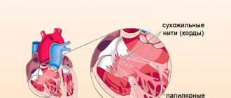
Cardiac thrombi are mixed, have a motley pattern, a characteristic appearance, and therefore are also called layered. According to their structure, they can be distinguished:
You can also read:What is cardiac myxoma
- head (more similar to a “white” blood clot);
- body;
- tail (looks like a “red” blood clot).
The head of the thrombus is attached to the inner wall (endocardial epithelium).
Some properties of intracardiac clots are important in the manifestation of symptoms.
Depending on the ability to move inside the cavity, blood clots can be:
- mobile - move freely in the atria or ventricles, most often formed in the left atrium;
- motionless - one end is attached to the wall, sometimes a “leg” is formed, like in polyps.
In addition, in relation to the cardiac cavity, there are:
- parietal thrombi - more often occurs during an inflammatory process in the endocardium with a transition to the valves, attached to the atrial appendages in case of chronic heart failure, ischemia, to the wall of the aneurysm, leaving a passage for blood flow;
- obstructive - completely block the lumen of the cavity, causing death.
As wall formations grow, they become capable of blocking blood flow
Comparative diagnostics allows us to classify this type as progressive and assume the imminent possibility of complete obturation. Thrombi of the left atrium gradually take on a complete volumetric shape and are called spherical.
With aneurysms, thrombus formation contributes to an increase in volume and expansion. Such a thrombus is called dilatational; it accelerates the thinning of the wall and its rupture.
How is left atrial appendage occlusion performed?
The procedure is performed using an endovascular, minimally invasive technique in a cath lab. By puncturing the femoral vein (usually on the right), the x-ray surgeon inserts a thin, flexible and long tube (catheter) into the right side of the heart. Next, puncture of the interatrial septum is performed and special instruments are carried out to the mouth of the left atrial appendage. During the entire procedure, X-ray images, as well as transesophageal echocardiography (TEE-CG) data, are used to monitor the implementation of instruments and their correct placement in the cavities of the heart.
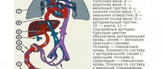
2Draining vessels
The superior and inferior vena cava are the two largest veins in the body, to which blood flows from all organs and tissues. Along with the vena cava, the smallest veins of the heart and the coronary sinus flow into the right atrium.
The smallest veins of the heart open into the atrium along its entire surface. The coronary sinus is a collector of the veins of the heart, which, with the help of an orifice, opens into the atrium cavity between the opening of the inferior vena cava and the atrioventricular opening.
Preparing for the study
Before the procedure, a consultation with a cardiologist and/or a neurologist is necessary, who will tell you in detail about all stages of the study, possible results and complications. A detailed allergy history is also collected to determine if there is an allergy to medications and/or contrast used during the procedure.
Your doctor will tell you which medications you are taking to stop taking on the day of your procedure. The patient should not independently decide to stop taking medications and can do this only after agreement with the cardiologist. It is advisable to avoid taking liquids and food several hours before the procedure.
What is the Amplatzer Cardiac Plug Occluder?
The Amplatzer Cardiac Plug is a device specifically designed for non-surgical, low-impact closure of the left atrial appendage.
The device is folded in a thin catheter (
4 mm in diameter) and is delivered folded to the orifice of the left atrial appendage. Next, the occluder is released from the catheter and takes on the shape as shown in the figure.
The occluder is securely fixed to the delivery cable and, if necessary, the x-ray surgeon can repeatedly remove the occluder again into the lumen of the catheter until he is sure that the occluder is securely fixed in the cavity of the ear. Only after this is the occluder disconnected from the delivery device.
Amplatzer Cardiac Plug is manufactured at the plant in Minnesota (USA) from a special alloy Nitinol (nickel-titanium alloy). Nitinol is absolutely not susceptible to corrosion, its strength exceeds both titanium and steel, and also has a special property of “shape memory”, when, when straightened, it acquires its original shape as shown in the figure.
What happens after the occluder installation procedure
Because the occluder procedure is minimally invasive, recovery is likely to be quick and easy. Many patients are discharged from the hospital within the next 48 hours, with subsequent medication recommendations to continue treatment and recovery in an outpatient setting. It is necessary to conduct a control TEE-CG 3 and 6 months after installation of the occluder to monitor the process of endothelialization of the installed device. Endothelialization is the growth of the occluder with connective tissue and, in fact, its ingrowth into the wall of the heart. This is a normal and desirable process. In 99% of cases, complete endothelialization of the occluder occurs within several months. The patient returns to his normal lifestyle within the first month.
Is it possible to travel with an implanted device? Will there be problems going through the metal detector at airport security?
The metal parts of the Amplatzer Cardiac Plug are very small and will not normally trigger an alarm in an airport metal detector frame. However, for your comfort and peace of mind, you will be given a special card confirming the fact that the occluder has been installed.
Will an MRI interfere with or disrupt the occluder?
Most modern devices do not in any way affect the operation of the occluder, and the presence of an occluder does not affect the operation of the devices. However, it is best to alert staff to the presence of implanted devices before undergoing any medical procedure. Magnetic resonance imaging (MRI) is acceptable and the Amplatzer Cardiac Plug will not affect the performance of an MRI in any way, even at 3 Tesla. It is necessary to inform the staff of the MRI department about the presence of an implant.
Is the procedure possible for pregnant women and nursing mothers?
The risk of exposure of a child to X-ray radiation and the benefits of treatment must be weighed, and the correct and most effective tactics must be adopted. If it is necessary to implant a device during pregnancy, all possible measures will be taken to minimize radiation exposure to the fetus and mother.
There is no evidence of the effect of installing an occluder on the lactation process in nursing mothers.
Possible complications associated with the Amplatzer Cardiac Plug installation procedure
There are some potential risks associated with the placement of an occluder, as well as additional risks associated with the vein puncture procedure itself. It is necessary to consult with a radiologist about the possible risks of implanting the device.
Potential risks include, but are not limited to the following:
- air embolism (an air bubble that can move through the vessels and block the work of some of them);
- allergic reactions to contrast;
- allergic reaction to anesthetic drugs;
- cardiac arrhythmias (the occurrence of irregular heart rhythms);
- bleeding;
- heart failure;
- cardiac tamponade (rupture of the heart muscle);
- death;
- fever;
- hypertensive or hypotensive reactions of the body;
- infections;
- multiple organ failure;
- myocardial infarction (heart attack);
- perforation of the heart cavity or vessel;
- pericarditis (excess fluid in the pericardial sac);
- renal failure/renal dysfunction;
- cerebrovascular accidents (temporary or permanent);
- arterial thrombosis;
- valvular regurgitation or insufficiency.
Appearance of the heart
If you examine the appearance of the heart, you will immediately notice that it is divided into two unequal parts by a certain groove.
The first part - the upper right - is located closer to the base of the heart, to its wide part, facing upward. This is the smaller part.
The second part - the lower left - is much larger in size, facing down and includes the apex of the heart.
The border between these parts of the heart is a groove, which is called the coronary groove. This groove surrounds the surface of the heart in a ring, interrupted only in front at the base, in the place where the aorta and pulmonary artery emerge from the heart.
The structure of the human heart
How does the heart work?
What is coronary circulation
All articles about the heart
In addition to the main or coronary groove, there are two more grooves on the surface of the heart:
- posterior interventricular groove
- anterior interventricular groove
Both the first and second depart from the coronary groove at right angles, rushing to the apex of the heart. These two grooves meet and merge into each other at the apex of the heart.
This is what the human heart looks like: a triangular pyramid, with rounded corners, with three surfaces, an apex and a base, twice braided with grooves: an oblique horizontal or coronal groove and an oblique vertical or two interventricular grooves.
These grooves contain blood vessels that supply the heart.