Almost any disease is accompanied by various symptoms that cause some discomfort, and depending on the intensity of the manifestations, they are forced to seek medical help. However, some pathological conditions are characterized by a completely asymptomatic course. For example, the detection of left ventricular hypertrophy on an ECG may come as a surprise for a young patient and, partly, a natural occurrence in an elderly one, although in both cases there may be no complaints about the heart.
Structure and functions of the heart
Every person knows what the heart is. However, in order to get a complete understanding of how cardiac activity is reflected in the electrocardiogram, it is important to have information about its anatomical structure and functions.
The main structural elements of the heart are 4 chambers (cavities):
- atria (left and right);
- ventricles (left and right).
Since the systemic circulation begins in the left ventricle and, accordingly, ends in the right atrium, the muscular wall of the ventricle has a significant thickness necessary to provide sufficient pressure to push out the entire volume of blood located in the cavity. Unlike the left, the right ventricle has a significantly smaller wall thickness, since the resistance to blood flow in the small circle is several times less and, accordingly, there is no need to provide the same high pressure as in the large circle.
Due to the fact that blood flow must occur in one direction, regulation is carried out using 4 valves that allow or prevent the movement of blood flow:
- mitral - bicuspid valve separating the left atrium and ventricle;
- tricuspid - separates the right atrium and ventricle;
- aortic;
- pulmonary.
The structure of the human heart
What it is?
The human heart includes 4 chambers. One of them is the ventricle on the left side. It begins a large circle of blood circulation. This part is located at the bottom of the heart, on the left. The volume of the left ventricle in newborns ranges from 6 to 10 cubic centimeters, and in an adult - from 130 to 210 cm.
Compared to the right ventricle of the heart, the left one is more elongated and developed in terms of muscle tissue. It has two sections:
- posterior - communicates with the atrium;
- anterior - communicates with the aorta.
Powerful myocardial muscles make the wall of the left ventricle thicker: it reaches 11–14 mm in thickness. On the inside of this part of the organ there are fleshy trabeculae (septa) that form projections and weaves. The task of the left ventricle is to push arterial blood filled with oxygen and nutrients to the organs. Thus, a large circle of blood circulation begins.
Causes
The causes of left ventricular hypertrophy (LVH) have a fairly wide range and depend on the following factors:
- patient's age;
- heredity;
- Lifestyle;
- presence of bad habits;
- state of the vascular system.
At a young age, the main cause of left ventricular myocardial hypertrophy is regular physical activity (sports, hard work), while for older people the main cause is atherosclerotic damage to the vascular system. In all cases, there is an increase in the thickness of the myocardium (heart muscle) designed to compensate for the increased need for oxygen (during sports) or with difficult hemodynamics (with atherosclerosis of the arteries).
Unlike the left, right ventricular hypertrophy (RVH) develops due to disruption of the valve system:
- congenital heart disease (usually found in children);
- mitral valve stenosis;
- lung diseases.
Important! In most cases, at an early stage, diagnosing RVH is difficult, since the size of the right ventricle is 3 times smaller than the left. Therefore, the diagnosis of RVH is made at stages when the size of the right ventricle exceeds the size of the left.
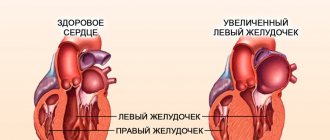
In the figure: on the left is a healthy heart, on the right is hypertrophy of the left ventricular myocardium (an increase in the thickness of the myocardium and a decrease in the ventricular cavity can be seen)
Prevention
The development of myocardial hypertrophy can be prevented by changing lifestyle and treating the underlying disease that loads the myocardium. Key important recommendations:
- Refusal to drink alcoholic beverages.
- To give up smoking.
- Fighting extra pounds. With a weight loss of even 3-5 kg, it is possible to lower blood pressure and reduce the load on the left ventricle.
- Avoid salt and smoked foods, which slow down metabolism and promote fluid retention.
- Exercise regularly. Loads should be measured, daily. Try to walk more, Nordic walking is encouraged. Yoga and fitness classes have a positive effect on the functioning of the cardiovascular system.
Principle of ECG research
An electrocardiogram is a graphical representation of changes in the electrical fields that occur when the heart operates. The contractile activity of the heart is initially regulated by an electrical impulse generated by the sinus node, located at the apex of the right atrium. The impulse quickly spreads through the muscular layer of the atria in the direction from top to bottom and to the left.
Having reached the atrioventricular junction, the impulse significantly reduces the speed and, now, in the opposite direction (from left to right), that is, it first covers the left ventricle, and then the right. All stages of the heart are reflected on the cardiogram in the form of teeth. Normally, an ECG has 5 waves: P, Q, R, S, T.
Table: Correspondence of the designations adopted in the ECG with the duration of the heart phases
| Prong designation | Process | The normal duration is seconds. |
| R | Atrial excitation | 0,16 |
| QRS | Ventricular excitation | 0,08 |
| ST | Complete ventricular excitation | 0,28 |
| straight line | State of rest | 0,23 |
| Total time | 0,75 |
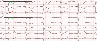
ECG normal
During operation, the heart creates an electric field, registering changes in which, you can obtain information about the processes occurring in it. A complete electrocardiographic examination is carried out by connecting 12 electrodes (leads).
Important! In practice, most often, only 6 leads are used to perform an electrocardiographic study, which often provides insufficient information about the dynamics of changes in heart rhythm.
Diagnostic methods
Most often, a comprehensive examination is performed to detect right ventricular hypertrophy, since the data does not always allow it to be identified. This is due to the fact that the mass of the left ventricle is larger, its potentials overlap the signs of enlargement of the right part. In the most typical cases, we can talk about an increase in the mass of the right ventricle if the following signs are present:
- deviation to the right of the electrical axis up to 100 degrees (on average);
- in three standard leads deep S;
- high R in V1 (occurs with congenital defects) or large amplitudes of S and R (with mitral disease and lung diseases);
- along with the increased amplitude of the ventricular complex, ST shifts and becomes negative.
Chest X-ray
To clarify the diagnosis, patients undergo a chest X-ray
, which provides information about the structure of the lung tissue, the presence of pathological changes in the respiratory tract and the expansion of the cardiac shadow due to myocardial hypertrophy.
Using ultrasound, the thickness of the walls of the heart chambers, the presence of valve defects, reverse blood flow, cardiac output, and myocardial contractility are determined. To detect changes in the liver, an additional ultrasound of the abdominal organs may be prescribed.
Candidates for cardiac surgery are recommended to undergo cardiac probing, ventriculography, angiography, etc. Also, when making a diagnosis and determining treatment tactics for inflammatory lesions of the myocardium and valves, the blood is examined for the level of leukocytes, ESR, C-reactive protein, and a coagulogram is prescribed. To study lung function, spirometry, blood gas analysis, and sputum analysis are performed.
Decoding
Myocardial hypertrophy, regardless of which part of the heart predominates, is always expressed in an increase in the excitation time and amplitude of the segments corresponding to the hypertrophied parts of the heart. For example, hypertrophy of the left atrium on the ECG is expressed by the growth and duration of P, and ventricular hypertrophy - by the QRS complex.
Blockade on the cardiogram - what is it?
In fact, myocardial hypertrophy is the main mechanism that compensates for increasing hemodynamic difficulties and develops when the blood-filled chambers of the heart are overloaded. Thus, the main signs characterizing hypertrophy are:
- an increase in myocardial mass, causing an increase in the electrical impulse, which as a result is expressed by deviations in the form of an increase in the cardiac vector in the direction of the hypertrophied ventricle (on the ECG this looks like an increase in the R wave);
- an increase in the number and length of myocardial muscle fibers, causing an increase in the transit time of the electrical signal along the hypertrophied wall of the ventricle, which on the ECG graph looks like an expansion or deformation of the QRS complex;
- uneven increase in muscle layer fragments disrupts repolarization processes (ST segment and T wave).
Depending on the place where the electrodes are fixed, all the teeth may have different directions. Therefore, when decoding the cardiogram, you should take into account the leads from which the data is read (thoracic region, left or right limb).
Table: Location of electrodes during an ECG study
| № | Lead | Electrode connection point | Electrode polarity |
| 1 | I | right upper limb | — |
| left upper limb | + | ||
| 2 | II | right upper limb | — |
| left lower limb | + | ||
| 3 | III | left upper limb | — |
| left lower limb | + | ||
| 4 | and ν R | right upper limb | + |
| 5 | and ν L | left upper limb | + |
| 6 | and ν F | left lower limb | + |
| 7 | ν1 | 4th intercostal space on the right | |
| 8 | ν2 | 4th intercostal space on the left | |
| 9 | ν3 | between ν1 and ν2 | |
| 10 | ν4 | 5th intercostal space at the level of the center of the clavicle | |
| 11 | ν5 | 5th intercostal space anterior axillary line on the left | |
| 12 | ν6 | 5th intercostal space mid-axillary line on the left |
Pulmonary stenosis
Stenosis is an isolated narrowing of the pulmonary artery. The narrowing of the exit to the pulmonary artery can be located at various levels:
- Subvalvular pulmonary artery stenosis is formed as a result of the proliferation of fibrous and muscle tissue in the infundibular portion of the ventricle.
- Stenosis of the fibrous ring is formed at the junction of the right ventricular myocardium into the pulmonary trunk.
- Isolated valvular stenosis is the most common heart pathology (about 9% of congenital heart defects). With this defect, the pulmonary valve is a diaphragm with a hole with a diameter of 2 to 10 mm. The division into valves is often absent, the commissures are smoothed.
With pulmonary stenosis, the pressure in the right ventricle increases, which increases the load on it. As a result, this leads to enlargement of the right ventricle.
Anatomy
The communication between the left ventricle and the left atrium is carried out through the left atrioventricular orifice; the ventriculus sinister is completely isolated from the right ventricle by the interventricular septum. The aorta emerges from this chamber of the heart, through which blood, enriched with oxygen, passes through smaller arteries to the internal organs.
The left ventricle looks like an inverted cone, and is the only one of all the chambers that takes part in the formation of the apex of the heart. Due to its larger size than the right ventricle, it is believed that the heart is located on the left, although in fact it occupies almost the center of the chest.
The walls of the left ventricle are ten to fifteen millimeters thick, which is several times greater than that of the wall of the right ventricle. This is due to the more developed myocardium on the left side due to higher loads. That is, the higher the amount of work performed, the thicker the heart wall. The left ventricle pumps blood into the systemic circulation, while the right ventricle supplies the pulmonary circulation. That is why, under normal conditions, the latter is less developed, and its thickness is correspondingly smaller.
The atrioventricular communication (orifice) on the left side is closed by the mitral valve, consisting of a posterior and anterior leaflet. In this case, the anterior one is located in close proximity to the interventricular septum, and the posterior one is located outside of it.
Chords extend from both valves - tendon threads that attach the valves to the papillary muscles. Due to these muscles, the valve performs its functions, that is, during systole, blood does not return back to the atrium.
The papillary muscles are attached to special myocardial protrusions (fleshy trabeculae), which are located on the inner plane of the ventricle. Such trabeculae are especially developed in the area of the interventricular septum and the apex of the heart, but their number in the ventricle on the left is less than on the right.
The length and number of chords of the left ventricle are individual.
At the exit of the aorta there is a semilunar valve, which prevents blood from returning from the aorta to the heart.
The nerve impulse to the left ventricular myocardium enters through the Hiss bundle (its left leg). It is worth noting that only to the left ventricle the impulse is sent through two branches - anterior and posterior.
Treatment
Drug treatment
Treatment of an enlarged LV is the treatment of the underlying disease. The earlier the patient’s treatment is started, the more favorable the prognosis and the higher the effectiveness of treatment. Treatment of an enlarged LV cannot occur without eliminating risk factors. Non-drug therapy is an important component in the treatment of cardiac diseases. The range of medications varies depending on the underlying disease. The general principles in the treatment of enlarged LV are to reduce the symptoms of the disease, slow the progression of pathological processes, and prevent complications.
In the treatment of an enlarged left ventricle, the following groups of drugs are used: angiotensin-converting enzyme inhibitors (ACE inhibitors), anigotensin receptor blockers, calcium channel blockers, beta-blockers. Treatment of an enlarged LV should be carried out immediately after diagnosis. The asymptomatic course of an enlarged left ventricle often becomes the reason for low patient adherence to treatment. A careless attitude towards one's health is a direct path to complications that the patient may encounter quite early. The main complications of an enlarged LV can be myocardial infarction, angina pectoris, heart failure, stroke, and sudden cardiac death.
Three sections of the left ventricle
The cavity of this chamber of the heart has an oblong-oval shape. It is longer and narrower than the same cavity on the right.
The entire space of this chamber is divided into three parts or departments:
- entrance department
- day off
- muscular
The inlet region is adjacent to the left atrium and the atrioventricular orifice.
The outlet section is adjacent to the aorta, which emerges from the left ventricle, and to the aortic orifice.