Normal values in adults and newborns
Myocardial mass is increased due to pathological processes leading to its overload:
- Arterial hypertension;
- Valve defects;
- Cardiomyopathy and myocardial dystrophy.
An increase in the mass of muscle tissue also occurs normally - with intense physical training, when intensive sports activities cause the growth of not only skeletal muscles, but also the myocardium, which supplies the organs and tissues of the trainee with oxygen-rich blood.
Athletes, however, risk over time becoming people with myocardial hypertrophy, which under certain conditions can become pathological. When the thickness of the heart muscle becomes greater than the coronary arteries can supply with blood, there is a risk of heart failure. It is this phenomenon that is most often associated with sudden death in well-trained and apparently healthy people.
It is impossible to determine uniform standards for the normal state of the heart muscle for men and women, for adults and children of different ages, for young and elderly patients. The figures below are average values and there may be slight differences in each case.
| Indicators | In adults | In newborns |
| Left ventricular wall thickness in systole | 10 – 16 mm | 4 – 5 mm |
| The same - in diastole | 8 - 11 mm | 3 – 4 mm |
| Right ventricular wall thickness | 3 – 5 mm | |
| Thickness of the interventricular septum in systole | 10 – 15 mm | 4 – 8 mm |
| The same - in diastole | 6 – 11 mm | 3 – 5 mm |
| Aortic diameter | 18 – 35 mm | |
| Left ventricular ejection fraction | More than 50% | 66 – 76% |
| Heart rate | 70 — 75 | 120 — 140 |
| Myocardial mass | 90 – 140 g – for women 130 – 180 g – for men |
The aortic valve in adults should open 1.5 centimeters or more, the opening area of the mitral valve in adults is 4 sq.cm. The volume of exudate (liquid) in the heart sac should not exceed 30 sq. ml.
- ischemic disease;
- myocardial infarction or pre-infarction condition;
- arterial hypertension and hypotension;
- congenital and acquired heart defects;
- heart failure;
- rhythm disturbances;
- rheumatism;
- myocarditis, pericarditis, cardiomyopathy;
- vegetative – vascular dystonia.
Ultrasound examination can detect other disorders or diseases of the heart muscle. In the protocol of diagnostic results, the doctor makes a conclusion, which displays the information received from the ultrasound machine.
These examination results are reviewed by the attending cardiologist and, if any deviations are present, he prescribes treatment measures.
Decoding a heart ultrasound consists of multiple points and abbreviations that are difficult for a person who does not have a special medical education to understand, so we will try to briefly describe the normal indicators obtained by a person who does not have abnormalities or diseases of the cardiovascular system.
Causes
The reasons leading to left ventricular hypertrophy include:
- arterial hypertension;
- various heart defects;
- cardiomyopathy and cardiomegaly.
The mass of the left ventricular myocardium in 90% of patients with arterial hypertension exceeds the norm. Hypertrophy often develops with mitral valve insufficiency or with aortic defects.
The reasons why myocardial mass may exceed the norm are divided into:
- genetic;
- biochemical;
- demographic.
Scientists have found that cardiac hypertrophy can be promoted by the presence or absence of several fragments in human DNA. Among the biochemical factors leading to myocardial hypertrophy, an excess of norepinephrine and angiotensin can be identified. Demographic factors for the development of cardiac hypertrophy include race, age, gender, physical activity, a tendency to obesity and alcoholism, and the body's sensitivity to salt. For example, men have higher myocardial mass than normal more often than women. In addition, the number of people with a hypertrophied heart increases with age.
Left ventricular myocardial mass index: norms and calculation examples
The study of the physical parameters of the myocardium is very important in the diagnosis and further treatment of patients suffering from diseases of the cardiovascular system. Cardiac muscle hypertrophy is a dangerous syndrome that can lead to dangerous complications and death. Therefore, this problem is relevant at the present time and requires careful consideration.
The myocardium is the muscular layer of the heart, which consists of mononuclear cells that have a special transverse arrangement. This ensures extreme muscle strength and the ability to distribute work evenly throughout the heart. The relative position of cells according to the type of intercalated discs determines the unusual properties of the myocardium. These include excitability, contractility, conduction, relaxation and automaticity.
It is possible to assess whether the heart is healthy using additional instrumental examinations. Normal indicators based on the results of echocardiography of the ventricular myocardium (one of the key methods for diagnosing the pathology of blood ejection) are as follows:
- left ventricle (LV): myocardial mass - 135-182 g, 95-141 g; mass index (LVMI) - 71-94 g/m2, 71-84 g/m2 in men and women, respectively;
- right ventricle (RV): wall thickness - 3 mm; size index - 0.75-1.25 cm/m2; the diastole value at rest is 0.8–2.0 cm.
The left ventricle takes on a greater functional load than any other part of the heart and, accordingly, is more often susceptible to pathological changes. Therefore, we will consider its parameters in more detail.
High blood pressure not only worsens well-being, but also provokes the onset of pathological processes that affect target organs, including the heart: with arterial hypertension, hypertrophy of the left ventricular myocardium occurs. This is explained by an increase in collagen content in the myocardium and its fibrosis.
However, myocardial hypertrophy is not a death sentence: people with a hypertrophied heart can live for decades. You just need to monitor your blood pressure and regularly undergo ultrasound of the heart to monitor hypertrophy over time.
Formula
Myocardial mass can be calculated according to the formula:
| 0.8 x (1.04 x (MZhP+KDR=3SLZh)x3 – KDRx3)+0.6 |
All measurements are taken in centimeters. Each abbreviation means:
| Abbreviation meaning | |
| MZhP | thickness of the muscular interventricular septum of the heart measured during echocardiography |
| CDR | left ventricular size during relaxation |
| SLJ | left ventricular wall thickness at rest |
The myocardial index can be measured using one of the formulas:
| MI = M/H2.7 |
| MI = M/S |
The meanings of the accepted abbreviations mean:
| Meaning of abbreviations | |
| MI | indicator that determines the myocardial index |
| M | the mass of the left ventricular myocardium, determined by the above formula |
| N | patient's height measured in meters |
| S | patient's body surface area |
In measurements, the area of the subject is used, because it is a more accurate value than body weight. This is due to limiting dependence on excess fat tissue. The surface area is calculated using a fixed formula, where the parameters change according to the patient’s age.
The myocardial index is most indicative in pediatrics. This is due to the fact that adult height remains unchanged when calculated over several survey years. The child’s growth is constantly changing, thanks to which pathologies in cardiac parameters can be accurately tracked.
In men, the average mass of the left ventricular myocardium (normal) is 135 g, and in women 95 g. At the same time, the upper limit, the excess of which is considered to be exceeding the norm for men is 183 g, and for women – 141 g.
The average value of the left ventricular myocardial mass index is 71 g/m2 in men and 62 g/m2 in women. The upper limit of this index is 94 and 89 g/m2, respectively.
The causes and mechanism of changes in left ventricular mass in various diseases are still poorly understood.
Myocardial hypertrophy is a fundamental mechanism of adaptation of the heart muscle to increased stress that occurs both during cardiovascular diseases and during physical exercise. The heart muscle, like any muscle, thickens when the load on it is increased.
The blood vessels supplying this organ cannot keep up with its growth, which is why heart tissue starves and various diseases develop. With myocardial hypertrophy, problems also arise in the conduction system of the heart, as a result of which zones of abnormal activity appear in it and arrhythmias appear.
The best method for studying the anatomy of the heart and its function is echocardiography. This method is superior to ECG in sensitivity to cardiac hypertrophy. Myocardial hypertrophy can also be detected using cardiac ultrasound.
Deviations from the norm and principles for interpreting results
As a result of echocardiography, the following pathologies of the development and functioning of the heart muscle and accompanying diseases can be detected:
- heart failure;
- slowing, accelerating or interrupting heart rhythm (tachycardia, bradycardia);
- pre-infarction condition, post-infarction;
- arterial hypertension;
- vegetative-vascular dystonia;
- inflammatory diseases: cardiac myocarditis, endocarditis, exudative or constrictive pericarditis;
- cardiomyopathy;
- signs of angina pectoris;
- heart defects.
The examination protocol is filled out by a specialist performing cardiac ultrasound. The parameters of the functioning of the heart muscle in this document are indicated in two values - the norm and the indicators of the subject. The protocol may contain abbreviations that are incomprehensible to the patient:
- LVMM – left ventricular mass;
- LVMI – mass index;
- EDD – end diastolic size;
- DO – long axis;
- KO – short axis;
- LA – left atrium;
- RA – right atrium;
- EF – ejection fraction;
- MK – mitral valve;
- AK – aortic valve;
- DM – myocardial movement;
- DR – diastolic size;
- SV – stroke volume (the amount of blood that is ejected by the left ventricle in one contraction;
- ТМВПд – thickness of the myocardium of the interventricular septum in the diastole phase;
- TMMVPs – the same, in the systole phase.
The determination of myocardial mass is calculated using numbers obtained during echocardiography. For accuracy and objectivity of measurement assessments, they are carried out in a combination of modes, comparing two- and three-dimensional images. The data is supplemented by the results of Doppler studies and indicators of ultrasound scanners, which are capable of displaying a projection of the heart in natural size on the monitor screen.
Calculation of myocardial mass can be done in several ways. Preference is given to two formulas ASE and PC, which use the following indicators:
- the thickness of the muscular septum separating the cardiac ventricles;
- directly the thickness of the posterior wall of the left chamber in a calm state, until the moment of its contraction;
- full size of the relaxed left ventricle.
The interpretation of values obtained from echocardiography should be considered by an experienced specialist in functional diagnostics. When evaluating the results, he will note that the ASE formula represents the left ventricle along with the endocardium (the cardiac membrane lining the chambers). This may cause distortion in the measurement of its thickness.
Formula
The mass of the left ventricular myocardium (calculation) is determined by the following formula:
- IVS – value (in cm) equal to the thickness of the interventricular septum in diastole;
- EDR is a value equal to the end-diastolic size of the left ventricle;
- LVSP is a value (in cm) equal to the thickness of the posterior wall of the left ventricle in diastole.
MI – myocardial mass index is determined by the formula:
MI=M/H2.7 or MI=M/S, where
- M – mass of the left ventricular myocardium (in g);
- H – height (in m);
- S – body surface area (in m2).
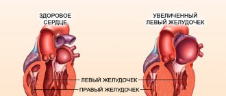
Hypertrophy
The thickness of the left ventricular myocardium is normally measured when it relaxes and is 1.1 centimeters. This indicator does not always remain this way. If it is elevated, then myocardial hypertrophy is noted on the left. This indicates excessive work of the heart muscle and can be of two types:
- Physiological (growth of muscle mass under the influence of intense training);
- pathological (enlargement of the heart muscle as a result of the development of the disease).
If the thickness of the wall of the left ventricle is from 1.2 to 1.4 centimeters, slight hypertrophy is recorded. This condition does not yet indicate pathology and can be detected during a medical examination of athletes. With intense training, skeletal muscles and at the same time myocardial muscles build up.
When the heart muscle changes up to two centimeters, states of moderate and significant hypertrophy are considered. They are characterized by the appearance of shortness of breath, a feeling of lack of air, pain in the heart area, disturbance of its rhythm and increased fatigue. If this change in the myocardium is detected in a timely manner, it can be corrected with medication.
This stage of myocardial pathology is life-threatening due to its complications. The treatment method is selected according to the individual situation.
How is ultrasound examination performed?
No special preparation is required for cardiac ultrasound. All that is necessary from the patient to obtain the most objective results: calm down and breathe evenly. Immediately before the examination, you should not overexert yourself physically, drink caffeine-containing drinks, or take medications (sedatives, etc.).
You can find out in detail how a heart ultrasound is performed on the Internet. On the websites of many medical centers, along with a description of the procedure itself and the price of cardiac ultrasound, visual materials are presented in the form of photographs and videos of cardiac ultrasound.
Before the heart examination, the patient undresses to the waist and lies down on the couch. All jewelry from the exposed area (chains, etc.) must be removed. The procedure is non-invasive. First, the subject lies on his back, then on his right side. The chest area is treated with gel. Afterwards, moving the sensor along the surface of the skin in the area of projection of the organ, the heart is examined. The whole procedure takes no more than 20 minutes. The monitor displays the heart and adjacent structures, which is made possible thanks to the property of ultrasound. It is reflected from the fabric, and, depending on their density, gives the corresponding picture.
Heart ultrasound allows you to diagnose pathologies that have not yet begun to manifest symptoms.
7 minutes Author: Irina Bredikhina 76254
A modern method of hardware diagnostics - echocardiography or ultrasound of the heart, is based on the use of oscillations of high-frequency sound waves. Through ultrasound examination, a medical specialist determines the cause of functional failures in the organ, identifies changes in the anatomical structure and histological structure of tissues, and determines abnormalities in the vessels and valves of the heart.
The prerogative aspects of ultrasound diagnostics are:
- absence of damage to the skin and penetration into the patient’s body (non-invasive);
- harmlessness. Ultrasonic waves are safe for health;
- information content. Clear visualization of the heart allows you to accurately determine the pathology;
- no contraindications to the use of the method;
- the ability to observe dynamic processes;
- relatively low cost of research;
- insignificant time costs for the procedure.
Ultrasound of the heart is performed by a doctor from the radiology department based on the direction and recommendation of a cardiologist. If you wish, you can go through the procedure yourself.
Treatment
The method of treating left ventricular myocardial hypertrophy depends on the cause that caused the development of this pathology. If necessary, surgery may be prescribed.
Heart surgery for myocardial hypertrophy can be aimed at eliminating ischemia - coronary artery stenting and angioplasty. In case of myocardial hypertrophy due to heart disease, valve replacement or dissection of adhesions is performed if necessary.
Slowing down the processes of hypertrophy (if it is caused by a sedentary lifestyle) in some cases can be achieved by using moderate physical activity, such as swimming or running. The cause of left ventricular myocardial hypertrophy may be obesity: normalizing weight while switching to a balanced diet will reduce the load on the heart. If hypertrophy is caused by increased loads (for example, during professional sports), then you need to gradually reduce them to an acceptable level.
Medicines prescribed by doctors for left ventricular hypertrophy are aimed at improving myocardial nutrition and normalizing heart rhythm. When treating myocardial hypertrophy, you should stop smoking (nicotine reduces the supply of oxygen to the heart) and drinking alcohol (many medications used for myocardial hypertrophy are not compatible with alcohol).
What allows examination (EchoCG)
Ultrasound of the heart allows the doctor to determine many parameters, norms and abnormalities in the functioning of the cardiovascular system, assess the size of the heart, the volume of the heart cavities, the thickness of the walls, the frequency of strokes, the presence or absence of blood clots and scars.
This examination also shows the condition of the myocardium, pericardium, large vessels, the mitral valve, the size and thickness of the walls of the ventricles, determines the condition of the valve structures and other parameters of the heart muscle.
After the examination (Echo CG), the doctor records the results of the examination in a special protocol, the decoding of which allows one to detect cardiac diseases, deviations from the norm, anomalies, pathologies, also make a diagnosis and prescribe appropriate treatment.
Ultrasound of the heart
Have you been struggling with HYPERTENSION for many years without success?
Head of the Institute: “You will be amazed at how easy it is to cure hypertension by taking it every day.
Ultrasound of the heart is one of the most informative diagnostic methods, which makes it possible to “see” the anatomical features of the heart muscle, pathology of the valve apparatus, changes in nearby structures: muscles, blood vessels. By visualizing the heart using ultrasound, the doctor also evaluates the functional parameters.
When is it necessary to do an ultrasound of the heart?
The clinical picture of many diseases (gastrointestinal tract, nervous system, respiratory organs) is similar to that of cardiac pathologies. To correctly make a diagnosis, it is necessary to do an ultrasound of the heart when the following symptoms occur:
Our readers successfully use ReCardio to treat hypertension. Seeing how popular this product is, we decided to bring it to your attention. Read more here...
- nausea accompanied by surges in blood pressure;
- persistent headaches; • dizziness up to loss of consciousness;
- weakness;
- persistent cough;
- dyspnea;
- swelling (legs, torso);
- cardiac arrhythmias;
- palpitations or a feeling of freezing of the heart muscle;
- pain of different localization: in the upper half of the abdomen, in the right hypochondrium, in the chest, under the shoulder blade on the left, behind the sternum;
- enlarged liver;
- cold extremities;
- pale, bluish-tinged skin;
- hyperthermia due to shortness of breath, chest pain and cyanosis, as well as the appearance of these symptoms after drinking alcohol;
- Noises are heard during auscultation.
It is this study that allows you to confirm or exclude heart damage.
Indications
There are a number of diseases in which the heart “suffers”. These include:
- scleroderma;
- angina pectoris;
- rheumatism;
- myocardial dystrophy;
- congenital anomalies and acquired defects;
- systemic pathologies (lupus erythematosus, etc.);
- history of myocardial infarction;
- arrhythmias;
- vascular aneurysm;
- tumor formations;
- arterial hypertension (including hypertension);
- heart murmurs of unknown etiology.
In the presence of these pathologies, ultrasound examination makes it possible to promptly notice the appearance of any deviations (both anatomical and functional) and take adequate measures.
Ultrasound of the heart is performed in cases where it is necessary to establish the cause of changes in the ECG, the type of heart failure, as well as to assess the functional state of the organ in athletes and people who have undergone heart surgery.
The procedure is safe and can be performed on patients of any age. No referral required. If the doctor recommends it, where to do an ultrasound of the heart, the patient himself must decide, based on his financial capabilities. The cost of a cardiac ultrasound varies from 1,200 to 4,500 rubles (depending on the level of the medical institution, the qualifications of the specialist and the scope of the required examination).
When should you do an ultrasound for your child?
An ultrasound of the child’s heart should be done when the following disorders occur:
- causeless loss of consciousness;
- deviations in the cardiogram;
- the presence of a heart murmur;
- frequent colds;
- hereditary burden (close relatives had cardiac pathologies);
- baby has difficulty sucking a bottle (or breast);
- the child talks about unpleasant and painful sensations in the chest area;
- in the baby (even at rest), the color of the skin around the mouth, as well as on the arms and legs, changes;
- With little physical activity, the child sweats a lot and gets tired quickly.
Stages and symptoms
In the process of increasing myocardial mass, three stages are distinguished:
- compensation period;
- subcompensation period;
- period of decompensation.
Symptoms of left ventricular hypertrophy begin to manifest themselves noticeably only at the stage of decompensation. When decompensated, the patient experiences shortness of breath, fatigue, palpitations, drowsiness and other symptoms of heart failure. Specific signs of myocardial hypertrophy include a dry cough and facial swelling that appears during the day or in the evening.
When should it be performed (Echo CG)
- periodic or frequent pain in the heart;
- rhythm disturbances: arrhythmia, tachycardia;
- dyspnea;
- increased blood pressure;
- signs of heart failure;
- previous myocardial infarction;
- if there is a history of heart disease;
You can undergo this examination not only with the direction of a cardiologist, but also with other doctors: endocrinologist, gynecologist, neurologist, pulmonologist.