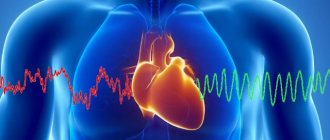
A decrease in myocardial contractility in coronary artery disease is accompanied by a significant increase in the risk of complications and death, so identifying a reversible impairment of contractility is very important.
Viable myocardium is myocardium whose contractility is reduced at rest but improves after revascularization.
1. Chronic ischemia leads to cellular changes, which ultimately result in irreversible disruption of the functioning of cardiomyocytes. 2. According to myocardial biopsy, chronic ischemia leads to fibrosis and a decrease in the number of contractile elements. It has been shown that if connective tissue constitutes more than 35% of the myocardium, restoration of its contractility is unlikely.
Stunning of the myocardium is a transient decrease in its contractility that occurs as a result of a short-term cessation of coronary blood flow. Most often this occurs in acute coronary syndrome.
Dormant myocardium is myocardium whose contractility is reduced due to metabolic disturbances resulting from adaptation to chronic or recurrent ischemia.
Treatment
Revascularization of viable myocardium has been shown to improve survival and quality of life. Along with surgical methods, drug treatment is being improved, so it is very important to select exactly those patients who need myocardial revascularization.
- Thrombolysis and emergency coronary angioplasty are used in acute coronary artery occlusion to restore coronary blood flow and limit the area of damage.
- Myocardial revascularization in the form of coronary artery bypass grafting or coronary angioplasty can improve local and global contractility of the left ventricle. In the presence of viable myocardium, myocardial revascularization is more effective than medical treatment. In contrast, in the absence of viable myocardium, myocardial revascularization has no advantage over medical treatment.
- Since revascularization does not benefit everyone, it is very important to select those patients who really need it. This avoids the dangers and costs associated with unnecessary interventions.
Epidemiology and classification
The disease is registered in both childhood and adulthood. The hereditary origin of the anomaly is confirmed in 18–20% of adults and in 40–50% of children, respectively.
The prevalence in the latter case is higher - about 1.3% of the total population of small patients; in adults, the incidence of the disease is lower - 0.014%. However, it is important to understand that the statistics shown should be considered relative, so the true numbers may be more meaningful.
Important. Non-compact myocardium of the left ventricle is diagnosed more often in males - 55-80% of all registered cases.
Pathology is formed during embryonic development, during the formation of myocardial layers in the fetus. The main sign of the anomaly is the development of deep trabeculae in the LV and interventricular septum, which negatively affects the systolic capacity of the cardiac chamber. In some patients, the painful process may also affect the tissues of the right side of the heart.
The anomaly can be either sporadic or familial. The latter is detected much more often, so the disease is classified as one that has a hereditary predisposition.
At the same time, a number of experts note that the pathology can also be considered a nasal form, since this may be a secondary consequence of other cardiac diseases. Currently, this issue remains controversial.
Methods for identifying viable myocardium
Figure: Isotope methods for detecting viable myocardium
Top row
– scintigraphy with thallium-201 at rest. Initially, an accumulation defect is noted, which disappears during the redistribution phase if the myocardium is viable, and remains if the myocardium is not viable.
Middle row
– scintigraphy with thallium-201, protocol with loading and repeated administration of the drug. During loading, an accumulation defect is noted; during the redistribution phase it is not filled, which may indicate its non-viability. However, upon repeated administration of the drug, the defect disappears, which means that the myocardium is still viable.
Bottom row
– positron emission tomography with the study of perfusion and metabolism. Decreased perfusion with preserved metabolism (as indicated by 18F-fluorodeoxyglucose uptake) indicates myocardial viability
18F-FDG - 18F-fluorodeoxyglucose
PET – positron emission tomography
RFP – radiopharmaceutical drug
Assessment of myocardial viability is carried out in patients with coronary artery disease with impaired contractility of the left ventricle at rest, if revascularization is possible in the future. Coronary angiography provides insight into coronary artery disease and whether revascularization is possible, but does not predict possible recovery of contractility after revascularization. Methods such as:
- single photon emission tomography
- positron emission tomography with metabolically active radiopharmaceuticals
- dobutamine stress echocardiography
- MRI with late contrast
All these methods are based on different properties of viable myocardium. When choosing a particular method, they usually proceed from the capabilities and traditions of the medical institution, although it would be better to proceed from the characteristics of the patient.
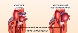
Signs of left ventricular hypertrophy
Symptoms of cardiomyopathy are not always obvious, and people are often unaware that there is a problem. If the fetus does not develop properly during pregnancy, there may be a congenital defect and hypertrophy of the left side of the heart.
Such cases must be observed from birth and complications should not be allowed. But if there are periodic interruptions in the heart's function and a person feels any of these signs, the walls of the ventricle may be abnormal.
The symptoms of this problem are:
- labored breathing;
- weakness, fatigue;
- chest pain;
- low heart rate;
- swelling of the face in the afternoon;
- disturbed sleep: insomnia or excessive sleepiness;
- headache.
Single photon emission tomography (SPECT)
This is the most commonly used method for detecting viable myocardium in the United States. It is based on the fact that the radiopharmaceuticals thallium-201 (201Tl) and technetium-99m (99mTc) are taken up only by living cardiomyocytes. Thallium has a long half-life and is produced in a cyclotron, while technetium has a short half-life and is supplied in the form of generators. Because single photon emission tomography with exercise or dipyridamole is also used to detect ischemia, this method is very convenient for large clinical centers. Synchronization with an ECG allows you to obtain information about the local contractility of the left ventricle, which is also very important when assessing vitality.
Thallium
Its properties are very close to potassium. It is actively captured by Na+,K+-ATPase and accumulates in cardiac myocytes. This allows one to distinguish viable myocardium from scar tissue. The half-life of thallium-201 is long (73 hours), so only small doses (75-150 MBq) can be administered. It produces X-rays with energies of 135 and 167 keV. The uptake of thallium-201 by the myocardium is directly proportional to blood flow, and this relationship persists during exercise.
Redistribution
After the initial accumulation of thallium in the myocardium, its redistribution begins between the intracellular and intravascular spaces. In this case, the initial accumulation defects gradually decrease. It is believed that this is due to the slow accumulation of the isotope in poorly supplied areas of the myocardium, along with its leaching from the well-supplied myocardium. Based on redistribution, several methods have been developed to detect viable myocardium using scintigraphy.
Protocols without load
Pictures are taken 30-60 minutes after administration of the radiopharmaceutical drug, and then after 4 hours. Storage defects that improve after 4 hours are considered to indicate viable myocardium. This protocol does not detect ischemia; in addition, it is less sensitive to viable myocardium than other thallium-201 and 18F-fluorodesoglucose positron emission tomography protocols.
Protocols with load
The radiopharmaceutical is administered after a pharmacological or physiological load, images are taken immediately and after 4 hours. Storage defects that do not improve after 4 hours indicate a scar, and those that are noted with exercise but disappear with rest indicate ischemic but viable myocardium. This protocol can detect ischemia and viable myocardium, but the sensitivity of the method is low because many storage defects that persist beyond 4 hours actually represent viable myocardium. Repeat imaging after 24 hours increases sensitivity by detecting “late redistribution” in viable myocardium, but reduces specificity.
Protocols with loading and repeated administration of the drug
These protocols involve repeated administration of thallium-201 at a dose of 37 MBq. This increases the sensitivity of the study. The drug is re-administered either after obtaining images in the redistribution phase (in this case, repeat images are taken immediately), or several hours after obtaining syncope at the height of the load (in this case, repeat images are taken after 18-24 hours). The sensitivity in both cases is approximately the same. With cicatricial changes, accumulation defects are noted during exercise and persist after repeated administration of the drug and redistribution. Viable myocardium is characterized by accumulation defects during exercise, which decrease after repeated administration of the drug.
Preparations based on technetium-99m
Preparations based on technetium-99m are absorbed by cardiomyocytes if the mitochondria are functioning, the integrity of the cell membrane is not compromised, and the cell’s energy metabolism is preserved.
The half-life of technetium-99m is shorter than that of thallium-201, being 6 hours. Thanks to this, it can be administered in large doses (370-410 MBq). It emits gamma radiation with an energy of 140 keV. The most commonly used are technetium-99m-isonitrile, technetium-99m-tetrofosmin, technetium-99m-teboroxime, technetium-99m-furifosmin and N-ethoxy-N-ethyldithiocarbamatonitrido-technetium-99m. The redistribution of drugs based on technetium 99 is much less pronounced than that of thallium-201, which reduces their value in assessing myocardial viability.
Quantification
Quantitative assessment of single-photon emission tomography results is more accurate than qualitative assessment. Quantitative assessment of perfusion not only provides a more accurate prediction of the recovery of myocardial contractility, but also better assesses the risk of cardiovascular complications.
Diagnostic accuracy
Single photon emission tomography with thallium-201 has been compared in a number of studies with positron emission tomography with 18F-fluorodesoglucose. Sensitivity was highest when quantitative analysis was performed and when repeat dosing protocols were used. Semi-quantitative analysis using loading and repeated administration protocols gives good results that compare well with positron emission tomography data. When comparing the results of single-photon emission tomography with the results of revascularization, less encouraging data were obtained. The sensitivity and specificity of protocols without loading were 90 and 54%, respectively, and protocols with loading and repeated administration of the drug were 86 and 47%. In myocardial segments where drug accumulation after redistribution is less than 60% of the average level, restoration of contractility after revascularization is extremely unlikely. Absorption of radiation by surrounding tissues may complicate the interpretation of thallium-201 single-photon emission tomography results. In addition, methods for quantitative assessment of its results have not yet been standardized. In a quantitative analysis, technetium-99m-isonitrile single-photon emission tomography was more informative than thallium-201 studies in terms of contractility recovery after revascularization. Other drugs based on technetium-99m (tetrofosmin, teboroxime, furifosmin and N-ethoxy-N-ethyldithiocarbamatonitrido-technetium-99m) are almost never used to assess viability; N-ethoxy-N-ethyldithiocarbamatonitrido-technetium-99m is redistributed like thallium-201 and may be used in the future. Technetium-99m-isonitrile is little used to assess myocardial viability due to its high cost.
Positron emission tomography
using metabolically active radiopharmaceuticals, particularly 18F-fluorodesoglucose, is considered the reference method for detecting viable myocardium. Positron emission tomography uses isotopes that emit positrons. When a positron collides with an electron, two high-energy photons (511 keV) are produced, flying in strictly opposite directions. Simultaneous registration of two photons makes it possible to establish the line on which the isotope is located. Positron emission tomography has higher spatial and temporal resolution than single-photon emission tomography. It allows quantitative assessment of metabolic activity and regional blood flow. 1. Radiopharmaceuticals used in positron emission tomography are produced in a cyclotron and have a fairly short half-life. Because of this, and the high cost of the positron emission tomography camera, it is used only in specialized centers. 2. Unlike single-photon emission tomography, positron emission tomography uses different radiopharmaceuticals to study perfusion and assess myocardial viability. 3. 15O-water, 13N-ammonia and rubidium-82 (82Rb) are commonly used to assess perfusion.
a.15O-water diffuses freely into tissues and is used to quantify blood flow. Blood flow less than 0.25 ml/g/min indicates scarring. Intermediate values of blood flow intensity are a very unreliable sign of myocardial viability. Based on positron emission tomography with 15O-water, a perfusion index has been developed, which allows a more accurate assessment of myocardial viability, but due to technical complexity it is calculated only in a few centers. b. Rubidium-82 is produced using generators. It is an analogue of potassium, it is actively taken up by cells and therefore accumulates in viable myocardium and is washed out of scar tissue. V. 13N-ammonia is produced in a cyclotron. It is also captured only by living cardiomyocytes. This drug is most widely used in positron emission tomography; it allows one to assess both perfusion and myocardial viability.
Metabolically active radiopharmaceuticals -
18F-fluorodeoglucose (18F-2-deoxyglucose), 14C-acetic acid and 14C-palmitic acid. 18F-fluorodesoglucose is produced in a cyclotron. The greatest clinical experience has been accumulated with this drug. Its relatively long half-life makes it easier to deliver to clinics. 18F-fluorodesoglucose is taken up by cells and then phosphorylated, which prevents its exit from the cell and leads to accumulation in the myocardium. In normal myocardium, energy needs are met mainly by free fatty acids, but during ischemia, glucose becomes the main source of energy. In diabetes mellitus, the uptake of glucose by cells is reduced, so in 10% of patients with diabetes mellitus, the interpretation of positron emission tomography with 18F-fluorodesoglucose is difficult. When studying perfusion and metabolism simultaneously, three results are possible.
1. Normal perfusion implies viability and no additional metabolic studies are required. 2. A decrease in perfusion with preserved metabolism (as indicated by the uptake of 18F-fluorodesoglucose) indicates myocardial viability. 3. Decreased perfusion and metabolic disturbances (18F-fluorodesoglucose uptake is reduced) indicate scarring.
Diagnostic value
18F-fluorodesoglucose positron emission tomography is the most reliable method for assessing myocardial viability. It has higher resolution than single-photon emission tomography; in addition, the 18F-fluorodesoglucose study is the only method for directly assessing metabolism. Positron emission tomography with 18F-fluorodesoglucose can identify those segments whose contractility will improve after revascularization; its sensitivity and specificity in this regard are 71-100 and 38-91%, respectively. The presence of viable myocardium as determined by positron emission tomography indicates low perioperative risk, improved exercise capacity after myocardial revascularization, and an improved prognosis with revascularization compared with medical treatment.
Types of perfusion
Depending on what it is needed for, perfusion is as follows:
- Complete perfusion (temporarily replaces the pumping function of the myocardium and the gas exchange function of the lungs) is a method in which all blood circulation is performed artificially in order to maintain gas exchange, metabolism, thermoregulation, and also supply the organs with the nutrients they need.
- Partial perfusion (partially replaces the functions performed by the myocardium) is an auxiliary method that helps saturate other organs with oxygen, and is also aimed at maintaining or correcting metabolic processes in the body, as well as detoxification.
- Regional perfusion is a method in which drugs are delivered to relatively isolated organs from the general bloodstream, such as the arms or legs, in order to concentrate the drugs at the site of the disease.
- Perfusion of isolated tissues and organs is a technique that is widely used in organ transplantation.
The first two types of perfusion are used by doctors during heart surgery.
Dobutamine stress echocardiography
Dobutamine stress echocardiography is a reliable way to predict improvement in myocardial contractility after revascularization.
Protocol
Low doses of dobutamine, up to 2.5 mcg/kg/min, are used to assess viability. Every 3 min. the dose is increased. If there are no indications to terminate the study (complaints, achievement of the target heart rate), the dose of dobutamine is adjusted to 20 mcg/kg/min. At each stage, echocardiography is performed. The information content of the study depends on the quality of the images obtained and the experience of the researcher.
Pathogenesis
Dobutamine has a positive inotropic effect on viable myocardium. Improvement in myocardial contractility in response to dobutamine administration is called contractility reserve. As the dose of dobutamine increases, the myocardial oxygen demand increases, which leads to ischemia and deterioration of contractility. Thus, a viable myocardium is characterized by a two-stage reaction: at low doses, contractility improves, and at high doses it returns to its original level or becomes even worse. It is believed that a two-step response is the most reliable indicator that myocardial contractility will improve after revascularization. Myocardium whose contractility is reduced at rest and does not improve with dobutamine infusion is scar tissue. A one-step response—an improvement in contractility with dobutamine infusion without worsening it with high doses of dobutamine—is typical for stunned myocardium not suffering from ischemia, for example, as a result of reperfusion after myocardial infarction. Stunned myocardium is a myocardium that has undergone severe ischemia and whose blood supply has subsequently been restored. A one-step response does not allow us to judge with complete certainty whether contractility will be restored after revascularization.
Diagnostic accuracy
Dobutamine stress echocardiography is the most specific method for detecting viable myocardium. This is partly due to the fact that echocardiography is usually used to assess improvement in contractility after revascularization, although myocardial metabolic activity is a more reliable indicator of viability. Dobutamine stress echocardiography, as a method for identifying myocardium whose contractility will improve after revascularization, has a sensitivity of 84% and a specificity of 81%.
Flaws
The disadvantages of dobutamine stress echocardiography include: uninformativeness of the study with low image quality, subjectivity of the assessment (even experienced cardiologists can evaluate the results of the same study differently), the possibility of severe ventricular arrhythmias during the study and lower sensitivity compared to isotopic research methods.
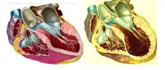
Causes of cardiomyopathy
Myocardial damage in cardiomyopathy can be a primary or secondary process due to systemic diseases and is accompanied by the development of heart failure and, in rare cases, sudden death.
There are three groups of main causes of the development of primary cardiomyopathy: congenital, mixed, and acquired. Secondary include cardiomyopathies due to any disease. As mentioned above, there are many causes for this pathology, but with the development of cardiomyopathy, the symptoms will be similar regardless of the cause that caused this condition.
Congenital heart pathology develops due to a violation of the formation of myocardial tissue during embryogenesis. There are many reasons, ranging from the bad habits of the expectant mother to stress and poor nutrition. Also known are cardiomyopathies of pregnancy and inflammatory cardiomyopathies, which essentially can be called myocarditis.
The secondary forms include the following types.
Storage or infiltrative cardiomyopathy. It is characterized by the accumulation of pathological inclusions between cells or in cells.
Toxic cardiomyopathy. The severity of damage to the heart muscle when interacting with drugs, especially antitumor drugs, varies: from asymptomatic changes on the ECG to fulminant heart failure and death. Long-term consumption of alcohol in large quantities can lead to the development of inflammation in the heart muscle (alcoholic cardiomyopathy); this cause ranks first in our country as the most frequently identified.
Endocrine cardiomyopathy (metabolic cardiomyopathy, dismetabolic cardiomyopathy) occurs as a result of metabolic disorders in the heart muscle, often leading to wall degeneration and impaired contractility of the heart muscle. The reasons are diseases of the endocrine system, menopause, obesity, unbalanced nutrition, diseases of the stomach and intestines. If cardiomyopathy develops as a result of thyroid disease and diabetes, hypertrophic cardiomyopathy occurs.
Nutritional cardiomyopathy is formed as a result of malnutrition, and in particular with long-term diets with limited meat products or fasting, the lack of consumption of vitamin B1, selenium, and carnitine affects the heart.
MRI
Recent advances in MRI have made it possible to obtain very high-quality images of the myocardium. MRI with late contrast with gadolinium-based drugs (at a dose of 0.2 mmol/kg IV) allows you to see myocardial infarction and distinguish dead myocardium from living one.
Pathogenesis
1. The mechanism of gadolinium uptake is not fully understood. It is known to diffuse into the interstitial space and accumulate in scar tissue. The degree of gadolinium contrast indicates the relative content of connective tissue in a particular segment of the myocardium. 2. The degree of contrast is divided into four quartiles. The degree of contrast of 0-25% corresponds to normal myocardium with minimal connective tissue content, 75-100% corresponds to scars. The degree of contrast is correlated with local contractility and perfusion. It has been shown that with weak contrast, contractility often improves after revascularization, and the degree of contrast greater than 75% indicates irreversible myocardial damage. Interestingly, the average degree of contrast at which contractility no longer improves is 41 ± 14%, which is consistent with biopsy data, according to which contractility after revascularization does not improve when the myocardium is replaced with connective tissue by more than 35%.
Diagnostic accuracy
Cardiac magnetic resonance imaging is a relatively new method that has good prospects in assessing myocardial viability. A very important advantage of MRI is the ability to determine the zone of transmural infarction. In addition, using MRI, you can see areas where the myocardium is not viable closer to the endocardium, and where it is viable closer to the epicardium. The information content of the method increases when contrasting is correlated with local contractility. When assessing myocardial viability, MRI results correlate well with positron emission tomography data. The disadvantages of MRI include high cost, low availability, and the inability to examine patients with implanted pacemakers and defibrillators.
Diet for left ventricular hypertrophy of the heart
To adjust your diet for cardiomyopathy, follow these tips:
- give up salt;
- eat often, about 6 times a day, but in small portions;
- stop smoking, drink less alcohol;
- choose foods that are lower in fat and cholesterol;
- limit the amount of animal fats;
- Fermented milk, dairy products, fresh vegetables and fruits are healthy;
- eat less flour and sweets;
- if you are overweight, follow a diet to lose weight and reduce the load on your heart.
sovets.net
Selecting a method
When choosing a method for identifying viable myocardium, the cost and availability of the method and the experience of a given medical institution are taken into account. Myocardial scintigraphy with thallium-201 and dobutamine stress echocardiography is much cheaper than positron emission tomography, but requires extensive experience to conduct the study and interpret its results. Cardiac MRI is a very promising method, but so far it is available only in a few clinics. In practice, the method is chosen based on the experience and equipment of the medical institution.
Forecast assessment
Identification of viable myocardium is a very important part of the evaluation of patients with left ventricular systolic dysfunction. In the presence of viable myocardium, revascularization has been shown to improve exercise capacity, reduce angina pectoris, and reduce mortality. Even if the contractility of the left ventricle does not change, the well-being of patients improves and mortality decreases. Restoring perfusion can reduce myocardial restructuring in the peri-infarction zone and the risk of recurrent infarction, improve left ventricular diastolic function, and eliminate the substrate of arrhythmias.
At what volume of viable myocardium is revascularization necessary?
It has been shown that revascularization is effective if the proportion of viable myocardium exceeds 18% on positron emission tomography or more than five viable segments are identified on dobutamine stress echocardiography. If the proportion of viable myocardium does not exceed 5%, revascularization is obviously ineffective. The decision to revascularize is made taking into account many other factors, in particular functional class and the presence of heart defects. When perioperative risk is high, the possible benefit of revascularization is assessed using several methods. A 2002 meta-analysis showed that in the presence of viable myocardium, revascularization significantly reduces mortality compared with medical treatment. This meta-analysis included studies that assessed myocardial viability using single-photon emission tomography, positron emission tomography, or dobutamine stress echocardiography. There were no significant differences in predictive value between these methods. In the presence of viable myocardium, mortality after revascularization was 3.2% per year, and with medical treatment - 15% per year. In the absence of viable myocardium, mortality was independent of treatment modality. The reduction in mortality after revascularization was inversely proportional to the severity of left ventricular dysfunction. This meta-analysis, although not without all the shortcomings inherent in meta-analyses, is nevertheless in good agreement with modern concepts and represents the most comprehensive review of data on the prognostic significance of viable myocardium. However, many questions remain. It is not yet clear which method of revascularization is preferable - coronary bypass surgery or angioplasty, how long after revascularization one should expect an improvement in myocardial contractility, and it is not completely known how revascularization improves survival. Literature: 1. B. Griffin, E. Topol “Cardiology” M. 2008