0
Author of the article: Marina Dmitrievna
2017.07.13
1 580
Treatment
Transesophageal electrophysiological study (TEPS) is aimed at studying the contraction function and heart rhythm by applying certain safe doses of electric current to the part of the myocardium that is close to the esophagus. In addition to the electrical effect on the heart through the esophageal wall, imaging methods, for example, echocardiography through the esophagus, are successful in cardiac diagnostics.
Advantages and disadvantages of the method
The advantages of this diagnostic method are the following:
- examination of rare and short forms of tachycardia of supraventricular origin;
- studying the exact place where it is possible to generate frequent signals or block the passage of impulses;
- can be used as an alternative to stress tests if the patient is unable to complete tasks for such studies;
- minor trauma;
- availability;
- no penetration into the vascular bed is required;
- There are no contraindications for repeated use.
Disadvantages include discomfort during insertion of the electrode.
We recommend reading the article about how phonocardiography is performed. From it you will learn about why auscultation is not enough, indications for phonocardiography, options for its implementation, contraindications and interpretation of the results. And here is more information about Doppler echocardiography.
Indications for cardiac PPE
The examination is indicated in complex diagnostic cases when the doctor must confirm or remove the following diagnoses:
- paroxysmal form of tachycardia;
- atrial fibrillation;
- bradycardia with weakness of the sinus node, blockades;
- the presence of additional pathways;
- myocardial ischemia, which could not be detected on the ECG (even with monitoring), or there are contraindications for stress tests.
Using TEE, you can monitor the success of treating arrhythmias, performing heart surgeries, and cauterizing the myocardium with radio waves; it is recommended to perform this before installing an artificial pacemaker.
Suspicions of a significant rhythm disturbance arise if the following symptoms are present:
- an attack of rapid heartbeat with a sudden onset and end, relieved by antiarrhythmic drugs;
- rare pulse with sensations of interruptions in the heart;
- syncope after excluding neurological or other causes.
Additional examination is prescribed for patients with complaints of arrhythmia, which could not be detected during a long-term ECG examination, or signs that require detailed detail have been identified.
Watch the video about when chest pain is cardiac and when it is anginal:
Indications
- Attacks of palpitations that are not recorded during daily monitoring of the electrocardiogram;
- episodes of rare pulse followed by attacks of palpitations;
- constant low pulse;
- fainting and dizziness, especially in young people;
- WPW syndrome;
- study of various characteristics of supraventricular paroxysmal rhythm disturbances with an established diagnosis;
- assessment of the effectiveness of antiarrhythmic treatment, including surgery;
- in some cases - diagnosis of coronary heart disease.
Contraindications
Myocardial stimulation with electrical impulses is not used for the following pathologies:
- severe rhythm disturbances detected on the ECG;
- complete blockade of pathways;
- atrial fibrillation;
- atrial or ventricular fibrillation;
- blood clot in the heart cavity;
- obstruction of the esophagus, tumors, narrowing, inflammation;
- infectious processes in the body;
- acute heart or coronary failure;
- heart attack or stroke;
- aneurysm;
- decompensation of the defect;
- increased body temperature;
- mental disorders.
Preparation for the event
The procedure has specific features; preparation for it begins 7-10 days in advance :
- discontinue medications for the treatment of arrhythmia or reduce their dose in consultation with the cardiologist;
- during angina attacks, only Nitroglycerin is left for sublingual use;
- before TEE they undergo ECG, EchoCG, Holter monitoring and measure blood pressure several times a day;
- give up smoking and alcohol at least a week in advance;
- Dinner should be very light, and on the morning of the examination you should neither eat nor drink.
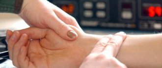
How is a transesophageal electrophysiological study performed?
TEE can be carried out in an outpatient or inpatient department of functional diagnostics. After measuring blood pressure and pulse rate, a baseline electrocardiogram is taken. Then the throat is treated with an anesthetic solution to alleviate discomfort when the probe is inserted.
TPE probe
A sterile tube with a tip that is equipped with an electrode for generating signals is passed through the nose or mouth. Sensors for a standard ECG are fixed on the chest. The depth of insertion of the device is 35 - 40 cm. If all stages were successful, then the supply of electrical impulses begins. During this time, there may be frequent heart contractions or a burning sensation in the chest.
The procedure itself lasts about half an hour. At this time, the doctor applies an electric current to the myocardium and takes readings of its activity with a cardiograph. During TPE, there is discomfort, but there is no severe pain.
In what cases does a doctor prescribe transesophageal pacing?
Transesophageal electrophysiological study (TEPS) is aimed at studying the contraction function and heart rhythm by applying certain safe doses of electric current to the part of the myocardium that is close to the esophagus. In addition to the electrical effect on the heart through the esophageal wall, imaging methods, for example, echocardiography through the esophagus, are successful in cardiac diagnostics.
Principle of the technique
The principle of transesophageal stimulation is based on the fact that the esophagus and the heart are anatomically close. It often happens that patients cannot detect heart rhythm disturbances using a traditional electrocardiogram or during daily monitoring of ECG and blood pressure. In this case, stimulation of the heart muscle is necessary. If the patient has arrhythmia, this will manifest itself during pacing through the esophagus. In other words, in certain cases, the myocardium needs to be “provoked” so that the diagnostician can correctly identify the disease, taking into account the characteristics of the conduction of electrical impulses.
Using an electrode inserted via the transesophageal method, impulses are sent to the heart muscle, and electrodes installed on the chest record the heart rate during the examination.
Pacing electrodes
In addition, when transesophageal pacing is performed, increased heart rate can provoke episodes of ischemic symptoms in patients who suffer from coronary artery disease that was not previously detected on the ECG.
Invasive research
In addition to transesophageal techniques, invasive research methods are used:
- Electrodes are inserted into the heart during a catheter-based invasive electrophysiological study.
- During an endocardial examination, a catheter with an electrode attached to the end is sent into the atrium or ventricle through a vein.
- An open electrophysiological study is performed during cardiac surgery. Stimulation is performed from the outside of the heart.
It is important to note that invasive techniques, compared to transesophageal ones, are more life-threatening.
Pros and cons of CPEFI
The undoubted advantage of this method of examination is the high accuracy of diagnosing heart rhythm, especially short-term supraventricular tachycardia. This means local diagnostics. Thus, during TPE in 95% of patients, it is possible to clearly identify the presence in the conduction system of the heart of conditions for the appearance of tachycardia or, conversely, for blocking the propagation of an impulse throughout the organ. Both types of rhythm disturbances are life-threatening, so accurate diagnosis using electrical stimulation can help maintain a healthy state and save the life of the patient.
There are no downsides to transesophageal echocardiography, other than discomfort. Other advantages include a low risk of injury, non-invasiveness, the ability to perform the procedure repeatedly, and the availability of the study for various patients.
Indications for electrical stimulation of the heart
The main indications for electrical pacing are various types of heart rhythm disturbances. Thus, TEE is recommended to clarify the diagnosis if the doctor suspects that a person has:
- Paroxysmal atrial or ventricular tachycardia.
- Paroxysmal atrial flutter (atrial fibrillation).
- Bradyarrhythmia, which is accompanied by a low heart rate (less than 50 beats per minute), for example, with weak sinus node syndrome.
- SVC or KL syndromes - with these types of tachycardia, the atria contract too often, and due to the presence of additional pathways between the ventricles and atria, tachycardia can develop in the ventricles. This pathology is life-threatening for the patient.
In some patients, examination is performed during the diagnosis of cardiac ischemia. For example, when it is not possible to record episodes of ischemic myocardial disease using standard electrocardiography methods or daily examination. In addition, TEE makes it possible to diagnose patients who have contraindications for performing procedures with additional physical activity.
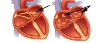
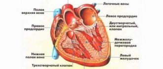
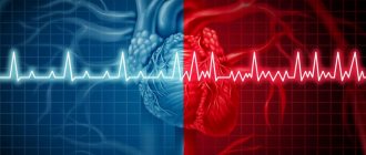
Heart rhythm disturbance
Repeated transesophageal electrical stimulation may be performed to monitor drug therapy for cardiac arrhythmias or in the postoperative period, for example, after ablation.
TEE is recommended before surgery to insert a pacemaker or defibrillator-cardioverter.
Symptoms indicating the need for TEE
The presence of pathological rhythm disturbances requiring electrical cardiac stimulation can be assumed based on the following clinical signs:
- Rapidly increasing heart rate, in which the pulse rises to 120 beats per minute for no apparent reason. Such attacks stop on their own or after the use of antiarrhythmic drugs.
- Irrhythmic heartbeat with weak pulse (less than 50 beats).
- Cases of loss of consciousness that do not occur against the background of neurological diseases or situational conditions (emotional outbursts, high air temperature, etc.), but are associated with heart rhythm disturbances.
Attention! When a patient complains to a cardiologist about the signs described above, he must prescribe an examination to accurately diagnose heart rhythm disturbances. And if the electrocardiogram does not reveal any types of arrhythmia, and the patient continues to experience its symptoms, it is necessary to resort to the method of electrical stimulation of the heart.
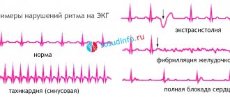
Research results
A test is considered normal if, when exposed to impulses supplied through the esophageal electrode, no rhythm disturbances are detected. If complaints of rapid heartbeat or attacks of loss of consciousness remain, and TPEFI is negative, then it is either repeated after 1 - 2 weeks, or an electrophysiological study is performed with an electrode inserted through the vascular network.
When rhythm disturbances are detected, its properties are recorded:
- type of arrhythmia,
- duration,
- appearance time,
- characteristics of electrical signals.
Example of PPEFI results
Indications for performing EPI and possible consequences of the study
There are a considerable number of pathologies associated with abnormalities in heart rhythm and conduction disorders. Some diseases are difficult to diagnose, so it is not always possible to quickly determine effective treatment tactics. Such pathologies can subsequently provoke more serious disturbances in heart rhythm and conduction with subsequent circulatory disorders. An electrophysiological study of the heart can detect a problem of this nature in a timely manner.
Problems associated with irregular heart rhythms are often difficult to diagnose
Types of EPI and the concept of PPEFI
There are two types of electrophysiological examination of the heart: invasive (endocardial, epicardial, combined) and non-invasive. The first involves surgical penetration through the natural membranes of the body, the second does not.
Endocardial EPI of the heart is performed without general anesthesia, only under local anesthesia. Vascular puncture is performed to guide the electrodes into the desired chambers of the heart. It is carried out through the femoral vein. The procedure can be considered rather minimally invasive. The endocardium has no pain receptors, so pain does not occur.
Epicardial EPS is performed during open-heart surgery with incision of the anterior chest wall.
There are several types of electrophysiological study of the heart
The invasive procedure can be either a stand-alone study or a surgical step in the treatment of arrhythmias, for example, through ablation. TEE is a non-invasive procedure. The essence of the method is as follows. If it is not possible to detect cardiac arrhythmias during an electrocardiogram or during daily monitoring, the heart is stimulated in such a way as to provoke it to a certain type of cardiac arrhythmia and already record this deviation. The heart is stimulated through electrical impulses.
TEE is performed more often, as it does not require high technical equipment, and also has the lowest number of complications and side effects when compared with invasive methods.
Pros and cons of CPEFI
Advantages of TEE - the electrophysiology of the heart is studied, which makes it possible to identify the cause of disturbing arrhythmias and their pathophysiology. The procedure is quite affordable, does not require powerful technical equipment, and can be performed in an outpatient setting.
TEE is a non-invasive and patient-safe study
The disadvantages are that discomfort may occur during the procedure. Local anesthesia is not performed; there may be discomfort in the nasal cavity.
The study has limitations - only the left side is examined due to anatomical features.
Indications for TEE
Patients with tachycardia refractoriness to AAP, with fainting states of unknown origin, and with paroxysmal tachycardia are referred for cardiac EPS examination. Transesophageal electrophysiological study of the heart is carried out for the following indications:
- sinus node dysfunction, the procedure allows for its assessment;
- WPW syndrome;
- bundle branch block;
- periodic loss of consciousness in the absence of neurological causes, or causes not found in non-invasive cardiac studies;
This procedure can be performed for bundle branch block.
- AV block;
- atrial fibrillation;
- sustained paroxysmal ventricular and supraventricular tachycardia.
How is it carried out?
Before understanding how TEE of the heart is performed, you should consider preparation for the procedure and preliminary examinations.
Preparation
All information about preparation should be obtained from your doctor in advance. Patients are referred for examination by a cardiologist, cardiac surgeon or arrhythmologist. Before cardiac EPI is performed, the patient fasts for at least 8-10 hours. A few days before the examination, you should give up caffeine, cigarettes and, especially, alcohol. It is also necessary to consult with a doctor about the advisability of temporarily discontinuing medications already taken.
It is very important to give up coffee, tobacco and alcohol before the procedure.
Before the cardiac EPI procedure, patients undergo a preliminary examination.
- ECG;
- stress tests;
- Holter monitoring;
- Ultrasound diagnostics of the heart;
In some cases, results of an electroencephalogram as well as an MRI of the brain may be necessary. All this is necessary to exclude syncope associated with neurological abnormalities. You may also need to consult an endocrinologist and other specialists.
It is also very important to do a cardiogram
Progress of the procedure
Transesophageal EPI is performed in the department of functional diagnostics. The procedure takes place in the morning. Its algorithm is as follows:
- Blood pressure measurement.
- ECG recording.
- A more complete explanation of what manipulations will be performed, how the patient will feel, and how he should behave.
- A probe is inserted, at the end of which there is an electrode. A tube is inserted either through the nose or through the mouth (rarely). The electrode promotes stimulation of the atrium through impulses of a given strength. It also allows you to record an electrogram. The probe itself is connected to equipment that initiates stimulation and analyzes the information received.
The transesophageal EPI procedure involves the insertion of a special probe that allows you to see the heart in maximum resolution
- An electrogram is recorded to identify rhythm disturbances.
- The probe is carefully removed along with the electrode through the nose. In case of getting stuck due to swelling of the mucous membrane - through the mouth.
- The information received is analyzed and a conclusion is drawn up for each type of arrhythmia, if any. Next, the results are sent to the attending physician.
- The procedure takes from half an hour to an hour.
- Artificially induced tachyarrhythmia disappears after a few minutes, but appropriate medications can be taken.
- If the examination is performed by an experienced specialist, then the discomfort will be minimal. A burning sensation in the chest is normal in this situation.
Transesophageal cardiac examination should be performed by an experienced specialist
Decoding the results
The norm indicates that the study did not reveal any arrhythmias under the influence of stimulation. If rhythm and conduction disturbances have been identified, each type is described separately. So, in order to identify the possible presence of myocardial ischemia caused by tachycardia, the ST segment is analyzed.
After the electrophysiological study has been completed, the results are interpreted by the doctor to determine further directions and treatment adjustments.
Possible complications
Most patients tolerate the examination well, as this technique is safe. But you still need to consider the possibility of the following complications:
- hypersensitivity to the anesthetic solution (swelling, laryngeal spasm, anaphylactic reaction, up to shock), therefore, if you are prone to allergies, tests are carried out first;
- myocardial infarction;
- attack of rhythm disturbance;
- pulmonary embolism;
- pain in the heart like angina pectoris.
To prevent such situations, it is necessary to undergo a complete cardiac examination before TEE..
Reasons why you shouldn't put off getting a heart exam
Carrying out the procedure
The procedure is carried out in a hospital setting if the specialist has a resuscitation kit, an oxygen inhaler, a saliva ejector, and equipment for determining blood pressure. During the entire examination, the patient is given a cardiogram.
Examination of the heart through the esophagus lasts no more than 15–25 minutes
During drug preparation, measures are taken to reduce:
- gag reflex during the procedure using local anesthesia of the oral cavity and pharynx (10% lidocaine solution in the form of a spray);
- excitement (Relanium 2 ml intravenously a few minutes before echocardiography);
- salivation and secretion of gastric juice (intravenously 0.1% atropine 1 ml).
Bacterial endocarditis in patients with valve implants is prevented by oral antibiotics.
The patient is placed on his left side, and an endoscope pre-lubricated with gel is inserted into his throat without excessive effort. After the subject makes swallowing movements, the device reaches the esophagus. The resulting feeling of discomfort felt at the beginning of the manipulation gradually disappears. All research data must be recorded on videotape.
After the procedure, the patient remains in a medical facility for several more hours under the supervision of doctors, and then can go home.
Minor injuries to the esophagus obtained during transesophageal echocardiography necessitate some restrictions at the end of the manipulation:
- the patient’s refusal to eat and eat for 2 hours;
- excluding tea and coffee drinks from the diet for several days, as well as eating food at room temperature.
Further consultation with a doctor is necessary if:
- sore throat that does not subside or gets worse;
- the appearance of pain in the sternum;
- difficulty breathing.
The results of the study are interpreted by qualified specialists, taking into account the Age, gender and physical parameters of the person. Based on the diagnostic conclusion, appropriate treatment is prescribed.