Portal vein thrombosis (PV) is the process of thrombus formation up to complete occlusion of the lumen of the vessel draining the bed of the gastrointestinal tract. Portal vein thrombosis is a rare vascular disease of the liver. It can be the result of a large number of different diseases and remain asymptomatic or manifest symptoms of the underlying disease [1-4]. Diagnosis of the disease is difficult. The prognosis for portal vein thrombosis is always serious and unfavorable, with deaths due to bleeding or hepatic coma often occurring.
The purpose of the work is to show, using clinical examples, the capabilities of ultrasound with color Doppler mapping (CDC) in diagnosing thrombosis in the portal vein system.
The lifetime diagnosis of thrombosis of the main trunk of the portal vein in Russia was first made by S.P. Botkin in 1862. In 1934 N.D. Strazhesko was the first in the world literature to describe intravital thrombosis of the right branch of the portal vein trunk and, based on his own research and literature data, developed the symptomatology of blockage of the right branch of the portal vein.
There is no reliable information on the incidence of portal vein thrombosis. According to sectional data, thrombosis is detected in 0.14-0.34% of all autopsies (L. Lissauer, 1908; L.T. Webster, 1921; E.S. Pallette, 1936). According to other data (A.I. Gritsyuk, 1973), during autopsies of those who died from major cardiovascular diseases (atherosclerosis, hypertension, endocarditis and heart defects), portal vein thrombosis was found in 0.56% of cases (in 10 out of 1763 deaths ), which accounts for 0.8% of all thromboembolisms developing in these diseases. It is known that portal vein thrombosis affects up to 30% of patients with hepatocellular carcinoma and up to 5% of patients with portal hypertension against the background of cirrhosis.
The occurrence of the disease, like other venous thromboses, can be explained by Virchow's triad, which includes the following elements.
- Injury to the vein wall during surgery.
- Reduced blood flow rate in the portal vein: compression of the vessel from the outside by a tumor, scars, echinococcal cyst, alveococcus; chronic heart failure; constrictive pericarditis; Budd-Chiari syndrome (hepatic vein thrombosis).
- Increased blood clotting or a change in the ratio of its cellular elements: in the postoperative period - especially in cancer patients, as well as after splenectomy; in inflammatory processes - like a donkey. /li>
The clinical picture depends on the location and extent of portal vein thrombosis, the speed of its development and the nature of the predisposing liver disease. The most severe manifestation of the disease is liver infarction or atrophy of its segment. However, in 1/3 of cases, thrombosis forms slowly, as a result of which collateral blood flow has time to develop, and the portal vein is recanalized over time and its cavernous transformation occurs. However, even with a relatively favorable course of the disease, portal hypertension develops [1-4].
When diagnosing portal vein thrombosis, you should pay attention to the coagulogram: an increase in fibrinogen content, the appearance of activated fibrinogen B, an increase in the prothrombin index, a decrease in blood clotting time [2].
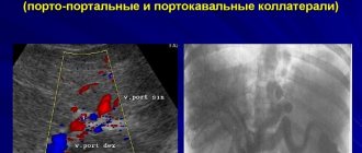
Ultrasound examination (ultrasound) in gray scale mode in the lumen of the portal vein can reveal a heterogeneous formation of increased or mixed echogenicity, with uneven, unclear contours, obstructing blood flow, the size of which can vary: from 0.5 cm to complete occlusion of the portal vein and/or its branches (Fig.
A). The echogenicity of a thrombus is often higher than that of the surrounding blood. However, in the early stages of formation, the echogenicity may vary so slightly that visualization of the thrombus is extremely difficult. An increase in the diameter of the vessel with blurred contours, an enlargement of the liver and a decrease in its echogenicity, and an enlargement of the spleen can be detected. The root causes of portal vein thrombosis can be found: hepatocellular carcinoma, metastases, liver cirrhosis, pancreatic neoplasms, etc. In pylephlebitis, liver abscesses are found [3].
With CDK, there is a complete or partial absence of Doppler signals inside the lumen of the vessel; in the case of partial thrombosis, the signal is detected near the wall around a thrombus that partially occludes the vein, or in a network of narrow collaterals (Fig. 1, b). In the case of partial thrombosis, a Doppler signal is determined with signs of turbulence against the background of increased blood flow velocity. In tumor thrombosis, blood flow can be pulsating or continuous [2, 3]. Small or large collaterals are visualized. During cavernous transformation of the portal vein in the CD mode, a smoothed Doppler curve with an average blood flow velocity of less than 8 cm/s is determined in the collaterals [5]. Cavernous vein malformations, spontaneous porto-portal, porto-caval and splenorenal shunts are possible.
Computed tomography (CT) reveals a thrombus as a filling defect in the lumen of the portal vein, which does not enhance the signal.
Magnetic resonance imaging (MRI) reveals areas of pathological signal in the lumen of the portal vein that do not differ in intensity from the surrounding tissue on T1-weighted images and have increased intensity on T2-weighted images.
Angiography is the method of definitive confirmation of the diagnosis. For safety reasons, the venous phase of superior mesenteric arteriography is more often examined, and splenoportography is performed less frequently. A filling defect is detected in the portal vein, or it is not contrasted at all.
Depending on the location of the thrombus, three types of portal vein thrombosis are distinguished: radicular form (thrombosis of the splenic vein and mesenteric vessels); terminal form (thrombosis of small branches and capillaries of the portal vein in the liver); stem thrombosis (in the very trunk of the portal vein) [1-3].
In addition, portal vein thrombosis can be acute (after splenectomy, with cirrhosis) and chronic (develops over a long period of time - from several months to several years) [1-3].
Stages of thrombosis
- Acute - echogenic thrombus. The portal vein may be enlarged.
- Subacute - a thrombus and small collaterals are visualized. The portal vein may be enlarged.
- Chronic - large collaterals in the projection of the obliterated portal vein (cavernous transformation of the portal vein). The portal vein is reduced or not visualized [1-3].
We present several clinical observations of patients with thrombosis of the portal vein and its branches.
Clinical observation 1
Patient Ts., 39 years old. There is a history of blunt abdominal trauma with subsequent development of hemorrhagic pancreatic necrosis. Repeated surgical interventions: drainage of the omental bursa. Complains of pain in the epigastric region.
Abdominal ultrasound
The liver is enlarged in size, its contours are smooth and clear, the structure is diffusely heterogeneous, mixed echogenicity, there are no signs of biliary hypertension. The trunk of the portal vein is visible as the formation of a heterogeneous structure with many echogenic partitions running parallel to the walls of the vessel. The presence of hyperechoic areas in the paravenous region of the portal vein (Fig. 2, a). With CDK, there are pronounced collaterals in the projection of the portal vein (Fig. 2, b). The gallbladder has been removed. The pancreas is enlarged in size, the head is up to 40 mm, the contours are uneven, unclear, the structure is diffusely heterogeneous, alternating areas of increased and decreased echogenicity are noted, the duct is visualized along its entire length, at the level of the body of the pancreas it is expanded to 10 mm. In the projection of the head of the pancreas there are pronounced parapancreatic collateral anastomoses (Fig. 3). The spleen is of normal size, its structure is homogeneous, of medium echogenicity. The splenic vein is 7 mm, its course is tortuous, deformed, in the projection of the hilum of the spleen there are pronounced collaterals (Fig. 4). Conclusion: hepatomegaly. Diffuse changes in the liver. Picture of chronic capitate pancreatitis. Thrombosis of the portal vein with subsequent recanalization and cavernous transformation of the portal vein, development of collateral anastomoses.
Fibrogastroduodenoscopy
Fibrogastroduodenoscopy without features.
Abdominal CT
Liver without signs of focal changes. The intrahepatic ducts are not dilated. The spleen is not changed. Varicose collaterals of vessels at the level of the hilum of the spleen, the hilum of the liver (Fig. 5). The area of the porta hepatis is not clearly differentiated; it is impossible to reliably judge the diameter of the portal vein. The pancreas is clearly heterogeneous and dense. The Wirsung duct can be traced throughout its entire length and is unevenly expanded (diameter at body level up to 1 cm). The diameter of the head of the pancreas is about 4 cm, the contours are unclear. Peripancreatic tissue at the level of the head and body is significantly compacted and fibrotic. It reveals cystic formations of irregular shape up to 1 cm. Conclusion: CT signs of chronic capitate pancreatitis. Portal vein thrombosis.
X-ray of the stomach
We pass the esophagus. The cardia is deformed; a filling defect with tuberous contours is noted in the subcardial region. There is a filling defect on the greater curvature and a rigid platform along the lesser curvature in the middle third with its shortening. The antrum is pulled up. When inflated with air, the walls of the stomach partially straighten, but the defect remains in the subcardial region. The duodenum is moderately deformed, the folds of the mucous membrane are unevenly smoothed due to edema, evacuation is timely. Conclusion: adhesions, perigastritis, gastric varicose veins.
Clinical observation 2
Patient B., 45 years old. Diagnosis: liver cirrhosis.
Abdominal ultrasound
The liver is enlarged in size, its contours are uneven, the structure is clearly heterogeneous, diffuse, with alternating areas of low and high echogenicity. Enlargement of the quadrate and caudate lobes of the liver. There are no signs of biliary hypertension. The lumen of the hepatic veins is not changed. The intrahepatic pattern is not disturbed. The typical tubular anechoic trunk of the portal vein is absent. The trunk of the portal vein is visible as the formation of a heterogeneous structure with many echogenic partitions running parallel to the walls of the vessel. In the paravenous region of the portal vein, a zone of increased echogenicity 68x26 mm with pronounced collaterals in this area is visualized; in the color flow mode, the blood flow in them is monophasic and low-frequency, Vps 10 cm/s, Ved 9 cm/s (Fig. 6). The venous pattern of the omentum is strengthened due to the expansion of the veins. The gallbladder is represented by a formation 45x18 mm, without lumen, collapsed. CBD 5 mm. The pancreas is without pathology. The spleen is enlarged in size, but structurally unchanged. In the Color Doppler mode, the diameter of the splenic vein is up to 11 mm, Vps 19 cm/s, Ved 18 cm/s, monophasic. Collaterals are visualized at the hilum of the spleen (Fig. 7). Conclusion: hepatosplenomegaly. Signs of subhepatic portal hypertension due to thrombosis of the portal vein with subsequent recanalization and cavernous transformation of the portal vein.
Abdominal CT
The liver and spleen were enlarged in size; no focal changes were detected in them. The pancreas is of normal size and shape and evenly accumulates contrast agent. The duct of Wirsung is not dilated. No focal changes in the pancreas were also detected. CT signs of varicose collateral vessels of the spleen. At the level of the liver gate, an additional formation of heterogeneous soft tissue density with polycyclic contours, measuring 5x6 cm, is determined (Fig. 8). Against the background of this formation, arteries are clearly visible (Fig. 9). The formation clearly but unevenly accumulates contrast agent in the venous phase (Fig. 10). In the delayed phase, the intensity of accumulation of the contrast agent decreases (Fig. 11). Against the background of this formation, the vessels of the hepatic hilum are not clearly differentiated. There are no CT signs of lymphadenopathy and free fluid in the abdominal cavity. Conclusion: hepatosplenomegaly. Portal hypertension (CT signs of portal vein thrombosis).
Fibrogastroduodenoscopy
In the lower third of the esophagus there are longitudinally dilated veins with areas of expansion (nodes). The mucous membrane of the stomach is thickened, the folds are large, elastic, and peristalsis is active. The pyloric sphincter and duodenal bulb are significantly deformed due to scars, the mucous membrane is edematous, hyperemic, and there are pinpoint hemorrhages. Conclusion: varicose veins of the esophagus. Chronic hypertrophic gastritis. Cicatricial-ulcerative deformation of the pylorobulbar zone. Hemorrhagic bulbitis.
Clinical observation 3
Patient K., 38 years old. Diagnosis: hereditary hemolytic anemia. History of splenectomy.
Abdominal ultrasound
The liver is enlarged in size, its contours are smooth and clear, its structure is diffusely heterogeneous, and its echogenicity is reduced. The diameter of the portal vein is 20 mm, the splenic vein is 12 mm, the superior mesenteric vein (SMV) is 10 mm. In the lumen of the portal vein and superior mesenteric vein, multiple hyperechoic formations with uneven contours are visualized (Fig. 12, a; Fig. 13, a). In the Color Doppler mode, there is a partial absence of Doppler signals inside the lumen of the vessels (portal vein, superior mesenteric vein), the Doppler signal is determined in the network of large collaterals (Fig. 12, b; Fig. 13, b). Common bile duct 5 mm. There are no signs of biliary hypertension. The gallbladder is of normal size, no stones were detected. The contours of the pancreas are smooth, clear, its structure is homogeneous, of medium echogenicity, the duct is 1 mm. The spleen has been removed. Conclusion: condition after splenectomy. Thrombosis of the portal vein, superior mesenteric vein.
Clinical observation 4
Patient T., 23 years old. Diagnosis: thrombosis of the splenic vein with advanced clinical signs of hepatitis B.
Abdominal ultrasound
The liver is enlarged in size, its contours are smooth and clear, its structure is diffusely heterogeneous, and its echogenicity is mixed. Portal vein 14 mm. Common bile duct 5 mm. There are no signs of biliary hypertension. The gallbladder is half asleep. The contours of the pancreas are smooth, clear, its structure is homogeneous, of medium echogenicity, the duct is 1 mm. The spleen is significantly enlarged in size, the contours are clear, the structure is homogeneous, of medium echogenicity. The splenic vein is dilated from 11 to 13 mm, deformed, its course is tortuous. In the lumen of the splenic vein, closer to the hilum of the spleen, a hyperechoic formation of irregular shape is visualized. In the Color Doppler mode, the absence of Doppler signals inside the lumen of the vessel is noted; the Doppler signal is determined in the network of large collaterals (Fig. 14). Some increase in volumetric velocities in the portal vein and splenic vein. A small amount of free fluid in the abdominal cavity. Conclusion: ascites. Hepatomegaly with diffuse liver changes. Significant splenomegaly. Thrombosis of the splenic vein.
conclusions
Thus, ultrasound with colorectal dosage is an extremely informative non-invasive method for diagnosing thrombosis in the portal vein system with good sensitivity and specificity.
Literature
- Zhestovskaya S.I., Grakova L.S., Aksenova N.A. Clinical application of duplex scanning in identifying collateral circulation in portal hypertension // Pervaya Krai. 2001. N10. pp. 10-12.
- Kuntsevich G.I. Ultrasound diagnostics in abdominal and vascular surgery // Minsk: Cavalier Publishers. 1999. 252 p.
- Mitkov V.V. Dopplerography in the diagnosis of diseases of the liver, gall bladder, pancreas and their vessels // M: VIDAR. 2000. 152 p.
- Scoutt LM, Zavin ML, Taylor KJ Doppler ultrasound clinical application // Radiology. 1990. V. 174. P. 309-319.
- Raby N., Meire HB Duplex ultrasound in the diagnosis of cavernosis transformation of the portal vein // Radiol. 1988. V. 61. P. 586-588.
Source: www.medison.ru
Features of blood supply to the liver
Such a system is an additional venous circulation circuit, which performs the function of purifying plasma from harmful microelements.
The department is formed from large vascular trunks that connect near the liver. Through the mesenteric veins, blood comes from the intestines, the splenic artery departs from the stomach, pancreas. At the porta hepatis, the main vessel divides into two branches that transport blood through the left and right lobes. Reaching the hepatic lobe, the veins weave around its periphery, then go deeper into the organ. After treatment with hepatocytes, the blood is sent to the central veins of both lobes. Such vessels combine into one larger one, transferring blood from the liver.
The portal vein system is isolated from other vascular beds. When problems arise with hemodynamics in this part of the circulatory system, other vessels are activated for normal circulation. But they cannot conduct the required volume of blood through themselves. Therefore their work is limited. The body can compensate for the obstructed blood supply in part due to hepatic parenchyma or portal vein obstruction.
The pressure in the vessels increases, blood begins to circulate through the anastomoses, and the vena cava flows.
Topography of the main bloodstream of the liver
The supply bloodstream of the liver, which supplies blood for cleansing of toxins, is the portal vein. The topographic anatomy of this organ is important for understanding the processes occurring in this most important organ. The large blood vessel that collects and transports blood for detoxification gets its name from its location in the vestibule of the liver, the so-called portal system. The portal vein of the liver, the anatomy of which is taken into account by a specialist when performing surgical interventions, is located deep in the hepatoduodenal ligament, located behind the hepatic artery and common bile duct. There are also nerves, lymph nodes and blood vessels. It is formed from the veins of the following organs located in the abdominal cavity:
- stomach;
- pancreas;
- spleen;
- large intestine, except for the blood vessels of the anal canal;
- small intestine.
Having found its true course, the portal vein goes up and to the right, passing behind the upper part of the duodenum and entering the hepatoduodenal ligament, where it passes between its leaves and reaches the porta of the liver.
Possible diseases
We list the common pathologies:
- Portal hypertension.
- Pylephlebitis.
- Blood clot formation.
- Cavernous transformation.
Causes of diseases:
- cirrhosis;
- malignant tumors of the gastrointestinal tract;
- the umbilical vein becomes inflamed when catheters are implanted in infants;
- infection in the digestive tract;
- internal organ injuries;
- poor blood clotting;
- infectious processes.
Rare provoking factors include prolonged use of oral contraceptives.
Signs of portal hypertension:
- severe abdominal pain;
- nausea;
- gagging;
- dyspeptic disorders;
- heat.
When chronic thrombosis develops, partial vascular patency is observed, and the following symptoms occur:
- A lot of fluid accumulates in the abdomen.
- The spleen swells.
- The lumen of the blood vessels of the esophagus increases.
Thrombosis is diagnosed by ultrasound examination. The monitor clearly displays a dense formation in the venous lumen. Computed tomography and MRI help determine the exact causes of the disease.
Deviations from the norm and pathology
When conducting research or operations and collecting the information obtained into a single whole, scientists found that in extremely rare cases, and only about 30 of them have been registered worldwide, a person completely lacks the portal vein. In most cases (about 70%), the variability of this bloodstream is manifested in various combinations of anastomoses and in the size of the vein itself. But in addition to the genetically determined characteristics of the portal system, pathological changes can develop in it, affecting the condition of the entire organism.
Many people suffer from thrombosis of the portal system, because blood and cholesterol clots are formed as a result of many reasons, such as poor nutrition, disturbances in the gastrointestinal tract, and excretory system. Pilethrombosis can occur in two forms:
- chronic progressive - the blood flow is partially blocked, the movement of blood is difficult, which affects the condition of the whole organism;
- complete thrombosis - the lumen of the portal vein is completely blocked, which causes a sharp deterioration in health, including death.
Signs of portal vein thrombosis include pain in the right hypochondrium, nausea, vomiting, fever, enlarged spleen (splenomegaly) due to increased blood flow in this organ. These symptoms appear simultaneously, worsening the general health of the patient. Complete thrombosis causes intestinal infarction.
The chronic course of pilethrombosis often does not have acute symptoms. This occurs due to the activation of compensatory mechanisms when other vessels take over the work of the portal vein. When the capabilities of the compensators are depleted, ascites, dilatation of the saphenous veins of the esophagus and the anterior abdominal wall, subtle abdominal pain and low-grade fever appear.
The consequences of chronic pilethrombosis are progressive chronic ischemia and cirrhosis of the liver (in cases where this disease was not the source of pilethrombosis).
The liver itself does not have nerve endings that can signal pain about existing problems. Therefore, preventive examinations should become a source of timely identification of potential problems.
Portal hypertension
The disorder occurs when pressure in the portal system increases as a result of the formation of blood clots. The normal value does not exceed 10 mm Hg. Art.
We list the factors that provoke this disease:
- Cirrhosis.
- Thrombosis of liver vessels.
- Hepatitis.
- Heart diseases.
- Metabolic problems.
- Blockage of the splenic vessels.
Symptoms:
- heaviness in the abdomen on the right under the ribs;
- jaundice;
- feeling of weakness;
- weight loss.
With portal hypertension, the spleen becomes enlarged due to stagnation of blood in the veins. Fluid accumulates in large volumes in the abdominal cavity.
Pylephlebitis
This is one of the most complex inflammatory processes occurring in the area of the portal vein. If left untreated, the tissue begins to die and the disorder is fatal.
Here are the signs of the disease:
- symptoms of poisoning;
- hemorrhage of blood vessels of the stomach, intestines;
- jaundice;
- chills.
During the examination, it is possible to determine an increase in the number of leukocytes, the intensity of erythrocyte sedimentation increases. This condition is a sign of purulent inflammation. The diagnosis can be confirmed by ultrasound, MRI, and computed tomography.
Inflammation
Complex pathological conditions arise due to the action of viruses. Irritation of the tissues of the vessels of the portal system is characterized by increased danger. Pyelonephritis is a suppuration that appears in acute appendicitis. If you provide assistance to the patient at the wrong time, pyelonephritis develops into an abscess. The likelihood of death increases. Fever is a characteristic sign of inflammation. The patient has a stomach ache, chills, and blood vessels in the digestive system bleed. Purulent cavities may form in the liver, and the skin turns yellow.
Diagnostics
We list the main ways to assess the condition of patients:
- Ultrasound scanning.
- Doppler.
- Magnetic resonance imaging.
- Angiography.
- Blood analysis.
A blood test is considered a classic way to detect vascular pathology. If a deviation from the norm is observed, the concentration of leukocytes and breakdown products of blood cells increases, specialists suspect the development of the disease. If significant deviations from the norm are observed, the doctor conducts an additional examination and selects an appropriate course of treatment to combat the disease.
Diagnosis of the syndrome
In addition to examining the patient and collecting anamnesis, diagnosis involves the use of visual research techniques:
- Ultrasonography;
- Doppler study;
- CT and MRI;
- X-ray with contrast agent;
- Portography with contrast agent;
- Angiography.
Another method that is used to determine the presence of pathology in the portal vein is portal scintigraphy. Its process uses a radiopharmaceutical sensor that is embedded in the human body. The sensor is fixed in the vessel.
But the main diagnostic method was and remains ultrasound with such an addition as Doppler study.
They allow you to see:
- How great is the dilatation (expansion) of the portal vein;
- Blood flow speed;
- Direction of blood flow.
Each of the pathologies listed above, with the exception of pylephlebitis, is quickly detected using ultrasound. Doppler ultrasound allows you to see the site of thrombus formation when the vessel is completely blocked.
Another great way to detect a blood clot is angiography. This method is one of the most reliable and accurate, along with CT and MRI.
Instrumental research methods are always accompanied by the results of blood tests and external symptoms.
Diagnostics Portal vein thrombosis should be suspected in any case of portal hypertension, combined with the usual results of liver biopsy. Coagulogram: increased fibrinogen content, appearance of activated fibrinogen B, increased PTI, decreased blood clotting time. Ultrasound: in the lumen of the portal vein it is possible to recognize an echogenic thrombus.
With color Doppler mapping, the signal from the blood flow is either absent, or it is detected near the wall near a thrombus that partially occludes the vein, or in a network of narrow collaterals. Cavernous vein malformations, spontaneous portacaval and splenorenal shunts are likely.
For safety reasons, the venous phase of superior mesenteric arteriography is more often examined, and splenoportography is performed less frequently. In the portal vein, a lack of filling is detected, or it is largely not contrasted.
The main method for diagnosing changes in the portal vein is ultrasound, the advantages of which can be considered safety, low cost and high accessibility for a wide range of people. The study is painless, does not take much time, and can be used for children, pregnant women and the elderly.
A modern addition to routine ultrasound is Doppler ultrasound, which allows one to assess the speed and direction of blood flow. On ultrasound, the PV is visible at the porta hepatis, where it bifurcates into horizontally located right and left branches. So the blood during Doppler is directed towards the liver. The norm on ultrasound is considered to be a vessel diameter within 13 mm.
When thrombosis occurs in the vein, hyperechoic content will be detected, heterogeneous, filling part of the diameter of the vessel or completely the entire lumen, leading to a total cessation of blood movement. Color Doppler mapping will show the absence of blood flow with complete obstruction by a thrombus or its parietal nature near the blood clot.
With LPG on ultrasound, the doctor will detect dilation of the lumens of blood vessels, an increase in liver volume, accumulation of fluid in the abdominal cavity, and a decrease in blood flow speed on color Doppler. An indirect sign of SPH will be the presence of cavernous changes, which can be confirmed by Doppler ultrasound.
In addition to ultrasound, contrast-enhanced CT is used to diagnose portal vein pathology. The advantages of MRI include the ability to determine the causes of changes in the portal system, examine the liver parenchyma, lymph nodes and other nearby formations. The disadvantage is the high cost and low availability, especially in small towns.
Angiography is one of the most accurate methods for diagnosing portal thrombosis. In case of portal hypertension, the examination necessarily includes an FGDS to assess the condition of the portocaval anastomoses in the esophagus, esophagoscopy, and possibly x-ray contrast examination of the esophagus and stomach.
Data from instrumental examination methods are supplemented by blood tests, which reveal abnormalities (leukocytosis, increased liver enzymes, bilirubin, etc.), and the patient’s complaints, after which the doctor can make an accurate diagnosis of damage to the portal system.
Step 1: pay for the consultation using the form → Step 2: after payment, ask your question in the form below ↓ Step 3: You can additionally thank the specialist with another payment for an arbitrary amount ↑
Liver fibrosis is formed during chronic inflammation of the organ, when reparative systems cannot cope with the restoration of damaged epithelium. Fibrosis may be reversible, unlike cirrhosis.
If hepatitis is stopped in time, the liver parenchyma is completely regenerated. High frequency of cirrhotic lesions - about 20% of all patients with hepatitis. The problem is the chronic course of inflammatory changes in the liver against the background of viral hepatitis and alcohol consumption.
Patients consult a doctor at the stage of subcompensation or decompensation, when clinical symptoms appear. At the initial stage, there are no signs of pathology, which is due to compensation of pathological changes by healthy hepatocytes.
Portal hypertension is diagnosed based on a medical examination, laboratory tests, and also using instrumental and endoscopic methods.
The esophagogastroscopic method is the simplest and most accessible way to detect vascular pathology in the stomach and esophagus. During the procedure, the specialist identifies dilated veins in these parts of the gastrointestinal tract, which becomes an absolute criterion for diagnosing portal hypertension syndrome.
With the first degree of dilation, the veins have a diameter of up to 3 mm, the second degree is determined when the diameter of the vessels increases to 5 mm. The third degree is spoken of when the lumen in the veins of the stomach and esophagus exceeds 5 mm.
Endoscopic examination allows you to accurately determine not only the degree of dilation of blood vessels, but also predict the likelihood of bleeding from them.
Despite the obviousness of the manifestations of portal hypertension syndrome and the high diagnostic capabilities of modern medical equipment, specialists sometimes have difficulty identifying this vascular pathology.
Inflammatory processes
Pylephlebitis is considered one of the rarest portal vein disorders. Symptoms and additional diagnostics show that the pathology can provoke blood clots. Pylephlebitis appears due to suppuration, after which appendicitis becomes inflamed. If the disease is not treated promptly, the patient will die. Due to the characteristics of the symptoms, it is difficult to diagnose such suppuration. It is impossible to determine the disorder without the use of MRI equipment. Modern technical devices help prevent deaths.
Therapy
Inflammation of the portal vein is treated using complex methods:
- Use of medications.
- Surgical intervention.
Drug treatment involves the use of the following drugs:
- Anticoagulants prevent the formation of blood clots and increase the patency of veins.
- Thrombolytics cut blood clots and cleanse the lumen of blood vessels.
- The medicine is prescribed only by specialists. The symptoms of the disease are always taken into account. If medications do not give the desired result, surgery has to be performed.
The following procedures are performed:
- Thrombolysis.
- Angioplasty.
The main complication after surgery is bleeding, ischemic disorder.
In case of such diseases, you need to contact qualified doctors.
The liver cleanses the body of toxins that enter the circulatory system from the gastrointestinal tract. Processes and removes breakdown products of red blood cells. The liver ensures blood clotting. Stopping the work of such an organ leads to death.
Source: zdravpechen.ru
Cavernous transformation of the portal vein in children
Treatment of bleeding from the upper gastrointestinal tract should always begin, regardless of its cause, with volume replacement, gastric intubation for emptying and lavage, and the use of measures aimed at stabilizing the patient's condition.
Sometimes tamponade with a Sengstaken-Blakemore probe is required. If balloon tamponade is used, intubation may be necessary to ensure normal airway function. Infusion of systemic vasopressin is indicated, which causes vasoconstriction of the arterial bed of the internal organs and thus reduces portal venous pressure. Initial doses are 0.2-0.4/1.73 m2/min, the drug is administered by continuous infusion.
In the past, esophagography was used to diagnose esophageal varices.
Currently, double Doppler sonography of the abdomen should be the main initial research method, which allows not only to determine the size and consistency of the liver and spleen, but also the caliber, vascular patency and direction of blood flow in the portal system and hepatic veins. The advantages of ultrasound over other methods include, in addition, its non-invasiveness and the possibility of use against the background of infusion therapy and resuscitation.
Portal venography and splenoportography in the diagnosis and evaluation of portal hypertension are nothing more than routine methods that can provide some help only in rare cases when bypass surgery is planned. Once the child's condition is stabilized, a Doppler ultrasound examination is performed. Esophagoscopy is carried out selectively, mainly for therapeutic purposes.
Esophageal endosclerosis
The revival of interest in esophageal endosclerosis to stop acute and chronic bleeding from esophageal varices in children is associated with (1) unsatisfactory results of shunt operations, (2) the development of hepatic encephalopathy after these interventions, (3) the desire to avoid surgical interventions that may complicate subsequent transplantation liver. Initially, the method of esophagoscopy was used under general anesthesia using a rigid esophagoscope.
Sequential sclerosis of varicose veins was carried out under eye control using 5% morruate (sodium morrhuate), after the introduction of which the injected varicose vessels were compressed by turning (rotating) the esophagoscope.
In children with acute variceal bleeding, endosclerosis is repeated every 2-3 days until the bleeding stops.
In patients with non-acute bleeding, endosclerotherapy is performed at intervals of 6 weeks to 3 months until all varicose veins in the area of the esophagogastric junction are obliterated.
Then esophagoscopy and endosclerotherapy, if required, are performed twice a year, and subsequently - once a year.
The results of using this method are excellent - bleeding can be stopped in all patients, and the rate of recurrent bleeding is very low.
Minor complications of this procedure include superficial ulceration of the esophagus, pleural effusion, atelectasis, fever, esophageal motility abnormalities, and development of esophageal stricture. Severe complications are rare.
These include esophageal perforation, spinal paralysis, mesenteric vein thrombosis with intestinal infarction, solitary brain abscesses, the formation of esophageal-bronchial fistula and, in adults, respiratory distress syndrome.
Currently, the method of endoscopic ligation of varicose veins with a thin ligature is used, which leads to their obliteration and avoids the injection of sclerosing substances.
Congenital anomalies of the portal vein are represented by aplasia and stenosis, which are the result of pathological obliteration of the vitelline veins and their ventral anastomoses.
Cavernous transformation of the portal vein (cavernoma) may represent a congenital malformation, but is usually the end result of postpartum thrombosis with subsequent recanalization and formation of new vessels (eg, omphalitis with portal vein thrombosis).
Vein phlebitis and its treatment
Have you been struggling with HYPERTENSION for many years without success?
Head of the Institute: “You will be amazed at how easy it is to cure hypertension by taking it every day.
Phlebitis of venous vessels, due to its causes, mechanism of development and consequences, is often combined with thrombophlebitis. “Pure” inflammation without thrombus formation lasts a very short period. Important importance in the origin of phlebitis is given to the development of local infection in the veins and the allergic mood of the body.
The role of infectious pathogens and increased vascular reactivity must be taken into account in treatment. The disease does not depend on age category. It is more severe in children and the elderly, which is associated with a decrease in immune defense.
Our readers successfully use ReCardio to treat hypertension. Seeing how popular this product is, we decided to bring it to your attention. Read more here...
Why does vein inflammation occur?
Causes of phlebitis include:
- Infectious agents - the greatest selectivity of action is characteristic of streptococci. Microorganisms enter the venous network during acute infectious diseases (scarlet fever, typhoid fever), pneumonia, septic endocarditis, tuberculosis, sepsis. In addition to external microbes, a weakened body is threatened by pathogens from chronic, untreated lesions. These include carious teeth, sinusitis, inflammation of the appendages, colitis, adenoids, tonsillitis. They constantly contain weakened bacteria, which, if the body loses resistance, can strike.
- Traumatization of the vein - damage to the walls of the veins accompanies bruises and fractures. Traumatic procedures include intravenous injections and taking blood for analysis, and the use of catheterization methods in the diagnosis and treatment of diseases. Phlebitis may appear in response to hirudotherapy.
- Transfer of inflammation from neighboring tissues - veins are affected in a similar way with abscesses, boils, pyoderma.
- Burning a vein with high temperature or a chemical substance in the treatment of varicose veins (radiofrequency ablation, sclerotherapy, laser therapy), if used inappropriately or with increased sensitivity of the patient, can cause an unexpected reaction.
- Varicose veins are the most common cause of phlebitis in the lower extremities.
Where are phlebitis located?
The localization of inflammation of the veins plays a significant role when affecting deep vessels located close to the central venous pathways directed to the head and heart.
The inflammatory process tends to quickly spread along the bloodstream or through neighboring tissues.
Phlebitis on the face can cause meningitis.
Inflammation of the vessels of the legs through the femoral vein infects the entire bed of the inferior hollow system. And phlebitis of the mesenteric veins, which occurs as a result of intestinal inflammation, brings infection to the portal vein of the liver.
The ulnar veins in the hands are most often affected. Their superficial subcutaneous location is convenient for medical procedures. Subclavian catheters are installed in hospitals for long-term administration of fluids and medications in case of poor access or collapsed veins of the arms. Decreased immunity of the patient and improper care of the catheter can cause phlebitis of the superficial veins of the neck area.
Classification
According to the degree of damage to the wall of a venous vessel, it is customary to distinguish:
- Endophlebitis - inflammation affects only the inner membrane. The most typical mechanism is manipulation of the catheter, long-term drip administration of hypertonic solutions (when the needle is constantly in the vein), sclerotherapy.
- Periphlebitis - the outer lining of the vessel becomes inflamed. Typically, such a lesion is typical for the transfer of infection from neighboring tissues (abscess, phlegmon).
- Panphlebitis - simultaneously affects all the membranes of the vein.
Clinical manifestations
According to the clinical course, acute and chronic forms are distinguished.
Signs of acute phlebitis:
- occurs suddenly;
- begins with severe pain at the location of the damaged vein;
- body temperature rises;
- if the inflammation is superficial, then there is swelling and redness of the skin, a painful compaction along the vein is palpated;
- When deep vessels are affected, the diagnosis is dominated by pain and the general severity of the patient.
Source: https://www.vam3d.com/kavernoznaja-transformacija-vorotnoj-veny-u-detej/
Possible diseases
We list the common pathologies:
- Portal hypertension.
- Pylephlebitis.
- Blood clot formation.
- Cavernous transformation.
Portal vein thrombosis is a complex disease in which blood clots appear in the lumen of the vessel, restraining the outflow after treatment of hepatocytes. If left untreated, the pressure inside the vessel increases. The result of thrombosis is portal hypertension.
Causes of diseases:
- cirrhosis;
- malignant tumors of the gastrointestinal tract;
- the umbilical vein becomes inflamed when catheters are implanted in infants;
- infection in the digestive tract;
- internal organ injuries;
- poor blood clotting;
- infectious processes.
Rare provoking factors include prolonged use of oral contraceptives.
Signs of portal hypertension:
- severe abdominal pain;
- nausea;
- gagging;
- dyspeptic disorders;
- heat.
When chronic thrombosis develops, partial vascular patency is observed, and the following symptoms occur:
- A lot of fluid accumulates in the abdomen.
- The spleen swells.
- The lumen of the blood vessels of the esophagus increases.
Thrombosis is diagnosed by ultrasound examination. The monitor clearly displays a dense formation in the venous lumen. Computed tomography and MRI help determine the exact causes of the disease.
Cavernous transformation
The disease develops on the basis of congenital abnormalities in the formation of blood vessels. A cavernoma appears in the area of the portal vein trunk. These are many small capillaries that compensate for the problems of blood supply in this area.
This transformation is diagnosed in children and indicates an anomaly in the formation of liver vessels. In adult patients, cavernous formations indicate another disorder developing due to cirrhosis or hepatitis.
Cavernomas appear in the liver as a result of various factors. The tumor begins to form in the capillaries. Such problems occur when the volume of blood passing through the liver decreases. Most often, tumors are detected near the trunk of the portal vein. This condition can develop as a result of congenital defects in the formation of the circulatory system, symptoms of hepatitis or cirrhosis.
Portal hypertension
The disorder occurs when pressure in the portal system increases as a result of the formation of blood clots. The normal value does not exceed 10 mm Hg. Art.
We list the factors that provoke this disease:
- Cirrhosis.
- Thrombosis of liver vessels.
- Hepatitis.
- Heart diseases.
- Metabolic problems.
- Blockage of the splenic vessels.
Symptoms:
- heaviness in the abdomen on the right under the ribs;
- jaundice;
- feeling of weakness;
- weight loss.
With portal hypertension, the spleen becomes enlarged due to stagnation of blood in the veins. Fluid accumulates in large volumes in the abdominal cavity.
An ultrasound scan reveals an enlarged liver and spleen; Dopplerography helps determine the size of the portal vein and the state of blood flow. With portal hypertension, the diameter of the vessel increases. The mesenteric and splenic veins also dilate.
Pylephlebitis
This is one of the most complex inflammatory processes occurring in the area of the portal vein. If left untreated, the tissue begins to die and the disorder is fatal.
Here are the signs of the disease:
- symptoms of poisoning;
- hemorrhage of blood vessels of the stomach, intestines;
- jaundice;
- chills.
During the examination, it is possible to determine an increase in the number of leukocytes, the intensity of erythrocyte sedimentation increases. This condition is a sign of purulent inflammation. The diagnosis can be confirmed by ultrasound, MRI, and computed tomography.
Inflammation
Complex pathological conditions arise due to the action of viruses. Irritation of the tissues of the vessels of the portal system is characterized by increased danger. Pyelonephritis is a suppuration that appears in acute appendicitis. If you provide assistance to the patient at the wrong time, pyelonephritis develops into an abscess.
Diagnostics
We list the main ways to assess the condition of patients:
- Ultrasound scanning.
- Doppler.
- Magnetic resonance imaging.
- Angiography.
- Blood analysis.
All pathologies of the portal system are determined using ultrasound. This examination method is characterized by accuracy and accessibility. Patients have no contraindications to ultrasound scanning. Other diagnostic methods are auxiliary. Doppler measurements allow you to determine the speed of blood flow. Angiography is used to determine portal thrombosis.
A blood test is considered a classic way to detect vascular pathology.
If a deviation from the norm is observed, the concentration of leukocytes and breakdown products of blood cells increases, specialists suspect the development of the disease.
If significant deviations from the norm are observed, the doctor conducts an additional examination and selects an appropriate course of treatment to combat the disease.
Inflammatory processes
Pylephlebitis is considered one of the rarest portal vein disorders. Symptoms and additional diagnostics show that the pathology can provoke blood clots. Pylephlebitis appears due to suppuration, after which appendicitis becomes inflamed. If the disease is not treated promptly, the patient will die.
Since pylephlebitis is a rare disease, few doctors check for suppuration in the portal vein area. Diagnostics allows for timely identification of infection, then CT, MRI, ultrasound, and Doppler sonography are performed.
Therapy
Inflammation of the portal vein is treated using complex methods:
- Use of medications.
- Surgical intervention.
Drug treatment involves the use of the following drugs:
- Anticoagulants prevent the formation of blood clots and increase the patency of veins.
- Thrombolytics cut blood clots and cleanse the lumen of blood vessels.
- The medicine is prescribed only by specialists. The symptoms of the disease are always taken into account. If medications do not give the desired result, surgery has to be performed.
The following procedures are performed:
- Thrombolysis.
- Angioplasty.
The main complication after surgery is bleeding, ischemic disorder.
In case of such diseases, you need to contact qualified doctors.
The task of the portal vein of the liver is to drain blood from the stomach, intestines, spleen, and pancreas. Inflammatory processes in the area where this vessel is located cause serious problems in the body. Full functioning of the liver is ensured due to its normal anatomical location. This is the largest gland in the body, its weight reaches 2 kg in normal condition.
The liver cleanses the body of toxins that enter the circulatory system from the gastrointestinal tract. Processes and removes breakdown products of red blood cells. The liver ensures blood clotting. Stopping the work of such an organ leads to death.
Source: https://znk-mos.ru/kavernoznaya-transformatsiya-vorotnoy-veny-detey/
Why does portal hypertension occur?
The vascular branches of the portal vein collect venous blood from the digestive system - the stomach, all parts of the intestines, the pancreas region; through the vessels, nutritional components are delivered to the liver for their processing for the needs of the body, as well as for the inactivation of toxins and other substances inside it. Under normal conditions, blood pressure in the veins is low, much lower than arterial pressure, and it flows slowly, and the walls of the veins are pliable and stretch greatly. Portal hypertension, by definition, is an increase in venous pressure in the area of the portal (or in other words, portal) vein. Pathology is indicated when the pressure inside the vessels in the area of the portal vein rises above 200 mm of water column.
Based on these data, several variants of portal hypertension are distinguished:
- The extrahepatic mechanism of portal hypertension is realized as a result of thrombus formation in the area of a large vessel - this is a mechanical obstacle to the passage of blood through the vessels. The structure of the portal vein may also be disrupted. In children, typical anomalies of its structure are congenital in nature or the development of a tumor inside the vessel.
- hepatic portal hypertension is associated with the formation of polycystic liver disease or cirrhotic changes in it, as well as as a result of the formation of hepatitis, damage to the organ by metastases.
- suprahepatic variant of portal hypertension. Its mechanism is associated with an excess of blood volumes inside the veins of the portal space. This phenomenon is associated with circulatory problems in the heart and lungs.
- a mixed form of hypertension is formed with the development of liver cirrhosis or severe hepatitis.
Structure of the portal system
The diameter of the portal vein is 1.2-1.5 cm, length - 6-8 cm, which makes it possible to determine it on ultrasound. The vessel collects blood from the unpaired organs of the peritoneal cavity - from the stomach to the upper rectum, spleen, gallbladder and pancreas. It includes 3 large veins - the superior and inferior mesenteric and splenic.
The portal system of the liver is called the “miraculous network.” This is due to the formation of a separate circle of blood circulation. The vessel enters the portal of the liver along with the artery and nerves and immediately divides into right and left branches, which are located in separate lobes of the liver. They are then divided into small veins. Afterwards, they form in a reverse ascending order into large vessels and exit as the hepatic vein, which goes to the inferior vena cava.
The journal Vessel Info published the findings of a study confirming that in 60 seconds there is up to 1500 ml of blood in the portal vein. This had an impact on the massiveness and size of the venous column.
Features of the lesion in children: liver involvement
During childhood, it is important to distinguish between portal hypertension, during the development of which the pathology involves the liver tissue and its blood circulation suffers, from the form in which excess pressure in the portal vein system does not affect the liver tissues themselves. In general, the problem is based on two influencing factors - blood flow through the veins of the portal zone is disrupted (outflow is hampered), and at the same time, blood flow to the liver is also increased.
If the liver tissue is not involved in the process , and the obstacle is located up to the area of the liver sinuses (branches of blood vessels inside its lobes), the functions of the organ itself are practically not impaired, and there are no problems with blood clotting and ascites (fluid accumulation in the abdominal cavity). There may be bleeding in the area of the esophageal veins and partially in the gastric veins, but they are insignificant and the prognosis for life and correction of pathology is good.
If liver tissue is involved in the process , when there is an obstacle already inside the venous vessels of the liver, then a dysfunction and structure of the organ is formed, the blood circulation of liver cells suffers, its main functions of protein synthesis and coagulation factors are disrupted, and this quickly leads, in addition to all the accompanying symptoms, to to liver failure.
Causes of pathology: congenital and acquired factors
If the outflow of blood from the liver is disrupted, Budd-Chiari syndrome occurs, which can also be called occlusive liver disease. Most often it forms as a congenital defect of the portal vein, less often it becomes the result of thrombosis in the area of the vena cava or hepatic vessels. Health problems arise against the background of increased pressure in the veins and, as a result, suffering from liver functions.
Causes of extrahepatic hypertension:
- Chronic liver diseases (fibrotic, cirrhotic lesions)
- Viral hepatitis, congenital biliary atresia
- Wilson-Konovalov disease
- Alpha trypsin deficiency
Against the background of these factors, in addition to liver suffering, jaundice, an increase in abdominal circumference due to dropsy, and significant developmental delays - physical and psychomotor - are observed.
Causes of extrahepatic hypertension:
- Violation of the lumen and patency of the portal vein
- Inflammation of the umbilical wound - omphalitis, placement of a catheter in the umbilical vein
- Sepsis, abdominal infections
- Dehydration
- Unknown factors.
If obstruction of a venous vessel is formed, bypass versions of venous vessels begin to form, through which blood flow is formed to bypass the inflamed and blocked vein. This is called cavernous transformation of the portal vessel. The veins in and around the liver are affected, and veins in the colon, spleen, and retroperitoneal area may be affected. These veins can return blood to the heart while bypassing the liver. It is especially dangerous when, against the background of such a pathology, the venous plexuses in the esophagus or stomach expand; dilated and torn veins can form serious bleeding.
Portal vein of the liver: anatomy and pathology, size norm on ultrasound
The portal (portal) vein of the liver is normal and abnormal.
Common diseases. Methods for detecting pathologies and methods for their treatment. The name of this vein comes from the word “gate”. It brings together blood from the gastrointestinal tract and delivers it to the liver. There the blood undergoes purification and returns to the blood circulation.
Purpose of the portal vein
The portal vein, together with the same vessels of the gastrointestinal tract, constitute the portal system. It can be called the third circle of blood circulation. It cleanses the plasma of toxins and destruction products.
The portal of the liver includes two vessels: an artery and a vein. Blood from these vessels enters the liver tissue, which serves as a filter, and then returns to its veins.
The portal vein system prevents harmful substances from spreading through the bloodstream. If this system did not exist, the body would be subject to severe poisoning by toxins.
Portal vein disorders
The size of a healthy portal vein: length 6-8 cm, diameter - 1.5 cm. The discrepancy between the vessel parameters and these indicators indicates the development of pathology.
The main pathologies of the portal vein include:
- Thrombosis
- Cavernome
- Hypertension
- Inflammation (catarrh) of the vein
Thrombosis
Thrombosis occurs due to:
- vein damage;
- inflammation of the peritoneal cavity;
- poor blood clotting;
- liver cirrhosis;
- oncology;
- infections.
Also, this disease can be inherited.
Constant thrombosis contributes to the accumulation of fluid in the peritoneum, enlargement of the spleen and dilation of its veins. May cause internal bleeding.
Signs of thrombosis:
- vomit;
- intestinal disorders;
- acute colic;
- enlarged spleen.
Cavernome
Cavernoma is a cavernous transformation of the portal vein. These are many small vessels intertwined with each other. They slightly compensate for the blood flow in this area.
As a rule, if such a transformation of the portal vein is found in a child, it is associated with birth defects.
Cavernoma in adults is caused by liver disease.
Portal hypertension
Portal hypertension occurs due to high blood pressure in the portal vein system. It can be caused by blood clots or serious liver disease.
Symptoms of portal vein system hypertension:
- gastrointestinal disorders;
- heaviness in the right hypochondrium;
- yellow skin and mucous membranes;
- enlarged spleen;
- varicose veins of the esophagus;
- weight loss and weakness.
Portal vein catarrh
The portal vein can become inflamed, for example, during acute appendicitis. This suppuration is called pylephlebitis. During pylephlebitis, the pressure in the blood vessels increases.
There is a risk of intracavitary bleeding. When infection enters the liver tissue, jaundice may develop.
This disease has no special signs, so it is quite difficult to detect.
The main signs of inflammation include:
- pain;
- fever and chills;
- high sweating;
- signs of poisoning.
Methods for detecting and studying pathologies of the portal vein system
The easiest way to identify pathologies of the portal vein is ultrasound examination (ultrasound). This is a popular and safe method that does not require significant costs.
Doppler ultrasound makes it possible to see a vein in a three-dimensional image, assess the diameter of its lumen and the intensity of blood through it.
Color Doppler can help identify cavernomas.
Using MRI, the causes of transformations and disorders in the portal system are determined. They also study the tissues and sizes of organs.
Treatment and prevention of diseases of the portal vein system
To treat thrombosis and varicose veins, drug therapy is used with drugs that reduce the intensity of blood clotting.
Antibiotics are used for inflammatory processes.
Intravenous injections are often used to treat these diseases.
For more severe and advanced forms of the disease, surgical intervention is used.
Diseases of the portal system lead to severe complications and death. Therefore, it is very important to consult a specialist in time and strictly follow the prescribed treatment.
It is also important not to forget about the prevention of diseases of the portal system: a balanced diet, exercise and the absence of bad habits.
Source: https://ipechen.ru/stroenie/vorotnaya-vena/
Extrahepatic form of portal hypertension in children
In the extrahepatic form of the pathology, the first manifestations of high pressure in the portal vein area form very early and quickly. The first of these is an increase in the size of the abdomen and constant causeless diarrhea, increased pressure of unknown origin, an enlarged spleen and the appearance of hemorrhages under the skin in the legs . When examined in the blood, a decrease in the number of all cells - red blood cells, platelets and leukocytes - is revealed, which is associated with dysfunction of the spleen when it enlarges.
Often the first symptom of portal hypertension is bleeding in the area of the esophageal veins and cardiac veins (in the area of the cardia of the stomach). It occurs in apparently healthy children and develops in the first six years of life in 85% of children. But during the examination there are no changes in liver parameters or they are insignificant. After bleeding, manifestations of ascites may occur - accumulation of fluid in the abdomen.
Possible complications
Among the main complications of liver vein pathologies is the transition of the disease to a chronic form. In this case, curing the disease becomes very difficult. Other consequences may include suppuration, infection in the blood, or infection. This process is extremely difficult to stop.
It is worth remembering that portal vein diseases are very serious and that is why it is necessary to contact a specialist immediately after the appearance of discomfort and pain in the liver area. In addition, doctors recommend undergoing a full examination at least once or twice a year to eliminate the possibility of pathologies.
Intrahepatic form of portal hypertension in children
Against the background of intrahepatic hypertension, the manifestations of high blood pressure are layered with the symptoms caused by cirrhosis of the liver. This usually occurs 5 years or more after the child suffered hepatitis. Thus, the leading ones will be liver damage with the development of weakness and rapid fatigue under habitual loads, weight loss and pain in the abdomen and right side, a feeling of heaviness in the epigastric zone, stool and digestive disorders, vascular bleeding, subcutaneous hemorrhages, and gum problems.
As the pathology progresses, both the size of the liver and its density change. In the initial period, it is slightly enlarged with a smooth surface and uniform density. As the changes progress, the liver increases sharply with an increase in its density and bumps on the surface. Also, against the background of liver enlargement, an enlargement of the spleen is typical with the formation of such a condition as hypersplenism (increased destruction by the organ of blood cells).
Closer to adolescence, bleeding begins to develop in the area of the esophageal veins and an increase in the size of the abdomen due to ascites. It is possible that the venous pattern on the abdomen may increase due to the expansion of the venous plexuses, as well as the formation of dark stools (melena) against the background of bleeding from the upper parts of the digestive tube.
Symptoms of portal vein diseases
All pathological processes are acute and chronic. In both forms of disease, the normal size of the hepatic portal vein may change. The acute form is characterized by the following manifestations:
- abdominal pain;
- heat;
- enlarged spleen;
- nausea;
- vomit;
- diarrhea.
If the disease is chronic, there may be no symptoms. In this case, the disease is diagnosed only during a general examination.
Methods for detecting childhood portal hypertension
The basis for diagnosing high pressure in the portal vein system is an ultrasound scan of the abdominal cavity and liver , spleen with simultaneous Dopplerography of all large veins branching from the digestive organs. Typical signs for the development of an intrahepatic form of pathology will be a liver that is completely normal in structure and special (cavernous) transformations in the area of the portal vein itself.
If this is a parenchymal form of hypertension , the liver will be compacted, and the portal vein area will be passable along its entire length.
Prehepatic hypertension may reveal an area of obstruction of the hepatic veins or the inferior vena cava area.
When conducting a Doppler examination of the venous vessels, a pronounced decrease in blood flow in the area of the venous vessels is noted. The indicators change sharply, which indicates problems with the delivery of blood and its outflow back.
of endoscopic examination is also indicated - examination of the mucous membranes of the esophagus and stomach to identify dilated and bleeding veins. A special type of gastropathy can also be identified - damage to the stomach against the background of impaired outflow through the veins of the liver. Often, ruptures of veins can occur in the area of the veins of the esophagus or the cardiac part of the stomach, which leads to sudden bleeding, which can be either minor or massive and life-threatening. Bleeding from other vessels of the digestive tract can be extremely rare.
If doctors doubt the diagnosis or are planning surgical treatment, it is important to perform angiography of the visceral vessels , and the techniques are selected individually in order to visualize the most actively affected area.
Additionally, a liver biopsy if cirrhosis is suspected or other liver lesions are identified that can lead to the development of hypertension.
During an external examination, attention is drawn to the presence of bruises and hemorrhages on the body, dilated veins around the navel, an increase in the size of the abdomen and general malaise, pallor. When palpating the liver and spleen, their enlargement, thickening and tuberosity may be noted. The presence of reduced platelets in a blood test may be one of the signs of pathology.
Extrahepatic portal vein obstruction
Such a system is an additional venous circulation circuit, which performs the function of purifying plasma from harmful microelements.
The department is formed from large vascular trunks that connect near the liver. Through the mesenteric veins, blood comes from the intestines, the splenic artery departs from the stomach, pancreas. At the porta hepatis, the main vessel divides into two branches that transport blood through the left and right lobes. Reaching the hepatic lobe, the veins weave around its periphery, then go deeper into the organ.
The size of the portal vein changes as a result of various pathologies. The normal length of such a vessel is 6-8 cm, the thickness of the lumen reaches 1.5 cm.
The portal vein system is isolated from other vascular beds. When problems arise with hemodynamics in this part of the circulatory system, other vessels are activated for normal circulation.
But they cannot conduct the required volume of blood through themselves. Therefore their work is limited.
The body can compensate for the obstructed blood supply in part due to hepatic parenchyma or portal vein obstruction.
The pressure in the vessels increases, blood begins to circulate through the anastomoses, and the vena cava flows.
What vessels are included
The most important components of the system:
- Superior mesenteric;
- Splenic;
- Inferior mesenteric.
Their characteristics
Superior mesenteric - performs the function of collecting blood from the small intestine;
Splenic - collects blood from the spleen, stomach and pancreas.
Inferior mesenteric - carries blood from the colon, sigmoid and rectum.
The portal vein goes directly to the liver. In a certain area, it divides into two branches of the portal vein - the larger right and left.
They, in turn, are divided into small venous vessels and entwine the liver outside and inside.
Thrombosis
This is an extremely serious disease, as a result of which the normal outflow of purified blood is disrupted due to the appearance of blood clots in the venous lumen. This leads to an increase in blood pressure.
The main symptoms of thrombosis are:
- severe abdominal pain;
- nausea, vomiting;
- stool disorder;
- elevated body temperature.
In the event of a transition from a chronic form of pathology to a progressive one, fluid appears in the abdominal cavity.
The veins of the esophagus become extremely dilated, which often leads to bleeding. The area of the left hypochondrium becomes painful, the spleen greatly increases in size.
When and who to contact
Specialized doctors treat such diseases. This includes a phlebologist. He will tell you about the norms of the portal vein and identify violations.
In addition, the doctor can give a referral for research by other specialists - a cardiologist, allergist, immunologist, etc. Also, in a huge number of cases, the help of a vascular surgeon is necessary.
It is very important to contact a medical facility if you suspect any disturbance in the functioning of the body.
Esophageal endosclerosis
The revival of interest in esophageal endosclerosis to stop acute and chronic bleeding from esophageal varices in children is associated with (1) unsatisfactory results of shunt operations, (2) the development of hepatic encephalopathy after these interventions, (3) the desire to avoid surgical interventions that may complicate subsequent transplantation liver. Initially, the method of esophagoscopy was used under general anesthesia using a rigid esophagoscope.
Sequential sclerosis of varicose veins was carried out under eye control using 5% morruate (sodium morrhuate), after the introduction of which the injected varicose vessels were compressed by turning (rotating) the esophagoscope.
In children with acute variceal bleeding, endosclerosis is repeated every 2-3 days until the bleeding stops.
In patients with non-acute bleeding, endosclerotherapy is performed at intervals of 6 weeks to 3 months until all varicose veins in the area of the esophagogastric junction are obliterated.
Then esophagoscopy and endosclerotherapy, if required, are performed twice a year, and subsequently - once a year.
The results of using this method are excellent - bleeding can be stopped in all patients, and the rate of recurrent bleeding is very low.
Minor complications of this procedure include superficial ulceration of the esophagus, pleural effusion, atelectasis, fever, esophageal motility abnormalities, and development of esophageal stricture. Severe complications are rare.
These include esophageal perforation, spinal paralysis, mesenteric vein thrombosis with intestinal infarction, solitary brain abscesses, the formation of esophageal-bronchial fistula and, in adults, respiratory distress syndrome.
Currently, the method of endoscopic ligation of varicose veins with a thin ligature is used, which leads to their obliteration and avoids the injection of sclerosing substances.
Congenital anomalies of the portal vein are represented by aplasia and stenosis, which are the result of pathological obliteration of the vitelline veins and their ventral anastomoses.
Cavernous transformation of the portal vein (cavernoma) may represent a congenital malformation, but is usually the end result of postpartum thrombosis with subsequent recanalization and formation of new vessels (eg, omphalitis with portal vein thrombosis).
Vein phlebitis and its treatment
Have you been struggling with HYPERTENSION for many years without success?
Head of the Institute: “You will be amazed at how easy it is to cure hypertension by taking it every day.
Phlebitis of venous vessels, due to its causes, mechanism of development and consequences, is often combined with thrombophlebitis. “Pure” inflammation without thrombus formation lasts a very short period. Important importance in the origin of phlebitis is given to the development of local infection in the veins and the allergic mood of the body.
The role of infectious pathogens and increased vascular reactivity must be taken into account in treatment. The disease does not depend on age category. It is more severe in children and the elderly, which is associated with a decrease in immune defense.
Our readers successfully use ReCardio to treat hypertension. Seeing how popular this product is, we decided to bring it to your attention. Read more here...
Why does vein inflammation occur?
Causes of phlebitis include:
- Infectious agents - the greatest selectivity of action is characteristic of streptococci. Microorganisms enter the venous network during acute infectious diseases (scarlet fever, typhoid fever), pneumonia, septic endocarditis, tuberculosis, sepsis. In addition to external microbes, a weakened body is threatened by pathogens from chronic, untreated lesions. These include carious teeth, sinusitis, inflammation of the appendages, colitis, adenoids, tonsillitis. They constantly contain weakened bacteria, which, if the body loses resistance, can strike.
- Traumatization of the vein - damage to the walls of the veins accompanies bruises and fractures. Traumatic procedures include intravenous injections and taking blood for analysis, and the use of catheterization methods in the diagnosis and treatment of diseases. Phlebitis may appear in response to hirudotherapy.
- Transfer of inflammation from neighboring tissues - veins are affected in a similar way with abscesses, boils, pyoderma.
- Burning a vein with high temperature or a chemical substance in the treatment of varicose veins (radiofrequency ablation, sclerotherapy, laser therapy), if used inappropriately or with increased sensitivity of the patient, can cause an unexpected reaction.
- Varicose veins are the most common cause of phlebitis in the lower extremities.
Where are phlebitis located?
The localization of inflammation of the veins plays a significant role when affecting deep vessels located close to the central venous pathways directed to the head and heart.
The inflammatory process tends to quickly spread along the bloodstream or through neighboring tissues.
Phlebitis on the face can cause meningitis.
Inflammation of the vessels of the legs through the femoral vein infects the entire bed of the inferior hollow system. And phlebitis of the mesenteric veins, which occurs as a result of intestinal inflammation, brings infection to the portal vein of the liver.
The ulnar veins in the hands are most often affected. Their superficial subcutaneous location is convenient for medical procedures. Subclavian catheters are installed in hospitals for long-term administration of fluids and medications in case of poor access or collapsed veins of the arms. Decreased immunity of the patient and improper care of the catheter can cause phlebitis of the superficial veins of the neck area.
Classification
According to the degree of damage to the wall of a venous vessel, it is customary to distinguish:
- Endophlebitis - inflammation affects only the inner membrane. The most typical mechanism is manipulation of the catheter, long-term drip administration of hypertonic solutions (when the needle is constantly in the vein), sclerotherapy.
- Periphlebitis - the outer lining of the vessel becomes inflamed. Typically, such a lesion is typical for the transfer of infection from neighboring tissues (abscess, phlegmon).
- Panphlebitis - simultaneously affects all the membranes of the vein.
Methods for treating pathology in children
How hypertension should be treated is decided individually, and both drug interventions and surgical correction can be used. This depends on the age of the children and the severity of hypertension and the disorders it causes in the baby’s condition. Typically, drugs are administered to stop bleeding or reduce pressure in the area of the portal vein, and eliminate changes in blood composition.
During surgical intervention, various intervention methods can be used, from endoscopic minimally invasive techniques to global abdominal surgery and, in severe cases, liver transplantation.
Medications will include the use of diuretics, which remove excess fluid from the body. This reduces pressure in the venous vessels and helps reduce ascites. Hemostatic drugs, beta blockers, nitrate compounds, and hormonal agents are also indicated.
To normalize the volume of fluid in the body, a strict drinking regime and a diet based on boiled vegetables and soups are indicated, which is necessary when being treated with medications or before surgery in order to stabilize blood pressure. Salt is excluded from food, the amount of protein is reduced to 30 g per day, which will help avoid complications during vascular surgery. In each case, the doctor leaves a list of prohibited and permitted foods, on the basis of which the child’s diet is compiled. Usually these are vegetable soups and main courses with a sharply reduced amount of meat or fish.
Alena Paretskaya, pediatrician, medical columnist
5, total, today
- Source: okeydoc.ru