X-rays of the heart began to be done relatively long ago, and today this research method is inferior to more modern high-precision tests, but despite this it is often prescribed. The main advantage is the low price and sufficient information content; the disadvantages include the high dose of radiation received by the patient.
X-ray examination of the heart allows you to find out the condition of the main blood vessels, visualize the chambers, and evaluate the contractility of the organ. As a rule, this technique is prescribed in addition to other tests.
Parts of the heart on x-ray
What does an x-ray show?
During the examination of the chest organs, this method determines the shadow of the heart - an oval outline, obliquely located on the left side of the sternum.
In people with a heavy build, the shadow is rotated in a vertical direction; in thin patients, it is located along a vertical line.
Picture of the heart. Changes in the aorta. Aortic aneurysm.
The great vessels are visualized closer to the skull. The space between these vessels and the oval outline is the depression that forms the waist.
The muscle fibers of the organ are represented by a dense shadow, which has a uniform structure and a pronounced, arched contour. Each visualized arc represents a chamber of the heart.
In the case of straightening of a certain section of this arc, we are talking about myocardial pathology, which progresses in the body of the subject.
In addition to the structures described, on the x-ray of the heart you can see:
- areas of calcification of blood vessels and valves;
- abnormalities in the structure of the aorta and pulmonary artery;
- changes in lung pattern;
- location of the diaphragm;
- increase in the volume of the pericardial sac.
What is cardiac radiography?
X-ray of the heart is a fairly old research method and is to a certain extent inferior to some modern diagnostic procedures. However, to this day, radiography is quite informative. It allows you to quickly and accurately find out the condition of the heart chambers and great vessels. You can also obtain data on the contractility of the organ. Heart disease can be detected without radiography; it is often prescribed as an adjunct. X-rays of the heart are possible in both public and private clinics. This diagnostic method is very common.
Why do you need a heart x-ray?
Fluoroscopy
Examination of the organ is carried out using radiography and fluoroscopy. In the first case, the doctor receives an image; in alternative diagnostics, he evaluates the functioning of the organ at the current time.
In some clinical cases, radiography of the heart in three projections is prescribed or the borders are contrasted.
Examination in three projections
To obtain an image in a direct projection, the patient is placed in a position in front of the screen. This type of procedure is provided for diagnosing diseases in the area:
- ascending aortic zone;
- left ventricle;
- atrial fusion;
- two angles between the shadow of the organ and the diaphragm;
- pulmonary artery.
If a picture of the heart is taken in an oblique projection on the right, the patient stands with the appropriate shoulder forward (45˚ angle) to the device. The equipment tube is placed behind the person. Research is carried out using this method:
- areas behind the sternum;
- conus arteriosus;
- contour line of the heart and all its components;
- pulmonary fields.
The principle of x-ray examination of the heart in a beveled position on the left is identical to that described above. Only in this case the patient stands with his left shoulder to the screen.
With the described type of examination, a specialist can assess the condition of the areas of the aorta, the posterior side of the ventricle on the left, and the trachea.
X-ray with contrast injection into the esophagus
Changes in the right cardiophrenic angle.
Pericardial cyst. The location of the esophagus is behind the heart. When the size of a certain cardiac chamber increases, the esophagus is pushed towards the spinal trunk along arcs of varying radii. In the process of diagnosing the local area, the size of this arc is determined.
If movement along a small radius arc is visualized in an oblique position on the right, a decrease in the size of the mitral orifice is suspected. We talk about valve insufficiency if movement of the esophagus is observed along an arc of a large radius.
This type of examination helps to identify the right placement of the aortic arch, an increase in the width of the pulmonary trunk and the lungs in particular.
Individuals who were examined complain of the following unpleasant symptoms:
- squeezing pain in the chest;
- enlarged liver;
- shortness of breath;
- prolonged cough;
- fever, etc.
Pathologies in which the need for an x-ray is obvious: cor pulmonale, hypertension, heart defects in adults, heart failure, vascular diseases, pericardial cyst (around the heart), etc.
Restrictions on the procedure
X-ray of the heart is contraindicated:
- pregnant women. The most dangerous is the first trimester, since ionizing radiation can irreversibly affect the formation of the internal organs of the embryo. In extreme cases, this procedure can be carried out, but with mandatory protection of the abdomen and pelvis with a lead apron and subsequent ultrasound examination of the fetus;
- in case of exacerbation of any type of disease accompanied by a serious health condition;
- people under 15 years of age.
The sensitivity of a young organism to ionizing radiation is several times greater than that of a mature one. An additional danger is that in a child the distance between organs is smaller - the risk of exposure to radiation on healthy organs is much higher.
Digital scanning fluorograph (the safest and most modern diagnostic method)
Progress of the examination
The procedure itself takes a few minutes and includes the following steps:
- The subject is in a special chamber and turns his front to the screen.
- To avoid overlapping the chest, the arms, bent at the elbow joints, must be raised above the head.
- If contrast is necessary, you should drink a barium solution.
- While holding your breath in various positions, the data is recorded as pictures. There are no unpleasant or painful sensations during the procedure.
- The result of the examination is images from which the radiologist draws conclusions regarding the presence of pathology.
Decoding the results
The heart on x-ray is assessed according to the following parameters: location, size, shape. Having assessed these indicators, the specialist draws a conclusion about the health status of the subject.
Digital code
Heart shadow
In the absence of pathologies, the organ is located in the anterior lower region of the left half of the sternum. When a person moves, the heart moves, but no more than 2 cm. An x-ray can demonstrate the following options for the organ’s shadow:
- localization on the right side;
- shift due to effusion into the pleural cavity;
- displacement under the influence of a diaphragmatic hernia or neoplasm;
- change in localization in case of lung shrinkage.
Cardiac configuration in pathologies
The cardiac configuration on x-ray indicates a certain type of damage to the valve apparatus and blood vessels of the heart:
| Cardiac configuration | Explanation |
| Mitral | The waist is smoothed, the angle between the right atrium and the vascular network is moved upward, the radius of the ventricular arch on the left side is increased, the pulmonary artery has elongated arches. A clinical picture is observed with pathologies in the structure of the mitral canal, a decrease in the lumen of the pulmonary trunk |
| Aortic | The waist is visualized, the size of the ventricle on the left is increased, and the presence of a wide aorta. Symptoms are observed in the case of Fallot's disease, aortic stenosis, arterial hypertension |
| Globular (trapezoidal) | The waist is defined, the arches of the atrium on the right side and the ventricle on the left are enlarged, the contour is smoothed. Signs are visualized in the inflammatory process of the myocardium, an increase in the lumen of the chambers, an abnormal structure of the septum |
Increase in organ size
The enlargement of the cavities of the heart and some vessels entails a change in the size of the organ. The development of a certain pathological process is judged by the location of such changes:
| Change area | Diagnostic explanation |
| Left atrium | The esophagus is moved back to the side, the appendage is enlarged, and an additional arch is visualized. Typical clinical picture of mitral valve disease |
| Ventricle on the left | The apical part has a rounded shape, the arc is enlarged. Symptoms of heart valve insufficiency, arterial hypertension |
| Aortic arch | The expansion of the first arch on the right is observed. Characteristic in case of development of hypertension, aneurysm |
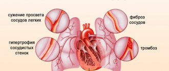
| Right atrium | Ambiguous signs of arch lengthening are visualized, and the vena cava is dilated. Indicates the development of pulmonary hypertension |
| Ventricle on the right | Lengthening and expansion of the pulmonary trunk, the upper part is rounded, “looks” upward, the cardiac diameter has an increased size, the space behind the sternum is not defined. Clinical picture of congenital defects of the septum and cor pulmonale |
The structure of the human heart
Muscle fibers (myocardium) are the predominant type of heart cells. They make up its bulk and are located in the middle layer. The outside of the organ is covered with epicardium. It wraps at the level of the attachment of the aorta and pulmonary artery, heading downwards. In this way, the pericardial sac is formed. It contains about 20 - 40 ml of clear liquid, which prevents the leaves from sticking together and being injured during contractions.
The inner membrane (endocardium) folds in half at the transition of the atria into the ventricles, the mouths of the aortic and pulmonary trunk, forming valves. Their valves are attached to a ring of connective tissue, and the free part moves with the blood flow. In order to prevent the parts from everting into the atrium, threads (chords) are attached to them, extending from the papillary muscles of the ventricles.
The heart has the following structure:
- three membranes - endocardium, myocardium, epicardium;
- pericardial sac;
- chambers with arterial blood - left atrium (LA) and ventricle (LV);
- sections with venous blood - right atrium (RA) and ventricle (RV);
- valves between the LA and LV (mitral) and tricuspid on the right;
- two valves separate the ventricles and large vessels (aortic on the left and pulmonary artery on the right);
- the septum divides the heart into right and left halves;
- efferent vessels, arteries - pulmonary (venous blood from the pancreas), aorta (arterial from the left ventricle);
- afferent veins - pulmonary (with arterial blood) enter the LA, vena cava flow into the RA.
We recommend reading the article about minor anomalies of heart development. From it you will learn about the causes of pathology in children, adolescents and adults, symptoms of the problem and diagnostic methods, treatment of the disease and prognosis for patients. And here is more information about the location of the heart on the right.
Cost of the procedure
The price of the procedure varies depending on a number of factors. The difference can be up to five times and depends on the region and the specific clinic. In the country, you can get an x-ray, paying from 750 to 3500 rubles.
The average cost of a one-time cardiac x-ray in Russia is accepted at the level of 2 thousand rubles.
Cardiac X-ray is one of the diagnostic methods that helps the cardiologist make an accurate diagnosis. The examination techniques used help to assess the structural components of the organ and determine the condition of the system as a whole. The procedure is easy to perform, takes little time, and is safe for the body. MRI is also an alternative method.
Preparation and carrying out diagnostics
You will only need to prepare if you need to undergo a cardiac X-ray with contrast. It involves taking tests to determine if you have an allergy to contrast, as well as visiting the office on an empty stomach, which will help avoid nausea.
To begin the procedure, you need to remove all jewelry or metal accessories that may affect the quality of the photo. The procedure is performed in 4 different projections:
- front;
- lateral left;
- oblique left (at an angle of 45 degrees);
- right.
Thanks to this, the description and conclusion of the heart x-ray will be as accurate as possible. The doctor will examine all planes of the organ and note the presence of pathological processes.