Cardiovascular diseases occupy a leading place in the structure of general morbidity and cause the death of hundreds of thousands of people around the world. Most often, disorders of the heart and blood vessels are diagnosed in people who have crossed the threshold of adulthood. Therefore, upon reaching 45 years of age, the following questions become relevant for men and women: “How to check the blood vessels?”, “What examination should I undergo?” Detailed information about methods for studying the bloodstream is presented in the text of this article.
Primary examination of the patient's heart
You can check your heart function at your nearest clinic. The first way to look for cardiovascular pathologies is physical examination. It is necessary to conduct an examination to determine external changes, and after tapping, the specialist will determine the boundaries of the organ. If pathologies arise, the boundaries are distorted. On a note!
Auscultation of the heart is a method of physical examination; with its participation, rhythm disturbances and murmurs can be detected.
A phonendoscope is used for the procedure. During the manipulation, listen to heart sounds:
- sound;
- sequence.
In a healthy organ, there are two constant tones, but in pathology, an additional one is detected. It is necessary to undergo regular examinations, tests and examinations. Most diseases are curable, the main thing is to suspect the pathology in time.
Rheoencephalography of cerebral vessels
REG (rheoencephalography) is a non-invasive method for diagnosing cerebral vessels, recording changes in the electrical resistance of vascular walls when a high-frequency pulse passes through them. Provides an opportunity to check the vessels of the head, their reactivity, tone, elasticity, level of resistance of vascular tissue, possible blockage and characteristics of blood supply.
REG is indicated for suspected diseases:
- vascular pathologies;
- circulatory disorders;
- atherosclerosis;
- stroke;
- vascular stenosis;
- clarification of the effect of drugs;
- traumatic brain injuries.
To perform rheoencephalography, rheographs with a number of channels from 2 to 6 are used, recording the corresponding number of vessels. The patient is sitting or lying down. Electrodes are installed on his head using rubber bands. A special paste can be applied underneath them to improve contact with the skin and reduce resistance. Electrodes are attached depending on the vessel being examined. So, if you need to check the internal carotid artery, the electrode is placed on the bridge of the nose and mastoid process.
Some electrodes send electrical signals, others receive them and transmit them to the rheograph. The latter processes and transmits to a monitor or print in the form of a graph - a rheoencephalogram. The procedure lasts several minutes.
During the examination, the patient may be given a small dose of nitroglycerin under the tongue, Papaverine, or asked to change body position or turn his head. These measures will allow you to see a change in tone and blood supply.
The advantage of rheoencephalography is its simplicity, safety for the patient, and the ability to monitor blood flow for a long time. Disadvantages include the inability to measure cerebral blood flow and determine the exact cause of circulatory disorders.
Before the study, you should not smoke, drink alcohol, coffee, or energy drinks. If the patient is taking medications, the doctor must be informed about this. Some may need to be stopped for a while.
Rheoencephalography is not prescribed for children under 7 years of age, as well as for wounds and injuries at the places where the electrodes are attached.
How to check your heart at home
Heart disease occurs unexpectedly. They do not manifest themselves in any way until a condition arises when it is impossible to do without a hospital. It is possible to check your heart at home. It only requires a stopwatch and some knowledge.
To measure your resting pulse, sit down and place your fingers on the radial artery in your wrist or on the artery in your neck. The number of strikes in 10 seconds is counted. The resulting number must be multiplied by 6. All data must be recorded.
Heart test on the stairs
The test was developed by the famous cardiologist N.M. Amosov. The bottom line: in 4 minutes you need to climb the stairs to more floors. At first glance, the test seems simple, but it very accurately reflects the state of the cardiovascular system.
On a note! You need to climb to the 5th floor at an average pace; if your pulse exceeds 140 beats per minute, then your condition is very bad. In this case, it is not worth checking the heart in this way.
The test shows fitness based on the number of floors completed:
- less than 7 floors – very poor training;
- exactly 7 floors – bad;
- 11 floors – satisfactory;
- 15 floors – good training;
- more than 16 floors – excellent.
Provided that such parameters are up to 30 years old, but also those over 50 years old, with good heart health, can show excellent results. It is important to pay attention: if during the experiment the heart beats are more than 150, then you need to slow down and move up the stairs at medium speed.
Martin Kushelevsky's sample
With the help of such a test, it is possible to find out at home not only the capabilities of the cardiovascular system, but also its strength. In 30 seconds you need to squat 20 times. Immediately after performing, check your pulse. Rest for 2 minutes and check the measurements again. If the result is the same as at rest, then everything is fine with the heart. If in 2 minutes everything has not had time to recover after the load, then this may indicate some kind of violation.
You can do a jump test. The normal heart rate is 60-70 beats at rest. Before performing, record the initial pulse. Afterwards you need to perform 60 jumps in 30 seconds. Take measurements again and evaluate the indicators.
Stange and Gench test
You can also examine the heart using the Stange method: for the experiment, you need to record your pulse for 30 seconds while standing, and then while sitting, hold your breath while inhaling. Record the time with a stopwatch. After holding your breath, measure your pulse again for 30 seconds.
If holding your breath is:
- less than 39 seconds, then this is bad;
- 40-49 seconds is normal;
- more than 50 seconds – a good result;
Then you need to divide the pulse after the test by the pulse before the test; if the numbers are more than 1.2, the cardiac system reacted negatively to the lack of oxygen in the blood.
There is also the Gench technique. You need to take your pulse before testing, and then stop breathing with a full breath. The time must be recorded using a stopwatch. After the experiment, you need to measure it, as before.
If a person can hold their breath:
- less than 34 seconds is not very good;
- 35 – 39 seconds – normal;
- more than 40 seconds is good.
The response of the cardiovascular system is calculated using the same formula as the Stange test.
Orthostatic test
This test is usually recommended for people who experience fainting for an unknown reason. And also, those who do not have heart disease. The patient is fixed to a table with a motor and left in a supine position for 15 minutes. Then they raise their head up at an angle of 60-80 degrees and observe for 45 minutes.
You need to monitor your blood pressure, weakness, and pulse. Sometimes the procedure can give a false positive result.
Electroencephalography of the brain
EEG is a non-invasive way to study the electrical activity of the brain, including under the influence of certain stimuli: light, sound, movement. Indirectly indicates a change in blood circulation, therefore it is not the main way to diagnose vascular disorders.
Indications:
- vascular lesions;
- epilepsy;
- neoplasms;
- traumatic brain injury;
- inflammatory diseases.
The brain examination is carried out using a special device - an encephalograph, capable of recording the frequency of electrical oscillations from 0.5 to 100 Hz. Electrodes are placed on the head to pick up minimal brain signals. The signals enter the amplifier, are magnified millions of times, and are transmitted to the computer monitor in the form of a graph - an encephalogram. In children under 3 years of age, the study is performed only during sleep or under light anesthesia.
The recording reflects the fluctuations and rhythms of the electrical process occurring in the nerve cells of the head:
- Alpha rhythm with a frequency of 8-14 Hz, characterizing a state of rest.
- Beta rhythm 13-30 Hz, indicating depression and anxiety.
- Delta rhythm 0.5-3 Hz, typical for sleep.
- Theta rhythm is 4-7 Hz, characteristic of an adult and a child in sleep.
The predominance of alpha and beta rhythms, the same electrical activity in both hemispheres and the occurrence of only a local reaction to the stimulus are signs of a normal encephalogram. EEG does not provide an anatomical picture of the structure of blood vessels.
Before performing an encephalography, you need to prepare. You should not eat 2 hours before the procedure, you should not drink coffee or energy drinks 12 hours before, and you should not smoke. Do not use hair styling products. The time the procedure may take is 45-120 minutes.
Electroencephalography is not used during periods of exacerbation of mental disorders, mental trauma, infectious diseases, injuries or wounds on the scalp. Unless absolutely necessary, it is not performed in children under 7 years of age.
Instrumental diagnostic methods
The technologies of our time can carry out various tests. For correct therapy, you need to know an accurate diagnosis. They are examined using standard methods: electrocardiography, echocardiography, angiocardiography. Sometimes they add: MRI, CT. Some of these methods are completely painless. Each procedure is carried out with equipment that may have a negative effect on the body.
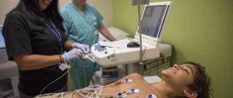
Electrocardiography
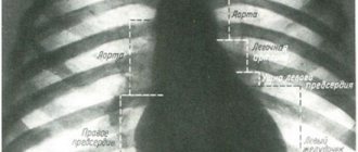
The most common way to examine the heart. Using ECG, most cardiovascular diseases are detected. An electrocardiograph records the electrical activity of the heart, and thermal paper is used for recording. If the test is carried out using electronic equipment, the results can be recorded in the system.
On a note! Electrical indicators are detected using a conductor, which displays all the results on a cardiograph.
Electrocardiography determines:
- presence of scars;
- disruption of blood flow;
- disruption of cellular metabolism;
- signs of myocardial muscle infarction;
- heart rhythm disturbance;
An ECG may sometimes fail to detect changes, so they use other studies that are based on it. Daily ECG monitoring is capable of detecting even mild heart rhythm disturbances. Such failures are difficult to detect with a standard ECG. The patient needs to wear a hand-held cardiograph for about a week, which also records changes.
ECG mapping or precordial mapping - with the help of a large number of electrical conductors, long-term diagnostics are carried out, as a result, even rarely identified diseases can be determined. Gives fairly accurate predictions.
Stress tests are prescribed to identify errors in the functioning of the heart that cannot be determined at rest. During exercise, all the factors that determine arrhythmia or other disorders appear. For example, when using bicycle ergometry you can determine:
- angina pectoris;
- coronary heart disease.
During the next heart test under load, you can understand whether the therapy has been chosen correctly and whether there is progress.
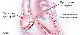
Intraesophageal electrocardiography - an electrical conductor is placed near the esophagus, located next to the heart. Often used to recognize rhythm disturbances. Vectorcardiography – builds a three-dimensional figure of the organ, which shows the electrical activity of the heart. With irregular rhythms, disturbances in the functioning of the cardiac system appear. Gastrocardiomonitoring - examination of the heart and acidity in the stomach. Used to identify gastrointestinal and cardiac problems.
Echocardiography
The method is more popularly known as cardiac ultrasound, or ultrasound diagnostics. This examination picks up signals that reflect the structure of the heart. The signal is not formed in the same way, but as a result it is possible to obtain the corresponding image. The method is used to identify heart lesions, congenital and acquired defects, as well as to assess the capabilities of the myocardium. A number of cardiac diagnostics have been created based on ultrasound:
- Doppler echocardiography - ultrasound shows intracardiac blood flow. This helps diagnose heart defects, shunts, chordae, and assess cardiac dynamics.
- One-dimensional echocardiography - makes it possible to see the heart in a plane. The procedure is needed to determine the mass of the organ, the thickness of the walls, the valve apparatus, and the contractile function of the heart.
- Two-dimensional echocardiography – this test provides more information compared to one-dimensional echocardiography. It is obtained through volumetric visualization.
This study is chosen when patients complain of weakness, dizziness, pain, fainting and tachycardia.
Angiocardiography
Diagnosis involves X-ray analysis, so a radiocontrast agent is used. With such cardiography, it is possible to examine the chambers of the heart and nearby veins and arteries from all sides. A catheter is used, it is connected to the heart and blood vessels in the femoral or subclavian artery, and then contrast is injected into the emptiness of the heart and blood vessels. All parts of the heart become visible and pictures are taken.
Attention! It is important to take a sedative or antihistamine before this test.
A similar study is carried out before operations to confirm the physical parameters of the heart. The method is accurate in establishing the following diagnoses:
- heart disease;
- disruption of the structure of large vessels.
After the popularity of echocardiography, angiocardiography became very rarely used. Only sometimes does it remain indispensable, because this procedure can accurately determine the structure of the heart. Angiocardiography is an invasive research method, so it is rarely used.
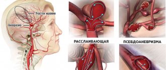
When is a blood vessel check needed?
There are rare signs of health problems, the appearance of which a person does not pay attention to. In hot weather, I lost my balance and had a severe headache - it’s okay, it will pass, it’s because of the heat. I stood up sharply, “spots” flew in front of my eyes - that’s okay too, I had to get up more slowly. The pressure has risen - of course it will rise, the weather is changing so quickly. There are explanations and justifications for everything, but any of these signs may indicate a serious pathology.
If you experience the following symptoms, you should immediately consult a doctor:
- The headache is constant and getting worse.
- Vision, speech, hearing deteriorate, and there are incidents with complete loss of these functions.
- Thought processes, memory, and attention are disrupted.
- Coordination of movements changes, loss of balance and the appearance of a shaky gait are possible.
- Convulsions appear.
- The person loses consciousness or experiences a faint state.
- Intracranial and blood pressure changes.
- “Floaters” appear before the eyes.
These symptoms indicate pathology of the cerebral vessels. To exclude it, a hardware medical examination may be prescribed. It includes both the basic procedure for checking vessels for patency (called angiography), as well as ultrasound, rheoencephalography, and electroencephalography.
These methods are used to clarify traumatic brain injuries, neck injuries, stroke, neoplasms, diseases accompanied by inflammation of the brain, thrombosis, and atherosclerosis. A head examination is performed before or after vascular surgery.
Depending on the pathology, either one type of study or several may be prescribed. Many methods have limitations, so they are prescribed when absolutely necessary.
Lab tests
Prescribed for diseases or the risk of their occurrence. In some heart disorders, the blood can secrete certain substances that can only be detected when tested in a laboratory. Laboratory tests used to detect heart failure include:
- Analysis of urine. The kidneys give a strong response to the health of one of the main systems of the body. With swelling, palpitations or inflammation of the inner lining, the abundance and composition of urine changes. In case of disturbances, red blood cells and protein decrease.
- Blood analysis. With heart defects, red blood cells often become enlarged. Such changes are due to lack of oxygen in heart failure. Inflammations often affect the inner layer of the membrane and the myocardium. As a result, the ESR increases.
- Sputum analysis. Prescribed when there is doubt about severe left ventricular failure. With this disorder, congestion occurs in the lungs and bloody-foamy sputum appears. Sometimes it may be colorless.
On a note! Sputum examination helps identify “cardiac cells”; their presence characterizes myocardial infarction and heart failure.
During any of the studies, it is important to comply with the doctor’s requirements. Any procedure must be prescribed by the attending physician. If you follow the rules, you can get a diagnosis (if there is one) and treatment. You need to cooperate with your doctor and then the treatment will be effective.
Ultrasound of the brain
Ultrasound (echoencephalography) is based on the principle of signal reflection from brain structures at different angles. Ultrasound is a non-invasive study of the brain, it allows you to obtain information about the characteristics of the walls, the patency of blood vessels, their diameter, the presence of atherosclerotic plaques, and check the speed of blood flow.
The study is indicated if the following disorders are suspected:
- cervical osteochondrosis;
- headache;
- dizziness;
- violation of movement coordination;
- stroke;
- diabetes;
- hypertension;
- elevated cholesterol levels.
Some time before examining the vessels, it is recommended to exclude coffee, strong tea, energy drinks, alcohol, and avoid strong emotional stress.
Before the procedure, jewelry is removed from the head and neck. The patient lies or sits. It is important to provide him with peace. A doctor or nurse checks the head for deformities, bruises, and ultrasound sensors are installed on the sides of the skin, which has been previously lubricated with gel.
The advantages include ease of implementation, non-invasiveness, safety and lack of contraindications. The main disadvantages are the inability to see dynamics, the lack of accurate information about blood circulation in small vessels and some vascular pathologies. In general, ultrasound cannot replace angiography.
Has no contraindications. The duration of the procedure is 30-40 minutes. Ultrasound examination of a child under 1 year of age is called neurosonography.
Ultrasound is usually combined with Doppler ultrasound. It is this combination that allows you to obtain not only a static picture, but also the sound characteristics of the blood flow.
Dopplerography
This diagnostic method is based on the reflection of an ultrasound beam from blood cells. A special transducer sends signals to the blood vessels, they are reproduced in frequencies corresponding to the blood flow and converted into sound signals. The changing frequency of the signal allows you to evaluate the flow rate, direction of blood, volume per second, obtain information about the lumen, size of blood vessels, their resistance, and the development of atherosclerotic stenosis.
During the procedure, the patient lies on the couch. A cushion is placed under the head. A gel is applied to the skin and a special sensor is passed over the head, viewing and measuring all the vessels. Typically, sensors are installed on the temples, above the eyes, and on the back of the head. The doctor may also ask you to hold your breath or make some movement to assess a possible reaction.
Duplex
This type of ultrasound examination combines two methods - direct ultrasound, which provides information about the anatomical structure of blood vessels, and Doppler ultrasound, which allows one to assess cerebral blood flow.
On the screen, a two-dimensional image of a blood vessel among the tissues is obtained, as well as signs of plaques, blood clots, thickening of the walls of blood vessels, and their pattern. The vessel can be viewed both longitudinally and in cross section. Duration – 30-45 minutes.
Triplex
The study includes 3 methods: ultrasound, Doppler and color Doppler mapping. In the process, the vessels are examined, the thickness of their walls, resistance, while the vessels are painted in different colors, depending on the speed of blood flow, and this is what distinguishes the study from duplex.