Intraventricular conduction disturbance is a type of cardiac arrhythmia. This changes the process of impulse transmission through the ventricular myocardium. The pathology may not have clinical manifestations and is discovered accidentally.
Also, conduction disturbances can develop against the background of another heart disease. Pathology is diagnosed using electrocardiographic examination. The article talks about intraventricular conduction disorders and the manifestation of this condition on the ECG. The causes of the pathology are described and the principles of treatment are indicated.
Ventricular conduction disorders are a type of cardiac arrhythmias.
The mechanism of pathology development
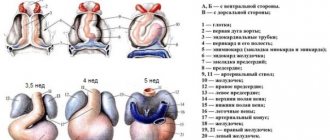
In order to fully understand the nature of the deviations within this form of the process, you need to refer to the anatomical and physiological certificate.
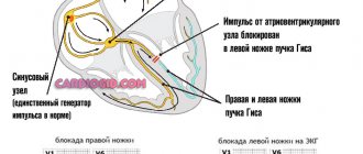
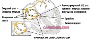
The generation of an electrical impulse in cardiac structures occurs in the natural pacemaker, the so-called sinus node. This is a cluster of special cardiomyocyte cells capable of spontaneous excitation.
The signal travels through fibers known as bundles of His to the ventricles and other structures, ensuring normal contractility of the entire organ. These pulse “conductors” are formed by two legs. Right and left. Then they branch within the cardiac structures.
If there is a partial or complete blockage due to injury or congenital malformations, the signal cannot travel further.
Some part of the heart is excluded from working because muscle fibers do not contract. In some cases, this does not pose a danger to life and health; in some situations, death is likely.
Everything depends on the localization of the process, and on the other hand, on the volume of the lesion and the duration of the condition.
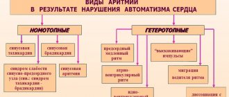
Classification
Based on the structure of the His bundle, a classification of intraventricular conduction pathologies is created.
Based on the number of blocked elements in the conductive system, existing types of violations are determined (arranged according to the degree of danger):
- Single-bundle blockade - inhibition of patency is observed in just one leg.
- Double-bundle pathology - affects two pathways.
- Three-bundle pathology - development occurs on all His bundles; impulses cannot reach the end point along any path.
According to the nature of the disease, the pathology can be persistent, transient (unstable signal transmission) and alternating, in which signs of different blockades may appear during examinations at different times. The division occurs into complete disruption of conduction and incomplete conduction, with inhibited conduction, but still delivering an impulse to the end point.
Classification of the phenomenon and its types
Three types of pathological process are observed within the ventricles.
- Leg block. A particularly common form of the disease. Accompanied by a minimal clinical picture when the right branch is affected. The left-sided option is much more dangerous, since the operation of the camera of the same name is disrupted. In the absence of an adequate signal, the systemic circulation suffers, generalized hypoxia affects all organs and tissues at the same time. In the long term, this leads to atrophy, multiple organ failure, cardiac arrest, stroke, and vascular dementia. Clinically harmless variants can last a lifetime without visible manifestations.
- Insufficient speed of movement of the electrical impulse along the fibers. The severity of the clinical picture and threat to life again depends on the nature of the pathological phenomenon. However, this is a less dangerous type compared to the classic complete block of the legs. Slowing of intraventricular conduction is the result of chest trauma, surgery, or congenital heart defects.
- Atrioventricular (AB) block. A rarer type. It is characterized by selective stopping of the movement of electrical signals along cardiac structures.
You can also classify the process on such a basis as the nature and degree of deviations in conductivity.
Then the following stand out:
- Complete blockade. The signal doesn't get through at all. This is the most dangerous clinical option. Outside the course of symptoms, cardiac arrest occurs in many cases.
- Partial ventricular nonconduction. The likelihood of lethal complications is present, the clinical picture is pronounced, but to a lesser extent compared to the first process.
- Slow conduction. Almost not noticeable until a certain point.
Finally, classification is possible based on the extent of the changes.
Thus, we distinguish:
- Generalized nonconductivity. Not only the ventricles suffer, but also the atria.
- Focal (local) conduction disturbances. One or two ventricles are involved. It proceeds relatively easily, without pronounced symptoms, the risks to life are determined by the localization of the process. If the left side is affected, the probability of death is almost three times higher.
A more accurate version looks like this:
- Single-beam nonconductivity. With blockade of a single branch.
- Bifascicular form.
- Full variety. When three fibers are blocked.
Clinical types are important for developing treatment tactics.
Study of heart disease in childhood
Studies have shown that about five percent of children who are healthy according to basic indicators have myocardial conduction disorders. And if we talk about first-degree blockade, then the situation will worsen with age. Complications can be caused by various heart pathologies and previous surgeries.
Symptoms of the disease in children are as follows:
- problems with memory and memorization,
- attention problems
- poor academic performance, if we are talking about a schoolchild,
- dyspnea,
- pain in the heart area,
- general weakness,
- rapid fatigue of the baby after minor stress (physical or emotional) and more.
The course of treatment for intraventricular conduction pathologies, as for adults, depends on the cause of the disease. The first stage of myocardial conduction does not require special treatment. Just an observation. If the situation has worsened to the second degree of severity, it is recommended to include supportive medications, and sometimes install a pacemaker. When the third stage of the disease is determined in a child, surgical intervention is required, which involves implantation of an pacemaker (artificial pacemaker).
Important! Children are the flowers of life. And it should not be surprising that with age, all symptoms and manifestations of the disease may completely disappear in a child. It's quite normal.
Reasons for violation
Factors in the development of a pathogenic phenomenon are always cardiac. It is extremely rare to find a different moment. Even so, it is indirectly related to the heart. Among the possible options:
Left ventricular hypertrophy
A classic type of organic change in cardiac structures against the background of long-term arterial hypertension. Moreover, if the process is active, without appropriate treatment the change is formed in a matter of years, sometimes faster.
It does not manifest itself clinically. Detected during echocardiography. It may be an incidental (random) finding.
Hypertrophy itself does not require correction; it is a permanent and irreversible deviation. However, if signs are present, symptomatic treatment is carried out aimed at eliminating the consequences.
Blockade of the legs of His or other types of conduction disorders are relieved in parallel, as part of a complex of manifestations.
Heart attack
Current, but more often transferred in the recent past. After emergency care and some time in the hospital, the patient may begin to experience arrhythmia, chest pain, shortness of breath during physical activity of the usual kind.
The problem is that at the moment of necrosis, the nerve conductive fibers also die. They are replaced by rough scar tissue.
The prospects for complete recovery depend on the extent of the process. The larger it is, the worse the forecasts. May be fatal in the short term.
Those who are luckier need to constantly take medications, and lifelong maintenance therapy is possible. Otherwise, there is a risk of adding to the sad statistics.
Arterial hypertension
At the first stage there are no deviations or they are minimal. The most threatening changes begin at stages 2-3 of the pathological process.
It is not so easy to adjust blood pressure levels, especially at the time of acquiring resistance. This is an inevitable outcome without treatment.
Coronary heart disease (CHD)
Inseparable from a heart attack. Only in this case, the pathological changes are not yet so great; this is a chronic phenomenon. It constantly makes itself felt, the symptoms increase gradually. Without therapy it leads to acute myocardial necrosis.
Cardiomyopathies
Heterogeneous group of diseases. They have one thing in common: a violation of the functional activity of the ventricles or atria. The muscle layer is not able to contract normally. This is a potentially fatal condition.
Atherosclerosis
And the subsequent processes of partial necrosis of cardiac structures. It is relatively rare. Mainly as a result of the long duration of the condition.
Without treatment it is potentially fatal.
Congenital and acquired heart and vascular defects
For the most part, they are asymptomatic, without any deviations in health. This is why they are dangerous.
To prevent sudden death, regular preventive examinations by a cardiologist are recommended. Once a year or a little more often.
Inflammatory pathologies of cardiac structures
Infectious or autoimmune origin, for example myocarditis or pericarditis. There is no fundamental difference in the clinical picture; the differences relate to prognosis and treatment methods.
Slightly less frequently, violations of VZhP are caused by diabetes mellitus, excessive production of thyroid hormones, COPD, bronchial asthma, smoking, alcohol abuse, and various intoxications.
A polyetiological process is possible, when many reasons fuel the onset of deviations at once. The only way to detect a problem early is to at least perform an ECG and listen to the heart sound (auscultation).
Characteristic symptoms
A great difficulty for diagnosticians and the patient himself is the complete absence of pathological manifestations from the body.
Of course, this does not mean that the clinical picture is absent. It will not be possible to simply detect signs without specific techniques. Only in the later stages or with a significant volume of blockade are the moments quite specific.
Among the symptoms:
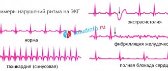
- Slowing (slow ventricular rhythm) or accelerating heart rate. Violations of contractility are manifested by other types of processes, for example, it is possible to change the time interval between each subsequent blow.
- Chest pain. Burning or pressing. They are characterized by inconstancy and paroxysmal course. The duration of the episode is minimal, from a couple of seconds to several minutes.
- Dyspnea. During periods of intense physical activity or in a state of complete rest. Depends on the extent of damage to cardiac structures.
- Weakness, drowsiness, apathy and reluctance to do anything. The patient not only does not want to. But he cannot perform everyday household duties. Although this option is relatively rare.
- Fainting and syncope.
But we need to make a reservation once again: such manifestations almost never occur.
As a result of severe deviations in the functioning of the organ, death is possible, sudden, without preliminary signs.
In such a situation, the cause of death is determined using the pathological method.
There are many more objective moments. They are determined in the process of diagnosis and verification of the suspected disease.
Also, the intensity of symptoms increases as a result of the development of emergency conditions (see complications). But these are special cases, for the most part it is a sneaky, silent disease.
Whether it is dangerous or not is difficult to say without an assessment; it depends on the “luck” of the patient. Some live for decades and suspect nothing.
The main factor in the development of the typical moments described above is a three-bundle, that is, a complete blockade of the legs of His, which is rare.
Symptoms
The clinical picture in this case depends on the factor that influenced the failure in the process of conducting the impulse. Most people are concerned about the following symptoms:
- the occurrence of swelling in the legs;
- manifestation of shortness of breath even without physical exertion;
- frequent dizziness due to disruptions in cerebral blood supply;
- feeling of a sinking heart;
- a feeling of lack of air, accompanied by fear and panic;
- memory impairment;
- fast fatiguability;
- attacks of bradycardia;
- constant mood changes;
- impaired coordination of movements;
- general weakness.
Sometimes a 1-2 degree blockade does not manifest itself in any way, unlike its full form. The following signs are typical for the advanced stage of the disease:
- sudden loss of consciousness;
- heartache;
- reduction in contraction frequency down to 30-40 per minute.
3rd degree block is manifested by complete separation of the ventricles and atria, which can be fatal. If we add a feeling of malaise and cold sweat to the stated symptoms, then such a clinical picture is often a harbinger of an imminent myocardial infarction. A patient can be helped by a timely visit to the clinic for hospitalization and treatment.
Diagnostics
Examination of patients with ventricular conduction disorders is the task of a cardiologist. To a lesser extent, other specialists. Their help may be required in the context of non-cardiac pathologies: diabetes mellitus, thyrotoxicosis, neurogenic problems.
Approximate scheme of events:
- Questioning the patient. It does not have great clinical significance, since in 90% of cases there will be no characteristic health complaints or they will be nonspecific.
- Anamnesis collection. Family history, lifestyle and habits, treatment, nutrition, physical activity, sleep are all important factors. Also somatic diseases diagnosed earlier.
- Measuring blood pressure and heart rate is not enough to establish the fact, but within the framework of identifying the etiology, this is exactly what is required.
- Daily monitoring as needed. Aimed at establishing the dynamics of cardiac activity during the day.
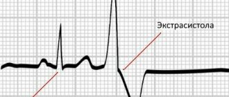
- Electrocardiography. Profile research. Usually the invasive type is indicated; for this purpose, a special probe is built into the heart. Intraventricular conduction disturbances on the ECG are manifested by changes in the QRS complex and P wave. Also by other features.
- Electrophysiological technique.
- Echocardiography. To identify organic abnormalities in cardiac structures. Allows you to see defects and hypertrophic phenomena. Carried out on a first-come, first-served basis.
- Angiography.
- CT or MRI. In especially difficult cases. Allows detailed visualization of tissue, but it is a static image. You can't see much of it.
It is impossible to single out any one study; a complex is needed. To ascertain the fact, ECG and ECHO-CG are used. Next, verification is carried out in other ways.
Explaining the concept
Surely you remember from your school anatomy course that special nerve tissues are responsible for the transmission of nerve impulses. Myocardial contractions are also caused by the transmission of impulses for which the conduction system of the heart is responsible. It consists of nodes and branches called the bundle of His. Anatomically, it is located in the interventricular septum; in its structure there are two legs - left and right, which conduct impulses to the corresponding ventricles. This:
- The right bundle branch is a single bundle of fibers that branches at the bottom of the right ventricle and is responsible for conducting impulses to this area.
- The left leg has two branches, left and right, each of which conducts an impulse to a specific wall of the left ventricle. The branches of the left leg have their own name, these are Purkinje fibers.
However, the impulse is not immediately conducted through the His bundle, forms its sinus node, from where the impulse is transmitted to the atrial conduction system, and only after its excitation through the Bachmann bundle enters the atrioventricular node. It is from the atrioventricular junction that the impulse enters the ventricular conduction system. Schematically, the conduction of an impulse looks like this:
Sinus node (Keith-Fluck node) - atrial system, Bachmann bundle - atrioventricular node (Aschoff-Tawar node) - bundle of His.
Conduction disturbances in any area are called blockades. In the modern classifier of diseases, nonspecific intraventricular block is assigned a code according to ICD-10 - I45.4.
Treatment
Mixed therapy is aimed at eliminating the underlying cause. On the other hand, to relieve symptoms. The doctor follows this dual path.
The primary measure is drug treatment. Even if the problem cannot be eliminated using a conservative method, no one will immediately prescribe surgery, except in emergency cases.
First you need to stabilize the patient's condition. This will reduce the risks of complications.
Medicines:
- ACE inhibitors and other antihypertensive drugs. The number of items is large, the combination is determined by a specialist.
- Antiprhythmic agents. Amiodarone and analogs.
These are preparatory activities. The main goal of treatment is to eliminate the etiological factor. Possible surgical techniques are aimed at correcting heart defects, prosthetics in case of destruction, restoring conduction through bypass paths, and installing a pacemaker.
However, therapy is not always required at all. The patient needs to be monitored for some time. If dynamic monitoring for 6-12 months does not indicate any negative movement, you can postpone supervision.
After 5 years, the patient does not require further observation and is considered conditionally healthy.
As part of self-help, it is recommended to give up smoking, alcohol, physical and emotional stress, prolonged sitting in one place, and uncontrolled use of medications.
Normalization of the diet is also included in the scope of measures. The nature of nutrition is adjusted with a nutritionist or independently.
Tables No. 3 and No. 10 are recommended; you can create the menu yourself, it’s not difficult.
Fractionation, small portion sizes, boiling or baking, these are the basic principles of the new “paradigm”. Salt no more than 7 grams per day, complete refusal is unacceptable. Sugar should also be present, not counting cases of diabetes.
What kind of disease is this?
Before you find out how to properly treat the disease, you need to find out what impaired intraatrial conduction means and what is the reason for its development. If even minor changes in the order of heart contractions occur in the body, this can negatively affect the functioning of each organ. This condition indicates that the process of blood flow into the aorta is difficult.
Doctors define several stages of the disease. Depending on the nature of the disorder, the following blockades can be distinguished:
- Incomplete. It can be detected at the initial stage of development of the disease. Its main symptom is the slow transmission of impulses transmitted through the fibers of the heart.
- Full. It is observed in patients who do not follow the doctor’s recommendations, or the therapy was chosen incorrectly. This type is most often incompatible with life and all because the passage of impulses is completely stopped.
The main reason for the disruption of intraatrial conduction may lie in damage to the heart muscles, the presence of defects and coronary artery disease. Smoking and alcohol abuse play an important role in the development of this pathology. An unhealthy lifestyle, stressful situations, and taking medications that stimulate the cardiovascular system have a negative impact on the functioning of the heart.
There are the following types of blockades depending on the location of the pathology:
- interatrial;
- sinoarterial;
- artioventricular;
- intraventricular.
Failures in the functioning of other organs and electrolyte disturbances caused by long-term use of diuretics can also lead to a slowdown in intraatrial conduction.
Possible complications
Possible consequences of acute or long-term conduction disturbances:
- Cardiogenic shock. A sharp drop in blood pressure combined with arrhythmia and generalized hemodynamic dysfunction. It is almost impossible to remove the patient from this condition; mortality is close to 100% in some forms. In other cases, the chances of survival are slightly higher (mortality rate is about 60%).
- Morgagni-Adams-Stokes attack. A drop in blood pressure due to impaired cardiac output. In itself, it is accompanied by fainting, and a stroke or heart attack may follow. Requires urgent treatment of the underlying disease. A pacemaker is installed.
- Cardiac arrest (asystole). This is the leading cause of sudden death in patients.
- Ischemia of the kidneys and liver. The result is dysfunctional disorders.
All of these phenomena are potentially fatal. What the risks are can only be determined through long-term observation.
How does the disease affect the functioning of all organs?
If the patient has only partial disturbances of sinoarterial conduction, then there will be no symptoms, but if complete, he may experience chest discomfort and dizziness; these symptoms are provoked by poor blood circulation in the brain.
The interatrial form of the disease is not life-threatening for the patient. But if it is, then the risk of developing atrial fibrillation increases several times, which is already a threat to health. A similar condition manifests itself in the form of shortness of breath at rest or while walking, swelling of the lower extremities, bluish discoloration of the skin, and discomfort in the chest area.
Atrioventricular disturbance of intraatrial conduction only manifests symptoms in some patients, but if bradycardia is additionally observed, then no one is immune from an attack of MAS. At this moment, the patient experiences weakness throughout the body. Dizziness with loss of consciousness and convulsions, which are associated with impaired blood flow in the brain, may occur. This condition requires urgent hospitalization and qualified assistance. If no measures are taken, the patient faces cardiac arrest.
The intraventricular form manifests itself due to difficulty passing blood through the bundle branches. Partial blockage does not always manifest symptoms, but a rare pulse, loss of consciousness and chest discomfort can indicate a complete blockage.
Forecast
Doctors assess prospects only based on the results of long-term monitoring of the condition. Something concrete can be said after 3-4 months. If the phenomenon occurred suddenly, then during therapy.
Based on practice, the following calculations can be made:
- With blockade of a single beam, mortality is no more than 3%, two - 32%, three - 70%.
- Right ventricular nonconduction of one fiber is not dangerous at all and does not even require treatment.
- In the absence of competent therapy, the risk of complications described above against the background of dangerous forms of the pathological process is almost 100%.
Favorable signs:
- Early start of supervision.
- Young age.
- Good response to therapy.
- Successful operation and no problems after the intervention.
- Improvement in well-being, disappearance of symptoms.
The older the patient, and the presence of a large number of somatic pathologies, the likelihood of a favorable outcome is several times lower.