In what days, in what week of pregnancy does the heart beat | heart of an embryo, fetus, child.
After the first emotions regarding the imminent addition to the family have subsided, future parents try to get as much information as possible about the baby’s condition. Is the pregnancy going normally, does its development correspond to the gestational age.
- A woman transfers her own idea of good health to her baby: does the baby eat well, when he sleeps, and when he is awake. However, the most important indicator of the condition of the fetus in the mother’s body is the beating of its heart.
Video: Fetal heartbeat in early pregnancy
- The venous and arterial sections, which have grown over time, are separated by a deep constriction, after which the formation of a two-chamber fetal heart begins. This happens in the 4th week of development.
- Now the baby has only a systemic circulation. After the lungs are formed, the pulmonary circulation will develop. The size of the heart is tiny and resembles a poppy seed, but it works clearly and rhythmically.
- At the 5th week or at the beginning of the 6th (this is the seventh obstetric week), the baby’s heart is already three-chambered due to the appearance of the interatrial septum.
Stages of Heart Development
- 6th week of development: the ventricular chamber is divided by a septum. At this stage, valve formation also occurs. The common arterial trunk is now divided. As a result of this division, the aorta and pulmonary artery appear. This is how the fetus develops a four-chambered heart. Heart rate is recorded: about 150 beats per minute. The heart rate of an adult is half that.
- At 6-7 weeks the fetal heart is almost formed. At this stage, the construction of the septum between the ventricles continues. Once the septum has formed, the left and right ventricles will be separated.
- At the 8th week, the baby’s heart is shaped like an adult’s heart, with the only difference being that inside the small heart there is an oval window located between the atria and the ductus arteriosus. This window connects the aorta to the pulmonary artery and ensures the supply of oxygen from the mother’s blood directly to the fetal organs. Since after the baby is born there is no longer a need for such a window, it closes.
Supply of nutrients and oxygen to the fetus
Heart (Mikhailov S.S.)
Heart development
The heart anlage appears in an embryo 1.5 mm long at the end of the 2nd week of intrauterine development in the form of two endocardial sacs arising from the mesenchyme. From the visceral mesoderm, myo-epicardial plates are formed, which surround the endocardial sacs. This is how two heart rudiments arise - cardiac vesicles, lying in the cervical region above the yolk sac. Subsequently, both cardiac vesicles close together, their inner walls disappear, resulting in the formation of one cardiac tube. From the layers of the heart tube formed by the myo-epicardial plate, the epicardium and myocardium are subsequently formed, and from the endocardial layer - the endocardium. In this case, the heart tube moves caudally and turns out to be located ventrally in the ventral mesentery of the foregut and covered with a serous membrane, which, together with the outer surface of the heart tube, forms the pericardial cavity.
The heart tube connects to developing blood vessels (see section Circulatory System, this edition). Two umbilical veins flow into its posterior section, carrying blood from the villous membrane, as well as two vitelline veins, bringing blood from the yolk bladder. Two primary aortas depart from the anterior part of the heart tube, which form 6 aortic arches (see section Circulatory system, this edition). Thus, the blood flows through the tube in one stream.
The development of the heart goes through four main stages - from single-chamber to four-chamber (Fig. 139).
Rice. 139. Embryonic development of the heart. a - three stages of development of the external shape of the heart; b - three stages of the formation of heart septa
Single chamber heart
. Due to the uneven growth of the heart tube, an S-shaped bend is formed, which is accompanied by a change in its shape and position. Initially, the lower end of the tube moves upward and posteriorly, and the upper end moves downward and anteriorly. In an embryo 2.15 mm long (3rd week of development), four sections can be distinguished in the S-shaped heart: 1) the venous sinus, into which the umbilical and vitelline veins flow; 2) the next venous section; 3) the arterial section, curved in the shape of a knee and located behind the venous; 4) arterial trunk.
Two-chambered heart
. The venous and arterial sections grow greatly and a deep constriction appears between them. Both sections are connected only through a narrow short canal, called the auricular canal, which lies at the site of the constriction. At the same time, from the venous section, which is the common atrium, two outgrowths are formed - future cardiac ears, which cover the arterial trunk. Both legs of the arterial section of the heart fuse with each other, the wall separating them disappears, resulting in the creation of one common ventricle. In addition to the umbilical and vitelline veins, two common veins flow into the venous sinus, formed by the fusion of the anterior and posterior cardinal veins. In the two-chamber heart of an embryo 4.3 mm long (4th week of development), there are: a venous sinus, a common atrium with two ears, a common ventricle communicating with the atrium by a narrow auricular canal, and an arterial trunk limited from the ventricle by a slight narrowing. At this stage of development there is only one large circle of blood circulation.
Three-chambered heart
. At the 4th week of development, a fold appears on the inner surface of the common atrium, growing downwards and forming a septum in the embryo 7 mm long (beginning of the 5th week) dividing the common atrium into two: right and left. However, a hole remains in the septum (foramen ovale), through which blood from the right atrium passes into the left. The auricular canal is divided into two atrioventricular openings.
Four chambered heart
. In an embryo 8-10 mm long (end of the 5th week), a septum growing from bottom to top is formed in the common ventricle, dividing the common ventricle into two: right and left. The common arterial trunk is also divided into two sections: the future aorta and the pulmonary trunk, which connect respectively to the left and right ventricles. At the same time, semilunar valves are formed in the truncus arteriosus and its two parts. Subsequently, the superior vena cava is formed from the right common cardinal vein. The left common cardinal vein undergoes reverse development and is transformed into the coronary venous sinus of the heart (see section Circulatory system, this edition).
Anatomical characteristics of the heart
Heart
, cor, is a hollow muscular organ having an irregular conical shape, flattened in the anteroposterior direction. It distinguishes between the base, basis cordis, directed upward, posteriorly and to the right, and the apex, apex cordis, directed anteriorly, downward and to the left. The base of the heart is represented by the atria and the beginning of large blood vessels. In front, at the base of the heart, are the exit points of the aorta and the pulmonary trunk. In the right part of the base there is the entry point into the heart of the superior vena cava, in the posteroinferior-inferior vena cava, in the left part - the left pulmonary veins, and somewhat to the right - the right pulmonary veins. The listed vessels are united by the concept of vessels of the heart root.
The heart has three surfaces: anterior - sternocostal, fades sternocostalis, lower - diaphragmatic, fades diaphragmatica, posterior - mediastinal, fades mediastinalis, and two edges: left - rounded, margo sinister, and right - sharper, margo dexter.
The sternocostal surface is formed over a large area by the right ventricle and over a smaller area by the left ventricle and atria (Fig. 140). The boundary between the ventricles is the anterior interventricular groove, sulcus interventricularis anterior, and between the ventricles and atria is the coronary groove, sulcus coronarius. Neurovascular bundles are located in the grooves: in the anterior interventricular - anterior interventricular branch a. coronariae sinistrae and the great vein of the heart, nerve plexus and efferent lymphatic vessels. In the anterior part of the coronary sulcus lie the right coronary artery, nerve plexus and lymphatic vessels.
Rice. 140. Heart (front view). 1 - brachiocephalic trunk; 2 - superior vena cava; 8 - ascending aorta; 4 - right coronary artery; 5 - right ear; 6 - right atrium; 7 - right ventricle; 8 - apex of the heart; 9 - anterior interventricular branch of the left coronary artery; 10 - anterior interventricular groove; 11 - left ear; 12 - pulmonary veins; 13 - pulmonary trunk; 14 - aortic arch; 15 - left subclavian artery; 16 - left common carotid artery
The diaphragmatic surface faces down towards the diaphragm. It is composed mainly of the left ventricle, partly the right ventricle and a small portion of the right atrium. On the diaphragmatic surface, both ventricles border each other along the posterior interventricular groove, sulcus interventricularis posterior, in which the posterior interventricular branch a. coronariae dextrae, middle vein of the heart, nerves and lymphatic vessels. The posterior interventricular groove near the apex of the heart connects with the anterior interventricular groove, forming the apical notch, incisura apicis cordis, on the right edge of the heart. The atria are separated from the ventricles on the diaphragmatic surface by the posterior part of the coronary sulcus, in which the right coronary artery is located, surrounding the branch of a. coronariae sinistrae, coronary venous sinus and small vein of the heart.
The mediastinal surface is posterior, it is adjacent to the mediastinal organs and is formed by both atria. The atria here are well delimited from each other by the interatrial groove, sulcus interatrialis.
The size of the heart varies individually. The length of the heart in an adult ranges from 10 to 15 cm (usually 12-13 cm), the width of the heart at its base is 8-11 cm (usually 9-10 cm) and the anteroposterior size is 6-8.5 cm (usually 6.5 -7 cm). The weight of the heart reaches 200-400 g, accounting for approximately 0.5% of the total body weight.
In children under 1 year of age, the length of the heart is 3-4.5 cm, width 3-5 cm, anterior-posterior size 2-3 cm. The heart has a spherical shape. Its weight increases 10-12 times.
The heart consists of 4 chambers: 2 atria and 2 ventricles. The atria receive blood flowing to the heart, and the ventricles, on the contrary, throw it into the arteries. Blood enters the right atrium from the veins of the systemic circulation and the veins of the heart. The right ventricle pumps blood into the pulmonary circulation located in the lungs, where it is purified and enriched with oxygen. From the lungs, blood flows into the left atrium, then into the left ventricle, which sends it throughout the body into the systemic circulation (Fig. 141).
Rice. 141. Cavities of the heart. 1 - superior vena cava; 2 - right ventricle; 3 - pulmonary trunk (cut and turned away); 4 - pulmonary veins; 5 - left ventricle; 6 — branches of the aortic arch
Right atrium
, atrium dexter, has a cubic shape. Below, it communicates with the right ventricle through the right atrioventricular orifice, ostium atrioventriculare dextrum, which has a right or tricuspid atrioventricular valve, valva atrioventricularis dextra s. valva tricuspidalis, which passes blood from the right atrium into the right ventricle and prevents its return. Anteriorly, the atrium forms a hollow process, the right cardiac appendage, auricula dextra. The inner surface of the right ear has a number of elevations - fleshy crossbars formed by bundles of pectineal muscles. On the outer wall of the atrium, the pectineus muscles end, forming an elevation - the border crest, crista terminalis, which corresponds to the border groove, sulcus terminalis, on the outer surface of the heart.
The inner wall of the atrium is the interatrial septum, septum interatriale, smooth. In its center there is an almost circular depression with a diameter of up to 2.5 cm - an oval fossa, fossa ovalis. Its edge, limbus fossae ovalis, is thickened, especially in front and above. The bottom of the fossa is formed, as a rule, by two layers of endocardium. In the embryo, in place of the fossa ovale, there is an oval opening, foramen ovale, connecting both atria. Often the foramen ovale does not close at the time of birth and remains functioning, causing the mixing of arterial and venous blood. This defect is corrected surgically.
From behind, the superior vena cava, v., flows into the right atrium at the top. cava superior, and below - inferior hollow, v. cava inferior. The mouth of the inferior vena cava is limited by the semilunar valve, valvula venae cavae inferiores, which is a fold of the endocardium up to 1 cm wide. The valve of the inferior vena cava in the embryo directs a stream of blood to the foramen ovale. Between the mouths of the vena cava, the wall of the right atrium protrudes and forms the sinus of the vena cava, sinus venarum cavarum. On the inner surface of the atrium between the mouths of the vena cava there is an elevation - the intervenous tubercle, tuberculum intervenosum. The coronary venous sinus of the heart, sinus coronarius, which has a small valve, valvula sinus coronarii, flows into the posterior lower left part of the atrium. The capacity of the right atrium of an adult ranges from 110-185 cm3, the wall thickness is 2-3 mm.
Right ventricle
, ventriculus dexter, has the shape of a triangular pyramid, with the base facing upward. According to its shape, it has three walls: anterior, posterior and internal - the interventricular septum, septum interventricular e. The ventricle has two parts: the ventricle itself and the right arterial cone, conus arteriosus dexter, located in the upper left part of the ventricle and continuing into the pulmonary trunk.
The inner surface of the ventricle is uneven due to the formation of fleshy crossbars, trabeculae corpeae, running in different directions. The crossbars on the inner wall - the interventricular septum - are very weakly expressed.
At the top, the ventricle has two openings: 1) on the right and behind - the right atrioventricular, ostium atrioventriculare dextrum; 2) in front and on the left - the opening of the pulmonary trunk, ostium trunci pulmonalis, containing valves (Fig. 142).
Rice. 142. Fibrous rings and valves of the vessels of the heart root. 1 - anterior semilunar valve of the pulmonary trunk; 2 - right semilunar valve of the pulmonary trunk; 3 - left semilunar valve of the pulmonary trunk; 4 - arterial cone; 5 - right semilunar valve of the aorta; 6 - left semilunar valve of the aorta; 7 - posterior semilunar valve of the aorta; 8 — mouth of the right coronary artery; 9 - right ventricle; 10 - left ventricle; 11 - septal leaflet of the right atrioventricular valve; 12 — front door; 13 - rear flap; 14 - fibrous ring of the right atrioventricular orifice; 15 - great vein of the heart; 16 - right fibrous triangle; 17 - left fibrous triangle; 18 — left fibrous ring; 19 — anterior leaflet of the left atrioventricular valve; 20 - posterior leaflet of the left atrioventricular valve
Atrioventricular valves consist of: 1) fibrous rings; 2) valves, cuspes, attached with their base to the fibrous rings of the atrioventricular orifices, and with their free edges facing the ventricular cavity; 3) tendon strings, chordae tendineae, running from the free edges of the valves to the wall of the ventricle - to the papillary muscles or fleshy crossbars; 4) papillary muscles, musculi papillares, formed by the inner layer of the ventricular myocardium (see Fig. 144).
The leaflets are folds of the endocardium. There are three of them in the right atrioventricular valve. Therefore, this valve is called tricuspid. The valves are distinguished according to the place of their attachment: anterior, cuspis anterior, posterior, cuspis posterior, and septal, cuspis septalis. A larger number of doors is possible.
Tendon strings are thin fibrous formations that run in the form of threads from the edge of the valves to the tops of the papillary muscles or to the fleshy crossbars. In the course from the papillary muscles to the valves, each string is divided into several threads.
The papillary muscles vary in location. In the right ventricle there are usually three of them: anterior, musculus papillaris anterior, posterior, musculus papillaris posterior, and septal, musculus papillaris septalis. The number of muscles, like valves, can be increased.
The pulmonary valve, valva trunci pulmonalis, prevents the backflow of blood from the pulmonary trunk into the ventricle. It consists of three semilunar valves, valvulae semilunares: anterior, right and left. In the middle of each semilunar valve there are thickenings - nodules, duli valvularium semilunar ium, which contribute to a more hermetically sealed closure of the valves. The capacity of the right ventricle in adults is 150-240 cm3, the wall thickness in the upper part is 5-8 mm, in the lower part - 3-5 mm.
Left atrium
, atrium sinistrum, like the right one, is cubic in shape, forms an outgrowth on the left - the left cardiac ear, auricula sinistra. The inner surface of the walls of the atrium is smooth, with the exception of the walls of the appendage, where there are ridges of the pectineal muscles. On the posterior wall there are the mouths of the pulmonary veins (two each on the right and left), between which there is a small depression - the venous sinus of the pulmonary veins, sinus venarum pulmonalium.
On the interatrial septum from the left atrium, the oval fossa is also noticeable, but it is less pronounced here than in the right atrium. The left ear is narrower and longer than the right, and is delimited from the atrium by a well-defined interception.
The capacity of the left atrium is 100-130 cm3, the wall thickness is 2-3 mm.
Left ventricle
, ventriculus sinister, conical in shape with the base facing upward, has three walls: anterior, posterior and internal - the interventricular septum. The anterior and posterior walls do not have a sharp demarcation due to the roundness of the left edge of the heart. There are two openings at the top: 1) on the left and in front - the left atrioventricular, ostium atrioventriculare sinistrum; 2) on the right and behind - the opening of the aorta, ostium aortae, which, as in the right ventricle, contains the corresponding valve apparatus: valva atrioventricular sinistra et valva aortae.
The part of the ventricle closest to the opening of the aorta is called the left conus arteriosus, conus arteriosus sinister. The inner surface of the ventricle, with the exception of the septum, has numerous fleshy crossbars, thinner than in the right ventricle.
The left atrioventricular valve usually contains two leaflets and two papillary muscles - anterior and posterior. Because of this, the left valve is called bicuspid, valvula bicuspidalis. Both the leaflets and the muscles are larger than those in the right ventricle.
The aortic valve, valva aortae, is formed like the pulmonary valve by three semilunar valves - posterior, right and left. The initial part of the aorta at the location of the valve is slightly expanded and has three depressions - the aortic sinuses (sinuses), sinus aortae. The capacity of the left ventricle is determined from 140 to 220 cm3, the wall thickness is 1 - 1.5 cm.
Topography of the heart
The heart is located in the lower part of the anterior mediastinum in the pericardial membrane between the layers of the mediastinal pleura. In relation to the midline of the body, the heart is located asymmetrically: about 2/3 of the heart is to the left of it, and about 1/3 is to the right. The longitudinal axis of the heart (from the middle of the base to the apex) runs obliquely from top to bottom, from right to left and from back to front. In the pericardial cavity, the heart is suspended, as it were, on the vessels of its root. Therefore, the base of the heart is the least mobile part of it, and the apex can move.
The position of the heart can be different: transverse, oblique or vertical. The vertical position is more common in people with a narrow and long chest, the transverse position is more common in people with a wide and short chest and a high diaphragm dome.
In a living person, the boundaries of the heart can be determined by percussion, as well as by radiography. In this case, the frontal silhouette of the heart is projected onto the anterior chest wall, corresponding to its anterior surface and large vessels. There are right, left and lower borders of the heart (Fig. 143).
Rice. 143. Projection on the anterior surface of the chest wall of the heart, leaflet and semilunar valves. 1 — projection of the pulmonary trunk; 2 — projection of the left atrioventricular (bicuspid) valve; 3 - apex of the heart; 4 — projection of the right atrioventricular (tricuspid) valve; 5 — projection of the semilunar valve of the aorta. The arrows indicate the sites of auscultation of the left atrioventricular and aortic valves
The right border of the heart, in its upper part corresponding to the right surface of the superior vena cava, runs from the upper edge of the second rib at the place of its attachment to the sternum to the upper edge of the third rib, 1-1.5 cm from the right edge of the sternum. The lower part of the right border corresponds to the edge of the right atrium and runs from the III to V ribs in the form of an arc, 1-2 cm from the right edge of the sternum. At the level of the V rib, the right border passes into the lower one.
The lower border is formed by the edge of the right and partially left ventricles and goes obliquely down and to the left, crossing the sternum above the base of the xiphoid process, to the VI intercostal space on the left and further, crossing the cartilage of the VI rib, reaching the V intercostal space 1.5-2 cm outward from the linea medioclavicularis.
The left border is formed by the aortic arch, pulmonary trunk, left cardiac appendage and left ventricle. It runs from the lower edge of the 1st rib at the place of its attachment to the sternum on the left to the upper edge of the 2nd rib 1 cm to the left of the edge of the sternum (corresponding to the projection of the aortic arch), then at the level of the 2nd intercostal space 2-2.5 cm outward from the left edge sternum (corresponding to the pulmonary trunk). The continuation of the same line at the level of the 3rd rib corresponds to the left cardiac auricle, from the lower edge of the 3rd rib 2-2.5 cm to the left from the edge of the sternum, the left border runs in a convex outward arc to the V intercostal space 1.5-2 cm outward from the linea medioclavicularis , corresponding to the edge of the left ventricle.
The orifices of the aorta and pulmonary trunk and their valves are projected at the level of the third intercostal space: the aorta - behind the left half of the sternum, and the pulmonary trunk at its left edge. The atrioventricular foramina are projected along a line drawn from the place of attachment of the V right costal cartilage to the sternum to the place of attachment of the III left cartilage. The projection of the right atrioventricular orifice occupies the right half of this line, the left - the left.
The heart is directly adjacent to the pericardial membrane on all sides and only through it is related to the organs surrounding it. The sternocostal surface of the heart is partially adjacent to the sternum and the cartilages of the left II-V ribs. The anterior surface of the heart is mostly in contact with the mediastinal pleura and the anterior costomediastinal pleural sinuses. The lower, diaphragmatic, surface of the heart is adjacent to the diaphragm. The posterior, mediastinal surface is in contact with the main bronchi, esophagus, descending aorta and pulmonary arteries.
Structure of the heart wall
The wall of the heart consists of three layers: 1) the splanchnic plate of the pericardial sac - epicardium, epicardium; 2) muscular layer - myocardium, myocardium; 3) the inner shell - endocardium, endocardium.
The epicardium is the serous membrane. It is thin and consists of several layers of connective tissue covered on the surface with mesothelium. The epicardium contains vascular and nerve networks.
The myocardium makes up the main mass of the heart wall, reaching 7/10 of its total thickness. It consists of striated muscle fibers of a special structure. The musculature of the ventricles is completely separated from the musculature of the atria by the right and left fibrous rings, anuli fibrosi, located between the atria and ventricles and limiting the atrioventricular openings. The internal semicircles of the fibrous rings turn into fibrous triangles, trigona fibrosa.
The muscular layers of the heart begin from the fibrous rings and triangles (Fig. 144).
Rice. 144. Direction of muscle bundles in different layers of the myocardium. Left ventricle. 1 - superficial longitudinal layer of the myocardium; 2 - internal longitudinal layer of the myocardium; 3 - 'whirlpool' of the heart; 4 — leaflets of the left atrioventricular valve; 5 - chordae tendineae; 6 - circular middle layer of the myocardium; 7 - papillary muscle
The muscular layer of the atria consists of a superficial - transverse and deep - loop-shaped layer, running almost vertically. The deep layer forms ring thickenings at the mouths of large vessels. Loop-shaped bundles protrude into the cavity of the atria and ears and are called pectineal muscles, mm. restinati.
The muscular layer of the ventricles is composed of three layers: the outer - longitudinal, the middle - circular and the inner - longitudinal. The outer and inner layers are common to both ventricles and pass directly into each other at the apex of the heart. The circular muscles form both common and isolated layers separately for the left and right ventricles. The inner layer forms fleshy crossbars and papillary muscles. The interventricular septum is formed over a larger area by muscles (pars muscularis), and at the top, in a small area, by a connective tissue plate covered on both sides with endocardium (pars membranacea).
The myocardium has a special system of fibers that have the ability to conduct impulses from the nervous system to all muscular layers of the heart and coordinate the sequence of contractions of the walls of the heart chambers. These specialized muscle fibers make up the conduction system of the heart, which consists of nodes and bundles (Fig. 145).
Rice. 145. Conducting system of the heart. 1 - sinoatrial node; 2 - atrioventricular node; 3 - atrioventricular bundle; 4 - left and right legs of the trunk of the atrioventricular bundle; 5 - fibers of the left and right legs of the atrioventricular bundle; 6 - superior vena cava; 7 - coronary sinus of the heart; 8 - inferior vena cava; 9 - interventricular septum; 10 - right ventricle; 11 - left ventricle; 12 - right atrium; 13 - left atrium; 14 - atrioventricular valves
Sinoatrial node
, nodus sinuatrialis, lies in the wall of the right atrium between the right ear and the superior vena cava. The node has a diameter of 1-2 mm; bundles extend from it, going into the atrial myocardium, to the mouths of the vena cava, as well as to the atrioventricular node.
Atrioventricular node
, nodus atrioventricular is, lying in the posterior part of the interatrial septum, oval in shape, up to 5 mm long and up to 4 mm wide. From it, the atrioventricular bundle, fasciculus atrioventricularis, up to 8 mm long, extends into the interventricular septum. The atrioventricular bundle is divided in the septum into the right, crus dextrum, and left, crus sinistrum, legs, lying under the endocardium or in the thickness of the muscular layer of the septum near its surfaces facing the cavities of the corresponding ventricles. The left leg of the bundle is successively divided into a number of branches up to very thin bundles that pass into the myocardium, the right leg, thinner, goes almost to the apex of the heart, where, dividing, it passes into the myocardium. Under normal conditions, the automatic mode of heart contractions occurs in the sinoatrial node. Impulses from the node spread along its bundles to the atrium muscles, to the atrioventricular node and further along the atrioventricular bundle, its legs and branches to the ventricular muscles. Excitation spreads spherically from the inner layers of the myocardium to the outer ones.
The endocardium lines the cavity of the heart, including the papillary muscles, tendinous strings, trabeculae and valves. The endocardium in the ventricles is thinner than in the atria. It consists, like the epicardium, of several layers of connective tissue covered with endothelium. Valve leaflets are folds of the endocardium containing a connective tissue layer.
Arteries of the heart
The blood supply to the heart is carried out, as a rule, by two coronary arteries - left and right, aa. coronariae sinistra et dextra, originating from the ascending aorta in the upper parts of the anterior aortic sinuses (Fig. 146). Rarely there is a larger number of coronary arteries - 3-4.
Rice. 146. Blood vessels of the heart. a — front view: 1 — superior vena cava; 2, 6 — aortic arch; 3 - brachiocephalic trunk; 4 - left common carotid artery; 5 - left subclavian artery; 7 - left pulmonary veins; 8 - left atrium; 9 - left coronary artery; yu - left ear; 11 - great vein of the heart; 12 - left ventricle; 13 - descending aorta; 14 - inferior vena cava; 15 - right and left hepatic veins; 16 - right ventricle; 17 - right atrium; 18 - right coronary artery; 19 - right ear; 20 - arterial cone. b — rear view: 1 — left subclavian artery; 2 - left common carotid artery; 3 - brachiocephalic trunk; 4 - azygos vein; 5 - superior vena cava; 6 - right pulmonary artery; 7 - right pulmonary veins; 8 - right atrium; 9 - inferior vena cava; 10 - small vein of the heart; 11 - right coronary artery; 12 - posterior interventricular branch of the right coronary artery; 13 - middle vein of the heart; 14 - left ventricle; 15 - coronary sinus of the heart; 16 - great vein of the heart; 17 - left pulmonary veins; 18 - left pulmonary artery; 19 - arterial ligament; 20 - aortic arch
The left coronary artery, upon its origin from the aorta, lies in the coronary sulcus and between the pulmonary trunk and the left appendage is divided into two branches: a thin one - the anterior interventricular, ramus interventricularis anterior, and a larger one - the left surrounding branch, ramus circujnflexus sinister. The first goes along with the great vein of the heart in the groove of the same name on the anterior surface of the heart to the apex, where it connects with the posterior interventricular branch of the right coronary artery. The left encircling branch passes in the coronary sulcus, where its terminal part anastomoses with a branch of the right coronary artery.
The right coronary artery passes from the aorta to the right and back and gives off the posterior interventricular branch, ramus interventricularis posterior.
The main branches of both coronary arteries give off secondary branches, among which are the atrial arteries, aa. atriales, heart ears, aa. auriculares, ventricular arteries, aa. ventriculares, anterior and posterior arteries of the septa, aa. septi anterior et posterior, papillary muscles, aa. papillares. These branches of the coronary arteries branch and, through multiple anastomoses, form a single intramural bed with networks of arteries located in all layers of the heart wall (Fig. 147).
Rice. 147. X-ray of the arteries of the heart (according to R. A. Bardina)
The left coronary artery supplies blood to the left atrium, the entire anterior and most of the posterior wall of the left ventricle, part of the anterior wall of the right ventricle and the anterior 2/3 of the interventricular septum. The right coronary artery vascularizes the right atrium, part of the anterior and entire posterior wall of the right ventricle, a small portion of the posterior wall of the left ventricle, the interatrial and posterior third of the interventricular septum.
However, such a distribution of arterial branches does not always occur. There are three types of blood supply to the heart: left coronary - with a predominance of the supply zone of the left coronary artery, right coronal - with a predominance of the supply zone of the right coronary artery, and uniform, in which the branching zones of both arteries are approximately the same.
In addition to the coronary arteries, the blood supply to the heart can partially occur due to the occasional additional arteries approaching the heart on its mediastinal surface, as well as a. thoracica interna by anastomoses between the arteries of the pericardium and the arteries of the heart.
Veins of the heart
The outflow of venous blood from the veins of the heart wall occurs mainly into the coronary sinus, sinus coronarius, which flows directly into the right atrium. To a lesser extent, blood flows directly into the right atrium through the anterior veins of the heart, vv. cordis anteriores, and through venous outlets called the smallest veins, vv. cordis minimae (see Fig. 146).
The coronary sinus is formed from the fusion of the following veins: 1) great vein of the heart, v. cordis major, which collects blood from the anterior parts of the heart and runs upward along the anterior interventricular groove and then turns to the left to the posterior surface of the heart, where it directly passes into the sinus coronarius; 2) posterior vein of the left ventricle, v. posterior ventriculi sinistri, collecting blood from the posterior wall of the left ventricle; 3) oblique vein of the left atrium, v. obliqua atrii sinistri, coming from the left atrium; 4) middle vein of the heart, v. cordis media, lying in the posterior interventricular groove and draining the adjacent parts of the ventricles and interventricular septum; 5) small vein of the heart, v. cordis parva, passing in the right part of the coronary groove and flowing into v. cordis media.
The system of veins of the coronary sinus carries out the outflow of venous blood from all parts of the heart, with the exception of the anterior wall of the right ventricle, from where the blood is drained through the anterior veins of the heart. The smallest veins are expressed differently; They mostly drain into the right side of the heart.
The lymphatic vessels of the heart are located in all its layers, where they arise from the intramural networks of lymphatic capillaries. The efferent lymphatic vessels generally follow the course of the branches of the coronary arteries and empty into the anterior mediastinal and tracheobronchial lymph nodes.
Innervation of the heart
It is carried out through the intramural cardiac plexuses, formed by branches of the cervicothoracic nerve plexus and clusters of nerve cells. Intramural nerve plexuses are located in all layers of the heart, but the most powerful plexus lies under the epicardium. The cervicothoracic nerve plexus is formed by cardiac nerves from the sympathetic trunk and cardiac branches from the vagus nerves.
X-ray anatomy of the heart
X-rays can provide various images of the heart. With the sagittal posterior-anterior direction of the beam, it is possible to obtain an orthodiagram of the heart with an accurate projection of its main parts onto the anterior chest wall.
When radiography, four projections are used: sagittal, 1st oblique position (the subject is positioned with his right shoulder forward), 2nd oblique position (the subject stands with his left shoulder forward) and frontal. With such projections, the contours of all parts of the heart and large vessels of the root, the position of the heart, its size and shape, the displacements that occur, and the expansion of the chambers are well determined. It is possible to determine the magnitude and nature of displacements of the heart during its contractions using the X-ray kymography method.
In modern conditions, wide opportunities for examining the heart are provided by the angiocardiography method, in which a contrast agent is injected into the heart and its distribution in the chambers of the heart is recorded through a series of high-speed X-rays. In this way, pathological communications between the chambers (non-closure of the interatrial and interventricular septa), developmental anomalies (three-chambered heart, etc.) are determined.
Finally, it is possible to place a probe at the mouth of the coronary artery and obtain an image of its branching in the wall of the heart, as well as determine the state of the vascular bed (narrowing, closure of the lumen by a sclerotic process, thrombosis, etc.).
Pericardium
Pericardium
, or pericardium, pericardium, is a closed serous sac in which the heart is located. It consists of two layers: the outer layer is fibrous, pericardium fibrosum, and the inner layer is serous, pericardium serosum.
The outer fibrous layer on the large vessels of the heart root passes into their adventitia, and in front is attached to the sternum by means of fibrous cords - the sternopericardial ligaments, ligg. sternopericardiacae.
The serous pericardial sac has two leaves or plates: parietal, lamina parietalis, and splanchnic, visceral, lamina visceralis, between which there is a pericardial cavity, cavum pericardii, which contains a small amount of serous fluid. Between the parietal and visceral plates of the serous pericardial sac a series of sinuses are formed - pericardial sinuses. One of them - the anterior sinus - is located between the anterior, sternocostal, and lower, diaphragmatic, parts of the pericardium. The other, the transverse sinus of the pericardium, lies behind the aorta and pulmonary trunk, the third, the oblique sinus, lies on the posterior surface of the heart between the ostia of the pulmonary veins.
The blood supply to the pericardium is carried out by the pericardial-phrenic arteries (branches aa. thoracicae internae). Between the branches of the arteries in the epicardium, anastomoses with the branches of the coronary arteries are formed. The veins of the pericardium form the pericardial veins, which flow into the vv. phrenicae superiores et v. azygos.
Lymphatic outflow from intraorgan networks occurs through drainage lymphatic vessels, following mainly along the course of the pericardial blood vessels to the anterior mediastinal, parasternal and tracheobronchial lymph nodes.
The pericardium is innervated by the intramural nerve plexus, formed by the branches of the cervicothoracic nerve plexus.
How and at what time can the baby's heartbeat be heard in the womb?
Ultrasound
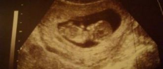
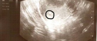
- A woman can hear her own baby's heartbeat in the early stages of pregnancy. This opportunity appears during the next examination using an ultrasound diagnostic device.
- With transvaginal (internal) ultrasound, the heartbeat can be heard at 5-6 weeks. At this stage, the embryo is only 3-4 weeks from the moment of conception.
- Transabdominal (external) ultrasound allows you to hear the baby’s heartbeat at 6-7 weeks.
Stethoscope
- Using a medical instrument called a stethoscope, a gynecologist listens to the beating of a tiny heart. This happens during an examination of a pregnant woman. The instrument used to perform auscultation (that is what this method is called) is a tube, usually made of wood.
- At the end of the tube there is a wide funnel, which the doctor places on the pregnant woman's stomach and listens to the tiny heart beating. Such listening is carried out not just out of curiosity: the doctor assesses the condition of the baby in the womb. Over long periods of time, the heartbeat can be heard very clearly.
- A stethoscope is used at the appointment after 18-20 weeks of pregnancy. Only at this stage can you use it to listen to the baby’s heartbeat.
How and at what time can a baby's heartbeat be heard in the womb?
Diagnosis and detailing of the fetal condition
The main indicator of normal fetal development is its heart rate.
Normal levels:
- 110-130 beats before the eighth week of pregnancy;
- 175-185 until the end of the first trimester;
- 145-160 before birth.
Malformations of the fetal cardiovascular system are indicated by:
- bradycardia or tachycardia;
- different time intervals between impacts;
- muffled sounds of heartbeat are a sign of oxygen starvation of the fetus.
A fetal heart defect on ultrasound becomes obvious when a defect in the organ chambers is visualized. The heart rate of the unborn child can be calculated already in the first trimester of pregnancy during a routine examination using an obstetric stethoscope. More complete information about the functioning of the fetal heart will be provided by CTG, which is prescribed after 32 weeks of pregnancy. Having assessed its results, obstetricians determine the parameters of the baby’s condition.
The main conclusions of the analysis of fetal condition indicators:
- below 1.0 is normal;
- 0.8-1.0 - borderline state;
- up to 2.0 - primary deviations. Repeated CTG within a week is indicated;
- up to 3.0 - severe deviations. Inpatient treatment is indicated;
- more than 3.0 - critical condition of the fetus, indicating severe suffering.
These are examination methods that are accessible to both the patient and the doctor. They do not require special preparation and do not take much time.
But Doppler echocardiography is considered the leader in the study of hemodynamic processes in the heart and blood vessels of the fetus. This method evaluates three main indicators - the direction, speed and nature of blood flow. The procedure is as follows: an ultrasound sensor is installed on the pregnant woman’s stomach. A color image is transmitted to the screen. Red indicates the flow of blood, which moves towards the sensor. Blue color indicates the volume of blood flowing in the opposite direction. The maximum intensity of a particular color indicates increased hemodynamics.
ECHO-CG provides extremely valuable information about the heart and the fetus itself. The test can be prescribed at any stage of pregnancy, but, as a rule, it is performed starting from the 12th week after receiving warning indications from the first screening.
This study will determine:
- direction and speed of blood flow in the vessels;
- patency of the bloodstream;
- the ratio of the volumes of blood entering and exiting the fetal heart;
- Heart rate.
It is best to carry out the procedure between 18 and 24 weeks, since it is during this period that the maximum visual effect is ensured.
Studies conducted earlier than this time will not be sufficiently objective due to the small size of the heart. Towards the end of pregnancy, ECHO-CG of the fetus is rarely used due to the large volume of the abdomen, which makes visualization of the organ difficult.
A pediatric cardiologist and cardiac surgeon are involved in resolving issues related to the results of examination of a fetus with congenital heart disease.
How many heartbeats per minute does a fetus have in the womb?
- In the early stages (up to 6 weeks), the fetal heart contracts slowly. This is explained by the initial stage of the formation of cardiac activity.
- When the connection between the circulatory and nervous systems is formed (which happens by the end of the 9th week), the heart rate is already 170-180 beats per minute.
- However, such heart rate indicators do not last long: by the second trimester, heart rate indicators decrease to 120-160 beats per minute. The second and third trimesters occur with the rhythmic pulsation of a small heart.
How to listen to the fetal heartbeat
Ventricular development
The primary ventricle begins to divide into two ventricles with the growth of the median ridge, the muscular interventricular (IV) septum with a superior free edge that arises from the base of the primary ventricle, closer to the apex of the heart. The expansion of the developing ventricles on either side of this septum is responsible for the initial increase in septal height. Further growth of the latter occurs due to ventricular myocytes located on both sides of the heart.
Between the upper free edge of this septum and the endocardial cushions there remains an opening called the IV foramen. Through it, blood continues to flow from the right ventricle to the left until complete closure at the end of the seventh week, when the left and right bulbar crests merge with the endocardial cushion, forming the membranous part of the IV septum. In the fifth week, the bulbar ridges are formed by the division of mesenchymal neural crest cells in the walls of the bulbus (cardiac bulb).
Membranous part IV of the septum occurs when the tissue on the right side of the endocardial cushion extends into the muscular part of the IV septum, eventually fusing with the aortic pulmonary septum and muscular IV septum. Once the fourth foramen closes and the membranous part of the fourth septum is formed, the aorta becomes the only outflow of blood from the left ventricle, and the pulmonary trunk is the only outflow of blood from the right ventricle.
As the ventricles develop, cavitation leads to the formation of muscle bundles. While some are preserved as columns of muscle on the inner surface of the ventricles (trabeculae carneae), others form the papillary muscles and chordae tendinae (heart strings) that connect the papillary muscles to the AV valves.
Posterior papillary muscle - lateral image left
Is it possible and how to listen to the fetal heart at home?
- If you can't wait to listen to your baby's heartbeat, a stethoscope won't help you with that. And even vice versa: while listening, various additional noises from the mother’s body join the beating of the little heart.
- Therefore, it is better to use another device - a portable ultrasound Doppler detector of the fetal heartbeat. The detector will record the baby's heartbeat, and you can hear it through headphones. Those who use this method of listening to the beating of a dear heart will have another opportunity: to record unique sounds, so that later they can listen to the heartbeat of an unborn baby on a computer.
Is it possible and how to listen to the fetal heart at home?
How is it diagnosed?
The only thing that will help determine the presence of a defect in the fetus is an ultrasound examination. The procedure allows you to identify the problem from the 17th to the 20th week of pregnancy. Therefore, every woman is prescribed a routine examination.
An ultrasound from a good doctor can make a diagnosis in 95% of cases. Determining the problem in the prenatal period will help you choose the method of delivery, assess the child’s condition and provide assistance to him in the first minutes after birth.
Ultrasound is also used to determine whether there is a need for surgical intervention. At 34 weeks of pregnancy, an echocardiogram is prescribed. During the study, the degree of development of the defect and its features before birth are assessed. Some heart problems are caused by chromosomal abnormalities. In this case, genetic karyotyping is performed.
The procedure allows you to examine the child’s cells in the prenatal period. The examination is prescribed for atrioventricular communications. These are the most complex abnormalities in the structure of the heart valves. In half of the cases, the pathology occurs in conjunction with Down syndrome.
Why is an ultrasound of the fetal heart done during pregnancy?
- Diagnostic procedures during pregnancy are prescribed for the timely identification of areas of danger and the ability to prevent the development of a critical condition.
- If doctors discover cardinal abnormalities in the later stages of pregnancy, then the baby will have a chance to survive outside the mother's womb. In some cases, a critical condition poses a serious danger and the child may die before the end of the pregnancy. It is important for doctors to be prepared for such a turn of events in order to have time to provide medical care.
- The doctor will determine which foods should not be consumed, what kind of stress is possible for a pregnant woman, and which medications are best to avoid for a while. If you delay visiting the ultrasound diagnostic room, you can create the illusion that there is no problem, which will lead to dire consequences.
External fetal cardiotocography
This non-invasive fetal test records the fetal heart rate for 10 minutes or between contractions.
The method allows the doctor to assess the condition of the fetus:
- before giving birth;
- during labor;
- in the interval between contractions;
- during a fight.
External fetal cardiotocography is an absolutely safe and painless procedure. It does not affect the condition of the fetus or labor. The study is carried out with the written consent of the pregnant woman.
The method can be combined with stress-free and stress tests. With the help of the study, it is possible to identify a state of fetal distress - a disruption of the normal function of organs under the influence of any factor. Fetal distress can occur during pregnancy and childbirth.
Manifestations of distress most often are signs of hypoxia (oxygen starvation of tissues) and asphyxia. Manifestations of distress include acceleration or deceleration of heart contractions and a decrease in the number of fetal movements.