For the smooth functioning of body systems, a number of conditions must be met. One of them is the absence of cardiac disorders. When exposed to a number of unfavorable factors, a cardiac pathology called impaired myocardial repolarization can develop.
What is myocardial repolarization?
Repolarization is one of the cyclic phases of the functioning of the heart muscle (myocardium), accompanied by restoration of the electrical membrane charge. In the absence of disturbances in the functioning of the heart, sodium ions return to their original state during the process of repolarization, due to which the membrane electrical charge is restored, and normal indicators predominate on the cardiogram (there are no significant deviations).
If the repolarization process is disrupted, cardiac activity is destabilized. Tissues and organs lack the oxygen and nutrients transported by blood required for normal functioning. As a result, health deteriorates and the likelihood of developing many diseases of various systems increases.
The primary diagnostic method is an electrocardiogram.
Normal indicators
With moderate intensity of the pathology, painful symptoms related to the functioning of the heart may not appear, so detection of deviations from the norm often occurs at advanced stages.
A cardiologist, conducting an examination if there is a suspicion of the development of disturbances in repolarization processes in the myocardium, studies the nature of the cardiogram waves and interval indicators.
Normal characteristics of teeth:
- Wave T. Directed upward (negative VR value).
- Wave Q. Normal value is 1/4 R (at 300 ms).
- R wave. Present in all leads.
- Prong S. Height – 2 cm.
- P wave. Positive value in the first two leads, negative VR value (100 ms).
Interval standards: QT – up to 400 ms, QRS complex – up to 100 ms, RR – 0.62/0.66/0.6 s, PQ – 120 ms.
In the absence of pathologies, the heart rate is from 60 to 85 beats per minute (sinus rhythm).
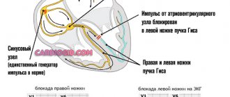
Heart rhythm disturbances
These are characteristic changes in the electrocardiogram that indicate that a person has problems with relaxation of the ventricles - the last phase of the heart rhythm. Such disorders can occur in both adults and children.
The younger the patient, the more benign these abnormalities are.
In children, sometimes they do not pose any health hazard at all. But in elderly patients, impaired myocardial repolarization indicates the development of dangerous pathological processes, such as heart attack, inflammation of the heart muscle.
The processes of its contraction are due to the presence of electrical impulses. Each stage of the cycle is responsible for a complex mechanism for the exchange of calcium, potassium and chlorine ions and their penetration into each cell.
During contraction of the cardiac muscle cell, depolarization occurs, and during its relaxation, repolarization occurs.
What is a normal cardiac cycle?
Normally, the cardiogram consists of several teeth:
- P is atrial contraction;
- complex Q, R, S - cycle of ventricular contraction;
- T - ventricular relaxation.
Danger of violations
This syndrome can cause so-called early coronary death in humans. Patients who have a history of syncope of cardiac origin are especially at risk.
In the presence of arrhythmias associated with the pathological condition of the right or left ventricle, and the development of diseases caused by hemodynamic disorders, early repolarization syndrome leads to rapid progression of cardiac ischemia.
Reasons for changes in myocardial repolarization
Progressive pathology is caused by:
- Coronary heart disease.
- Thickening (hypertrophy) of the heart.
- Overstrain of the cardiac ventricles.
- The presence of additional ventricular chords.
- Electrolyte (calcium, potassium, magnesium) imbalance.
- Hypersympathicotonia (disorders related to repolarization processes in the myocardium are explained by increased concentrations of norepinephrine, adrenaline, and tissue hypersensitivity to hormones).
- Cardiomyopathy.
- Abuse of medications (taking medications not prescribed by a doctor, exceeding the prescribed dosage).
- Regular consumption of alcoholic beverages.
- Complications of diseases of the neuroendocrine system involved in the regulation of the functioning of the heart and blood vessels.
- Hormonal imbalances.
- Impaired functioning of the thyroid gland, diabetes, and other diseases affecting the endocrine system.
- Severe menopause, pregnancy. During pregnancy, the cardiovascular (as well as other) systems of the body are susceptible to the effects of negative factors, therefore, at the first symptoms of impaired repolarization processes in the myocardium, you should consult a doctor.
- Being in a state of chronic stress.
- Intense physical activity, professional sports activities.
- Negative consequences of exposure to low temperatures.
- Age factors.
- Heart defects (congenital, acquired).
- Tumor diseases.
- Having suffered a stroke.
- Traumatic brain injuries.
- Hereditary predisposition to cardiovascular diseases.
Risk factors specific to childhood
Pathological repolarization found in children is explained by intensive growth, anatomical abnormalities, deterioration of aortic blood transportation, overload (emotional, physical), and poor resistance to stress.
The list of unfavorable factors includes hypersympathicotonia, accompanied by excess concentrations of norepinephrine and adrenaline in the blood.
The likelihood of developing pathology in childhood increases with asthma, pneumonia, neurosis, myocarditis, chronic tonsillitis, anemia, over or underactive thyroid gland.
To identify the exact causes (risk factors) of impaired repolarization processes in the myocardium, it is necessary to seek help from experienced specialists in the field of cardiology.
Possible deviations
One of the possible deviations is hypoxia, which occurs due to impaired breathing and circulation. Changes in repolarization are one of the first signs of the disease. To store energy, the synthesis of adenosine triphosphoric acid is required, and at the same time oxygen is needed, which is not supplied in the required quantity.
n
nrn (function(w, d, n, s, t) {rn w[n] = w[n] || [];rn w[n].push(function() {rn Ya.Context.AdvManager.render ({rn blockId: "RA-258969-11",rn renderTo: "yandex_rtb_R-A-258969-11",rn async: truern });rn });rn t = d.getElementsByTagName("script")[0 ];rn s = d.createElement("script");rn s.type = "text/javascript";rn s.src = "//an.yandex.ru/system/context.js";rn s.async = true;rn t.parentNode.insertBefore(s, t);rn })(this, this.document, “yandexContextAsyncCallbacks”);rn
Ischemia appears and the first sign will be a violation of repolarization processes on the ECG. Severe hypoxia causes arrhythmias and affects heart rate. Ischemia is a reversible process. An electrocardiogram shows all the changes, which will allow the doctor to make an accurate diagnosis.
Hypoxic diffuse conditions vary in duration, cause changes in the teeth, and appear in certain disorders - with persistent anemia, myocarditis and other metabolic disorders. Repolarization is influenced by conditions affecting cellular ions. Violation of the cellular electrolyte balance entails various changes - in the teeth, segment, etc.
Conditions such as hypoxia and oxygen starvation negatively affect ECG readings
.sj-r96 .q-zr-:hover {n background: #d26522!important;n }SlickJump®
What to do if you have insomnia?
Magnerot® can help with sleep and falling asleep disorders
n Find out more…SlickJump® HAS CONTRAINDICATIONS, CONSULT A SPECIALIST RU.MAGN.20.30 n
Deviations in the repolarization process are sometimes nonspecific. They are asymptomatic and detected completely by chance. Mostly such cases occur in teenagers and athletes. This phenomenon is called early repolarization.
Often diffuse deviations appear against the background of central nervous system overload. Hormonal changes greatly affect membrane restoration. All these deviations can be seen on the electrocardiogram graph and another differentiated examination will be required to make a diagnosis.
nnnrn (adsbygoogle = window.adsbygoogle || []).push({});rnnnn nnrn (adsbygoogle = window.adsbygoogle || []).push({});rn
Acute pericarditis on the ECG presents a picture similar to a repolarization disorder. To verify this, it is necessary to trace the dynamics.
One of the most serious pathologies leading to deviations in repolarization is hypersympathicotonia.
Hypersympathicotonia is manifested by high levels of adrenaline
n nrn (function(w, d, n, s, t) {rn w[n] = w[n] || [];rn w[n].push(function() {rn Ya.Context.AdvManager. render({rn blockId: "RA-258969-10",rn renderTo: "yandex_rtb_R-A-258969-10",rn async: truern });rn });rn t = d.getElementsByTagName("script")[ 0];rn s = d.createElement("script");rn s.type = "text/javascript";rn s.src = "//an.yandex.ru/system/context.js";rn s. async = true;rn t.parentNode.insertBefore(s, t);rn })(this, this.document, “yandexContextAsyncCallbacks”);rn
It appears in childhood. A feature of the pathology is an increased level of adrenaline. Often the causes of deviations are stress and constant hard work. Violations appear against the background of any hormonal abnormalities.
Symptoms of the disease
Pathological processes accompanying disturbances in myocardial repolarization are manifested:
- Decreased ability to work, fatigue, weakness.
- Painful sensations in the heart area.
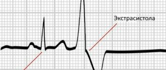
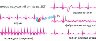
- Arrhythmias (ventricular, supraventricular, tachyarrhythmias).
- Instability of pulse rate.
- Shortness of breath observed with increased physical activity.
- Irritability, mood instability.
- Cardiogenic shock, hypertensive crisis, pulmonary edema (with cardiac dysfunction).
The symptoms of the pathology in children and adolescents are supplemented by tachycardia and neurocirculatory dystonia. Also, a violation of repolarization processes in a child is manifested by increased tone of the vagus nerve.
A disease that affects the myocardium is often discovered by chance during medical examinations, which is explained by the asymptomatic course of the disease.
Early ventricular repolarization syndrome
For the first time, such an electrocardiographic phenomenon as early ventricular repolarization syndrome was discovered in the middle of the 20th century.
For many years, it was considered by cardiologists only as an ECG phenomenon that does not have any effect on the functioning of the heart. But in recent years, this syndrome has begun to be increasingly detected in young people, adolescents and children. According to world statistics, it is observed in 1-8.2% of the population, and the risk group includes patients with heart pathologies, which are accompanied by cardiac disorders, patients with dysplastic collagenosis and dark-skinned men under 35 years of age. It was also revealed that this ECG phenomenon is detected in most cases in people who are actively involved in sports.
A number of studies have confirmed the fact that early ventricular repolarization syndrome, especially if it is accompanied by episodes of syncope of cardiac origin, increases the risk of sudden coronary death. Also, this phenomenon is often combined with the development of supraventricular arrhythmias, deterioration of hemodynamics and, with progression, leads to heart failure. That is why early ventricular repolarization syndrome has attracted the attention of cardiologists.
In our article we will introduce you to the causes, symptoms, methods of diagnosis and treatment of early ventricular repolarization syndrome. This knowledge will help you to adequately identify it and take the necessary measures to prevent complications.
What is early ventricular repolarization syndrome?
This ECG phenomenon is accompanied by the appearance of the following uncharacteristic changes in the ECG curve:
- pseudocoronary elevation (elevation) of the ST segment above the isoline in the chest leads;
- additional J waves at the end of the QRS complex;
- shift to the left of the electrical axis.
Based on the presence of concomitant pathologies, early repolarization syndrome can be:
- with damage to the heart, blood vessels and other systems;
- without damage to the heart, blood vessels and other systems.
According to its severity, the ECG phenomenon can be:
- minimal – 2-3 ECG leads with signs of the syndrome;
- moderate – 4-5 ECG leads with signs of the syndrome;
- maximum – 6 or more ECG leads with signs of the syndrome.
In terms of its persistence, early ventricular repolarization syndrome can be:
- permanent;
- transient.
Causes
While cardiologists do not know the exact cause of the development of early ventricular repolarization syndrome. It is detected both in absolutely healthy people and in persons with various pathologies. But many doctors identify some nonspecific factors that may contribute to the appearance of this ECG phenomenon:
- overdose or long-term use of adrenergic agonists;
- dysplastic collagenoses, accompanied by the appearance of additional chords in the ventricles;
- congenital (familial) hyperlipidemia, leading to atherosclerosis of the heart;
- hypertrophic obstructive cardiomyopathy;
- congenital or acquired heart defects;
- hypothermia.
Research is currently underway on the possible hereditary nature of this ECG phenomenon, but so far no data on a possible genetic cause have been identified.
The pathogenesis of early ventricular repolarization is the activation of additional abnormal pathways that transmit electrical impulses and disruption of the conduction of impulses along the pathways that are directed from the atria to the ventricles. The notch at the end of the QRS complex is a delayed delta wave, and the shortening of the PQ interval observed in most patients indicates activation of abnormal nerve impulse transmission pathways.
In addition, early ventricular repolarization develops due to an imbalance between depolarization and repolarization in the myocardial structures of the basal sections and apex of the heart. With this ECG phenomenon, repolarization becomes significantly accelerated.
Cardiologists have identified a clear relationship between early ventricular repolarization syndrome and nervous system dysfunction. When carrying out dosed physical activity and a drug test with Isoproterenol, the patient experiences normalization of the ECG curve, and during night sleep the ECG indicators worsen.
Also during the tests it was revealed that early repolarization syndrome progresses with hypercalcemia and hyperkalemia. This fact indicates that electrolyte imbalance in the body can provoke this ECG phenomenon.
Symptoms
This ECG phenomenon can exist for a long time and not cause any symptoms. However, this background often contributes to the occurrence of life-threatening arrhythmias.
Many large-scale studies have been conducted to identify the specific symptoms of early ventricular repolarization, but all of them have been inconclusive. ECG abnormalities characteristic of the phenomenon are detected both in absolutely healthy people who do not make any complaints, and among patients with cardiac and other pathologies who make complaints only about the underlying disease.
In many patients with early ventricular repolarization, changes in the conduction system provoke various arrhythmias:
- ventricular fibrillation;
- ventricular extrasystole;
- supraventricular tachyarrhythmia;
- other forms of tachyarrhythmias.
Such arrhythmogenic complications of this ECG phenomenon pose a significant threat to the health and life of the patient and often provoke death. According to world statistics, a large number of deaths caused by asystole during ventricular fibrillation occurred precisely against the background of early ventricular repolarization.
Half of the patients with this syndrome have systolic and diastolic cardiac dysfunction, which leads to central hemodynamic disturbances. The patient may develop shortness of breath, pulmonary edema, hypertensive crisis, or cardiogenic shock.
The syndrome of early ventricular repolarization, especially in children and adolescents with neurocirculatory dystonia, is often combined with syndromes (tachycardial, vagotonic, dystrophic or hyperamphotonic) caused by the influence of humoral factors on the hypothalamic-pituitary system.
ECG phenomenon in children and adolescents
In recent years, the number of children and adolescents with early ventricular repolarization syndrome has been increasing. Despite the fact that the syndrome itself does not cause significant heart problems, such children must undergo a comprehensive examination, which will identify the cause of the ECG phenomenon and possible concomitant diseases. For diagnosis, the child is prescribed:
- urine and blood tests;
- ECG;
- ECHO-KG.
In the absence of heart pathologies, drug therapy is not prescribed. The child's parents are advised to:
- clinical observation by a cardiologist with ECG and ECHO-CG once every six months;
- eliminate stressful situations;
- limit excessive physical activity;
- Enrich your daily menu with foods rich in heart-healthy vitamins and minerals.
If arrhythmias are detected, the child, in addition to the above recommendations, is prescribed antiarrhythmic, energy-tropic and magnesium-containing drugs.
Diagnostics
Electrocardiography is the main method for diagnosing early ventricular repolarization syndrome.
The diagnosis of “early ventricular repolarization syndrome” can be made on the basis of an ECG study. The main signs of this phenomenon are the following deviations:
- displacement above the isoline by more than 3 mm of the ST segment;
- prolongation of the QRS complex;
- in the chest leads, simultaneous leveling of the S wave and increase in the R wave;
- asymmetrical high T waves;
- shift to the left of the electrical axis.
For a more detailed examination, patients are prescribed:
- ECG with physical and drug stress;
- 24-hour Holter monitoring;
- ECHO-KG;
- urine and blood tests.
Once early repolarization syndrome is identified, patients are advised to continually provide their physician with past ECG results, as ECG changes may be mistaken for an episode of coronary artery disease. This phenomenon can be distinguished from myocardial infarction by the consistency of characteristic changes in the electrocardiogram and the absence of typical radiating chest pain.
Treatment
If early repolarization syndrome is detected, which is not accompanied by heart pathologies, the patient is not prescribed drug therapy. It is recommended for such people:
- Avoiding intense physical activity.
- Prevention of stressful situations.
- Introduction to the daily menu of foods rich in potassium, magnesium and B vitamins (nuts, raw vegetables and fruits, soy and sea fish).
If a patient with this ECG phenomenon has cardiac pathologies (coronary syndrome, arrhythmias), then the following medications are prescribed:
- energy-tropic agents: Carnitine, Kudesan, Neurovitan;
- antiarrhythmic drugs: Ethmozin, Quinidine sulfate, Novocainamide.
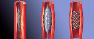
If drug therapy is ineffective, the patient may be recommended to undergo minimally invasive surgery using catheter radiofrequency ablation. This surgical technique eliminates the bundle of abnormal pathways that cause arrhythmia in early ventricular repolarization syndrome. Such an operation should be prescribed with caution and after eliminating all risks, since it may be accompanied by severe complications (PE, damage to the coronary vessels, cardiac tamponade).
In some cases, early ventricular repolarization is accompanied by repeated episodes of ventricular fibrillation. Such life-threatening complications become the reason for an operation to implant a cardioverter-defibrillator. Thanks to progress in cardiac surgery, the operation can be performed using a minimally invasive technique, and implantation of a third-generation cardioverter-defibrillator does not cause any adverse reactions and is well tolerated by all patients.
Detection of early ventricular repolarization syndrome always requires complex diagnostics and follow-up with a cardiologist. Compliance with a number of restrictions in physical activity, correction of the daily menu and exclusion of psycho-emotional stress is indicated for all patients with this ECG phenomenon. When concomitant pathologies and life-threatening arrhythmias are identified, patients are prescribed drug therapy to prevent the development of severe complications. In some cases, the patient may be indicated for surgical treatment.
How to decipher a cardiogram of the heart? The formation of a conclusion on an electrocardiogram (ECG) is carried out by a functional diagnostics doctor or cardiologist. This is a difficult diagnostic process,...
Ventricular extrasystoles: causes, signs, treatment Ventricular extrasystoles (VES) are extraordinary contractions of the heart that occur under the influence of premature impulses that originate from the intraventricular...
Right atrial hypertrophy: causes, symptoms, diagnosis Right atrial hypertrophy (RAH) is a term that refers to the enlargement of this part of the heart. Let us remember that venous blood enters the right atrium...
Left ventricular hypertrophy on the ECG: recommendations from a cardiologist The left ventricle is the part of the heart, during the contraction of which blood is ejected into the aorta. This is the main chamber of the heart, providing blood flow throughout the organ...
Source: tahikardiya.lechenie-gipertoniya.ru
How is disease identified on an ECG?
The cardiogram reveals changes in the T waves (shape distortion, widening of the base, asymmetry), P, R (positive), Q, S (negative). The ST line rises above the isoline by 1-3 mm, and a notch appears before the increase in ST. The ST shape becomes rounded or convex, pointing downward.
Failures related to the processes of repolarization of the left ventricular myocardium are identified by the complex of QRS waves: Q, S - negative, R - positive. The ST segment rises from the J point, and notches are noticeable in the descending segment of the R wave.
For a more detailed study of disorders and monitoring of the patient’s condition, diagnostic procedures are periodically repeated, supplemented by auxiliary measures.
Additional examinations
Diagnostics is supplemented by:
- Ultrasound examinations (heart, other internal organs).
- Daily ECG monitoring.
- Electrophysiological examination.
- Coronary angiography.
- Load tests.
- General, biochemical tests of urine and blood (allow to identify metabolic failures, inflammatory diseases).
- Endocrinological consultation.
Before carrying out diagnostic measures, it is necessary to exclude physical activity in order to avoid distorting the results.
Treatment of the pathological process
Therapeutic procedures include the use of:
- Mineral and vitamin complexes (help meet the needs of vital organ cells for essential substances).
- Cocarboxylase hydrochloride (ensures normalization of carbohydrate metabolism, prevents neurological disorders, improves the condition of the heart and blood vessels).
- Corticotropic hormonal drugs (due to the content of cortisone, cardiac pathologies are eliminated).
- Beta blockers (eliminate concomitant ailments affecting the heart).
To increase the effectiveness of therapy for diffuse disorders of repolarization processes in the myocardium, it is advisable to:
- Minimize the consumption of fried, fat-rich foods, and give up alcohol.
- Fortify your diet.
- Optimize your daily routine (eliminate physical overload, eliminate sleep disturbances).
- Avoid stress, stabilize the psycho-emotional background.
In the absence of severe symptoms, as prescribed by the doctor, the treatment and prophylactic course is limited to organizational measures; potent medications are not used.
If the preconditions are created for the development of unsafe ventricular tachyarrhythmias, the short QT interval syndrome progresses, and conservative methods do not lead to improvement in dynamics, there is a need to install an electrical pacemaker.
Forecast
The prognosis for disturbances in repolarization processes in the myocardium is determined by a list of negative factors and accompanying symptoms.
With heart disease, heart attack, ventricular diseases, or an unfavorable medical history, the likelihood of irreversible consequences reaches a maximum. The benign course of pathology detected at an early stage is characterized by insignificant risks of irreversible pathologies. There is no serious threat to the life of the body.
To avoid complications, you should optimize (improve) your nutrition system, work and rest schedule, and give up bad habits. With strict adherence to the doctor’s recommendations, the normal functioning of the myocardium is restored, and favorable dynamics are observed, confirmed by periodic examinations.
What methods of preventing heart disease do you know? Which ones are most effective? Share your opinion by leaving a comment.
Classification and risk groups
There is the following classification of early repolarization syndrome:
- with damage to the heart muscle and blood vessels;
- there is no lesion.
The syndrome is also classified according to the degree of severity on the electrocardiogram into 3 classes:
- Minimal (observed in a small number of leads, from 2 to 3).
- Moderate (the number of leads increases from 4 to 5).
- Maximum (6 or more leads).
However, most often the disease appears during pregnancy or menopause in women, since at this time the sensitivity of the body increases significantly and the general hormonal background changes. The disease is detected, as a rule, during routine examinations, in case of any complaints about well-being.
Professional athletes who experience constant physical activity and people who have suffered from hypothermia are also at risk. And some doctors even claim that the disease is hereditary.