4 / 5 ( 3 voices)
Neoplasms in the heart are the rarest among the general population of malignant pathologies. Most likely, this is explained by the fact that the organ is well washed with blood, intense metabolic processes occur in its tissues, which are constantly contracting, and the cells are characterized by high functional activity.
Tumors are predominantly benign (myxomas); heart cancer accounts for less than 1% of all registered oncological pathologies.
Cancerous growth in the heart
More about the disease
Heart cancer can refer to a tumor growth inside the heart chambers or damage to the heart muscle. The heart is a muscle in the form of a hollow organ through which blood is pumped through the circulatory system. It is located between the lower parts of the lungs.
The rarity of diagnosing the pathology is explained by the fact that blood circulation and metabolism in the tissues of the organ are at a high level due to the functional purpose of the chambers and other components of the heart. Due to the difficulty of diagnosis, the presence of this disease does not make itself felt until the final stages.
Tests and diagnostics
Diagnosis of heart cancer presents certain difficulties due to the multiplicity of morphological forms of heart tumors and the variability of clinical symptoms.
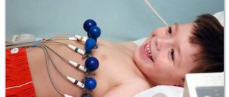
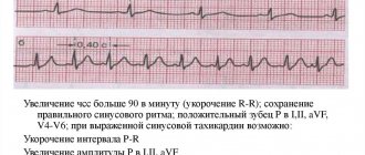
ECG data are low specific and polymorphic. Ischemic changes, rhythm and conduction disturbances, and signs of hypertrophy of the cavities of the heart can be recorded.
X-rays reveal signs of pulmonary hypertension and an increase in heart size. The most informative method for diagnosing cardiac tumors is echocardiography:
- transthoracic echocardiography can detect ventricular tumors;
- Transesophageal echocardiography clearly visualizes atrial tumors.
If the results of the diagnostic search are uncertain, MSCT and MRI of the heart, ventriculography, probing of the cardiac cavities and radioisotope scanning are performed. A biopsy is performed through catheterization and diagnostic thoracotomy to verify the histological structure of the tumor. In the exudative form of pericarditis, important diagnostic information is provided by a cytological examination of the fluid obtained by puncture of the pericardium.
It is necessary to carry out differential diagnosis with the following diseases:
- pericarditis;
- cardiomyopathy;
- myocarditis;
- congenital heart defects ;
- amyloidosis of the heart.
Leading clinics in Israel
Assuta
Israel, Tel Aviv
Ikhilov
Israel, Tel Aviv
Hadassah
Israel, Jerusalem
Typically, cancer is formed from degenerated epithelial cells when they begin to divide uncontrollably. Due to the virtual absence of epithelium in the heart (there is only a single-layer endothelial lining), cardiac tumors are a rare occurrence. One of the endothelial cells may begin to mutate, and a benign or malignant tumor begins to form.
On a note! Heart tumors occur in only 0.25% of all cardiac diseases.
Diagnostics and its cost
The tumor is characterized by vague symptoms, which are found in most diseases of this organ. Therefore, to determine the tumor, the following additional examinations will be required:
- Ultrasound of the heart. Such an examination will primarily indicate an increase in size and the presence of a compaction in the heart. There are two options: the first is a transesophageal examination, which allows you to examine the atria; the second is transthoracic ultrasound, intended to study the cardiac ventricles. The cost of the procedure is from 280 rubles.
- An electrocardiogram, which will show only the abnormal electrical activity of the organ. And this is a sign of dysfunction and serves as a reason for further examination. The procedure will cost at least 100 rubles.
- Echocardiography (EchoCG), which gives a complete picture of the size and location of the tumor formation in the heart. This study is based on the action of ultrasonic waves, which transmit images to the screen using a special sensor. The starting price for echocardiography is 3050 rubles.
- Tomography – computed tomography and magnetic resonance imaging. These diagnostic methods by visualizing the heart will determine the location and size of the tumor. You will have to pay at least 8,800 rubles for an MRI.
- Catheterization, which involves injecting an illuminating substance into the circulatory system to further examine x-rays for abnormal areas. The average price in the Russian Federation is 15 thousand rubles.
- Radioisotope organ scanning and probing. The cost of the procedure is from 7 thousand rubles.
- A biopsy of tumor material, which is removed during catheterization or exploratory thoracotomy. Minimum cost – 1500 rubles.
- Histological studies of pericardial fluid. The lowest price for the procedure is 1600 rubles.
Tumor diagnosis
Such diagnostic methods are preceded by an examination by a cardiologist, analysis of the nature of complaints, listening to noises and standard tests.
If the tumor is malignant, then the entire body is examined to determine possible metastasis (if the tumor is primary) or to find out where it metastasizes from (if secondary).
Classification of the disease
Heart pathology can be located in different places and tissues, and have different manifestations.
Heart tumors are divided into primary and secondary:
- primary are those that were formed in the heart itself, they can have various forms;
- secondary – appear as a result of oncology in the following organs: thyroid gland,
- Mammary gland;
- Lungs;
- Stomach;
- Kidneys.
The spread of cancer cells occurs through the lymph and circulatory system. These cells end up in the heart, growing into the organ.
Primary neoplasms
What forms of primary tumors are there? Primary heart cancers are divided into the following forms:
- lymphoma . Rarely encountered;
- sarcoma _ This type of tumor is more common. Sarcomas are formed from mesenchyme (mesoderm parenchyma) - embryonic connective tissue (mesoderm), usually in the right parts of the heart - endocardium or pericardium. They develop on the outer surface of the heart muscle; they can occur in the cavity of one (several) chambers of the heart or inside the tissues of the muscle itself. Sarcomas occur in men and women over the age of 30. The tumor tends to spread quickly. Tumor cells grow through the tissue of the heart and affect neighboring organs. If vessels and valves occur along the path of a growing tumor, they are also damaged by the developing tumor. Cancer cells grow into all major arteries, layers of the heart muscle and veins. Metastases affect the lungs, lymph nodes and even the brain.
Sarcomas, in turn, are divided into several subtypes:
- liposarcoma . It is diagnosed in adulthood and is considered a rare subtype of the disease. The tumor body consists of lipoblasts, the tumor itself is located in the cavity of the heart and is similar in appearance to a myxoma. It affects the atrium and metastasizes to the liver, bones, and lungs. It comes in myxoid and pleomorphic forms. Histiocytomas and schwannomas are even less common. The formation has a massive soft body of a yellowish color. This pathology responds well to treatment procedures;
- rhabdomyosarcoma . Its development occurs in muscle tissue, usually in the thick muscle layer of the myocardium. This type of tumor is diagnosed in every fifth patient out of the total number of primary tumors. Rhabdomyosarcoma occurs more often in men than in women. The formation is usually soft to the touch and white in color. In a microscope in a tumor node, you can see cells of various types: Round;
- Fusiform;
- Oval and others.
- Sarcomatous cancer (angioendothelioma);
Secondary neoplasms
Secondary tumors are often benign. Metastases of tumors of the kidneys, lungs, thyroid and mammary glands, and stomach form secondary heart cancer. Such tumors develop in the heart 25 times more often than primary ones.
Malignant melanomas, leukemia, and lymphoma have a high level of metastasis. The pericardium is most often affected, less often the myocardium of the heart chambers, and rarely the endocardium and heart valves.
Secondary tumors, even in the form of hard, small nodes, tend to diffuse infiltration, especially with hematological tumors or sarcomas.
The following neoplasms occur:
- jelly-like myxoma of the heart. Usually occurs in adults. It is located in the left or right atrium, has a leg that is attached to the septum;
- Rhabdomyomas are tumors with a structure similar to striated muscle tissue. Diagnosed in children after one year and in newborns.
Causes
Causes and factors that provoke the formation of a heart tumor:
- Genetic predisposition. Due to hereditary factors, the patient may be predisposed to the formation of cancer cells in the body. Despite the fact that the pathology manifests itself in old age, in medical practice there are increasingly cases of heart cancer in adolescents under 18 years of age. It is quite possible that in such children a hereditary factor served as a catalyst for starting the oncological process in the heart.
- Toxic effects. Working in hazardous industries, radiation, and prolonged exposure to negative factors can provoke mutations in cells.
- Infectious diseases. In some cases, the oncological process is detected after an infectious disease. The presence of infectious processes in the body indicates reduced immunity , which cannot protect the body, detect mutations and resist the development of tumors.
- Degeneration of a benign tumor into a malignant one. After surgery, a myxoma may develop on the organ. The transition from one type of tumor to another is facilitated by negative factors (smoking, alcohol abuse, chronic intoxication).
- Negative impact of ecology . Genetic failure in the body can be caused by poisons that accumulate in the body, entering it from polluted air, land and water poisoned by pesticides, and agricultural products grown on such land.
- Wrong diet. With excessive consumption of carcinogenic foods, fast food, cholesterol on the walls of blood vessels, and the functioning of the cardiovascular system is disrupted. Even seemingly minor disruptions in the functioning of the heart can lead to such serious consequences.
- Abuse of alcoholic beverages . Alcohol can change the anatomy of the epithelium of the mucous membranes and cause irreversible changes in the cells of any organ, including the heart.
- Smoking. Carcinogenic substances enter the body along with cigarette smoke.
- The presence of malignant neoplasms in the body. The heart can be damaged by a metastatic attack.
Causes
The exact causes of heart cancer are not known, but there are factors that can trigger the occurrence of primary formations in the heart:
- degeneration of myxoma (benign tumor), which can develop after surgical intervention in an organ, as a result of toxic effects, as a consequence of an infectious disease, due to harmful effects caused by drinking alcohol and smoking;
- genetic predisposition;
- impaired immune defense of the body;
- the appearance of aberrant growths;
- chemical, radiation, ultraviolet exposure.
Secondary tumors arise as a result of the spread of cancer that has formed in other organs.
Don't waste your time searching for inaccurate cancer treatment prices
*Only upon receipt of information about the patient’s disease, a representative of the clinic will be able to calculate the exact price for treatment.
Symptoms of malignant neoplasms
Compared to benign tumors, malignant tumors provoke a more severe and rapid deterioration in health and can spread to the spine, nearby soft tissues and vital organs.
Symptoms of cancers that start directly in the heart include:
- sudden heart failure;
- accumulation of fluid in the mucous membrane of the main organ due to blockage of blood flow;
- various arrhythmias.
If the formation has spread from another part of the body, then the diagnosis records:
- rapid enlargement of the heart, change in shape on x-ray;
- heart rhythm disturbances;
- heart failure for no reason.
Most babies with tumors diagnosed in the hospital usually do not survive their first year.
Symptoms and signs of heart cancer
Depending on the location of the tumor and its size, the picture of the disease may differ. Some symptoms are stronger, while others are completely absent. In the initial stages, the disease can be practically asymptomatic.
The following symptoms may indicate the development of heart cancer:
- the appearance of pain in the chest, joints;
- dyspnea;
- signs of oppression of the vena cava;
- detection of hemorrhagic effusion in the pericardium;
- enlargement of the chambers of the heart, disruption of the rhythm of the organ;
- elevated temperature;
- fast fatiguability;
- tamponade;
- swelling of the facial muscles, numbness of the limbs;
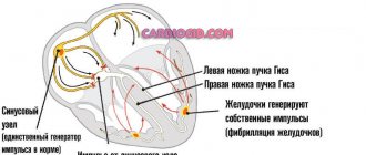
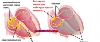
- disturbances in the activity of the conduction system;
- severe weight loss.
It is difficult to suspect heart cancer due to the fact that these symptoms of the disease can be confused with manifestations of other diseases:
- fever or cough;
- joint pain;
- Raynaud's phenomenon - fingers turning blue when pressed;
- swelling of the abdomen, ankles, legs;
- swelling of the veins of the neck due to poor pumping of blood from the atrium or obstacles that prevent blood from freely flowing from the vessels into the heart.
Patients may also experience:
- difficulty breathing when lying on your back or side;
- decreased blood pressure;
- dizziness, fainting;
- feeling of a lump in the chest.
Metastases and secondary forms of oncology give the following symptoms:
- shortness of breath with minor exertion;
- acute pericarditis;
- cardiac tamponade;
- systolic murmur;
- heart rhythm disturbance (tachycardia);
- heart failure;
- an increase in the area of the heart line (visible on an x-ray).
Clinical picture
Cardiac carcinogenesis in a photo taken using magnetic resonance imaging
A serious problem that arises during diagnosis is that there are no specific signs of heart cancer. In this case, the symptoms depend more on the location of cancer formation rather than on its type.
For example, neoplasias that cause blood flow disturbances manifest themselves earlier, in contrast to those that grow into neighboring tissues. The second table shows locations that can be identified based on the emerging clinical picture.
Table 2. Symptoms of heart cancer, depending on its location:
| Damage area | Characteristic signs |
| Damage to valves, embolism of the pulmonary and coronary arteries, as well as in the vessels of the brain. The presence of various non-specific signs. |
| Violation of the rhythm of the heartbeat, innervation, abnormal increase in the size of the cavity (in the X-ray photo), development of signs of failure, atrial flutter. The ECG registers abnormal changes (rise of the ST segment above the isoline, inversion of the T wave is observed in half of the patients, and in the case of damage to the left part of the ST and the T wave, as a rule, remain unchanged). |
| The presence of effusion in the heart sac and accumulation of fluid between the leaves, pericarditis, arrhythmias, chest pain. Quite often, this area is affected by metastases from tumors of the lungs and mammary glands. With rhabdomyosarcoma, pleural pain, shortness of breath and symptomatic manifestations. |
| Heart attack and angina. |
| Stenosis, heart failure, heart disease and unexpected death (SCD). |
Note. With any type of cancer, patients complain of general malaise, weakness, headaches, loss of appetite and body weight, causeless fever and other nonspecific symptoms that should be alarming. At the slightest suspicion, it is advisable to consult a doctor, and in some cases insist on diagnosis.
With heart cancer, signs can be expressed in the form of inflammation of the vascular walls or changes in connective tissue. The blood reveals signs of anemia, a slight increase in leukocytes, platelets and ESR.
All four valves can be affected by cancer at the same rate. As a rule, in such a case, the clinical manifestations have fairly clear features. With tumors of the mitral valve, neurological symptoms are more pronounced (aortic - less), which can cause sudden death.
The note. In the presence of confirmed primary oncogenic foci, metastasis to the heart tissue or coronary vessels cannot be excluded, but signs of their presence may be absent.
Stages of disease development
Depending on the stage at which the disease was detected, treatment will be prescribed, and the prognosis of the disease depends on this.
There are the following stages of heart cancer:
- 0 or precancerous stage, it has no tendency to recur;
- Stage 1, the emerging tumor is less than 2 cm;
- Stage 2, the neoplasm measures 2-5 cm, metastases are possible;
- Stage 3, tumor size is more than 5 cm, there are metastases in the lymph nodes and nearby areas;
- Stage 4, the size of the tumor is significant, active metastasis occurs.
Reasons for the appearance of formations
Modern medicine cannot yet accurately determine the factors that influence the development of primary tumors. This is due to the fact that the pathology affects people with completely different medical histories.
While the reasons for the progression of secondary neoplasms have been identified. The disease develops when cancer of the kidneys, stomach, breast or thyroid gland metastasizes. With this process, metastases occur in pathological foci located in other parts of the body and enter the heart through the bloodstream.
Diagnosis of the disease
Several examination methods are used to diagnose heart cancer. First, the doctor analyzes the patient’s complaints and listens to the heart to identify murmurs characteristic of valve damage.
This is followed by instrumental and laboratory examination methods:
- X-ray examination is used to obtain data on the size of the heart and its parts;
- angiocardiography. It is used to study chambers, chest veins, large vessels;
- ECG - detects heart rhythm disturbances;
- MRI talks about the condition of the heart chambers, surrounding tissues and organs. CT scan details the data obtained;
- EchoCG - clarifies the location of the tumor, determines its size, shows the presence of fluid in the pericardial area;
- echoscopy with color Doppler examination;
- coronary angiography;
- radioisotope ventriculography;
- blood test to determine the levels of hemoglobin and platelets, leukocytes, ESR, C-reactive protein;
- analysis for tumor markers;
- histological analysis.
Diagnostics
Diagnosing heart cancer is difficult. This happens for several reasons. Firstly, the similarity of the symptoms of heart cancer with the symptoms of other diseases. Secondly, the absence of any symptoms in the first stages of the disease.
If you experience any symptoms indicating heart problems, you should consult a cardiologist. Diagnosis of heart disease includes:
- Blood analysis. It is done primarily to identify deviations from the norm in indicators of anemia (decreased hemoglobin), an increase in ESR and other indicators that reveal the presence of inflammatory processes.
- Electrocardiography (ECG). With its help, the doctor determines changes in the rhythm of the heart, enlargement of its areas and blood conductivity.
- Cardiac echocardiography (EchoCG). Makes it possible to determine the size of the tumor, its location and the presence of fluid in the pericardium.
- Chest X-ray. Detects an increase in the size of the heart muscle as a whole or its parts.
- Magnetic resonance imaging (MRI). Shows the condition of the heart chambers and tissues located nearby. Using MRI, the doctor determines the presence of fluid in the tumor.
- CT scan. It is considered one of the most effective studies of malignancy. It reveals not only the location of the formation, but the direction of its increase.
- Angiocardiography. With its help, the chambers of the heart, blood vessels and thoracic veins are studied.
Additional research methods:
- echoscopy;
- radioisotope ventriculography;
- tumor marker analysis;
- coronary angiography;
- histological analysis;
- biopsy.
Treatment of heart cancer
The main treatment methods for heart cancer are:
- chemotherapy;
- irradiation;
- maintenance therapy.
These treatment methods can stop the development of cancer, reduce metastasis and improve the patient’s quality of life.
In most cases, heart cancer cannot be treated with surgery. This is due to the fact that the disease is most often diagnosed when other organs are already affected by cancer. Radiation therapy is often used alone or in combination with chemotherapy.
This combination allows you to reduce the severity of the disease and stop its progression. In this way, the patient’s life expectancy can be increased by an average of 5 years. In addition to this, symptomatic treatment is prescribed to relieve the patient of unpleasant symptoms.
The stage at which the disease was diagnosed and the type of cancer determine the type of treatment. At the zero stage, a complete cure of the patient is possible. In the first or second stages, a small tumor is surgically removed and this is supplemented with appropriate therapy, which gives a chance for a complete cure. But after treatment, the patient is constantly examined for the appearance of metastases.
With primary myxoma, the tumor is removed along with its attachment site.
Excision of single tumors: rhabdomyomas, fibromas, lipomas, teratomas, pericardial cysts is not performed if they do not interfere with the functioning of the heart.
Surgery is performed using a gamma knife or brachytherapy.
Sometimes a transplant of an organ damaged by a tumor is required. In this case, all risks associated with this (graft rejection) are taken into account. To reduce the risk of side effects and relapse, doctors are testing heart autotransplantation. To do this, the organ is removed, the formation is removed and restored to its original location. This method is considered safer, since there is no need to prescribe immunosuppressants, which can contribute to cancer recurrence.
If secretions accumulate in the tumor and interfere with the functioning of the heart, drugs are administered that prevent the development of the disease. If there is a risk of cardiac tamponade, a therapeutic puncture of the pericardium is performed (in order not to perform it repeatedly, an operation is performed to establish drainage from the pericardium to the pleural area).
Liposarcoma is treated with a combination of chemotherapy and radiation therapy. For secondary tumors, palliative treatment is used.
Benign tumors
Benign neoplasms of the heart are of primary origin and develop from cells of one’s own tissue. The causes of this disease have not been determined, but provoking factors can be identified as follows:
- hereditary predisposition;
- concomitant diseases (Carney or Lamb syndrome);
- nicotine addiction;
- work related to hazardous production (use of cadmium, benzene, nickel and other substances);
- viral and bacterial infections.
In some cases, a neoplasm may develop against the background of arterial embolism: peripheral or pulmonary.
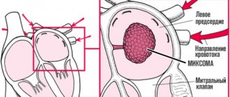
Myxoma
This benign neoplasm is detected in 37% of cases, of which the most likely location is the left atrium (up to 80%). In 15%, myxoma occupies the right atrium, and in 5%, both. Its average size is about 4 cm (but can reach 8 cm), the structure is loose, lobular, there is a high probability of small fragments breaking off, which can provoke an embolism.
As the myxoma grows, it fills the entire cardiac chamber and interferes with the valve, impairing the emptying and filling of the atrium. This is dangerous due to dysfunction of the circulatory system in both circles.
Myxoma
Papillary fibroelastoma
Such a tumor, as a rule, has a leg and forms on the valves: aortic, mitral on the left and tricuspid on the right. But this neoplasm does not disrupt the functioning of the valves; it is only dangerous by tearing off the papillae, which can lead to embolism.
Papillary fibroelastoma
Rhabdomyoma
This benign formation is characteristic of childhood and often (70%) goes away on its own. It forms in the walls of the septum or left ventricle (most often) and grows simultaneously from several foci. The risk of developing rhabdomyoma is damage to nerve fibers, increased wall thickness and changes in the shape of the heart, which leads to arrhythmia and failure of this organ.
In medical practice, there are also the following types of benign neoplasms: fibroma, lipomatosis, teratoma, hemangioma, lipoma. They are detected much less frequently, which does not make them any less dangerous.
Rhabdomyoma
Disease prognosis
How long do people live with a diagnosis of heart cancer? The prognosis in the initial stages may be optimistic. If heart cancer is detected before metastases occur, the patient's life can be extended to five years. In the last stages, from the moment the disease is diagnosed, the patient dies within a year, despite the therapy.
The 2-year survival rate was found to be:
- at stages 0 and 1 - 8.3%;
- at stage 2 - 3%;
- at stages 3-4 - 0.9%.
For sarcomas, life expectancy from the moment of diagnosis is as follows:
- 6-11 months (angiosarcoma);
- 1 year - rhabdomyosarcoma stages 1 and 2, for stages 3 and 4 - less than 1 year after removal of the tumor, concomitant radiation and a course of chemotherapy;
- 6-8 months (liposarcoma).
Video on the topic:
Treatment of cardiac oncopathology
Heart cancer progresses very quickly, and in the initial stages it appears extremely rarely. Because of this, surgical treatment is not always effective. The operation is possible only if the detected tumor is located within the organ, and the process has not spread more distantly.
We recommend reading Salivary gland cancer - causes, symptoms, treatment
Methods and methods of treatment are determined by the size of the tumor. Endoscopic surgery is performed for small tumors. Large cancerous lesions are removed using open-heart surgery. In clinics in Israel, Germany, and some other countries, cancer patients undergo donor organ transplantation.
Important! Symptomatic treatment is prescribed to improve the patient’s general well-being and relieve pain.
The method of conservative treatment depends on the stage of the process. Patients are prescribed radiation therapy in case of inoperable form of the disease. Brachytherapy is prescribed - the introduction of a radioactive source directly into the tumor, and a cyberknife is also used. With its help, the tumor is burned out. To do this, X-ray machines are used that direct a beam of high-frequency rays to the desired area.
Metastases in late stages can appear anywhere. To stop their spread and reduce the size of the cancer, patients are prescribed chemotherapy with cytotoxic drugs.
Prevention for heart cancer
To prevent heart cancer, the following measures should be taken:
- play sports (to train the heart muscles and strengthen the immune system);
- treat acute infectious diseases and prevent their development into a chronic form;
- maintain optimal weight with proper nutrition;
- stop smoking and drinking alcohol;
- reduce cholesterol and blood sugar levels by eating fruits and vegetables and eliminating fatty, spicy, salty, fried foods;
- control blood pressure;
- undergo regular preventive examinations with specialists.
Prevention
To prevent the development of malignant tumors in the heart, doctors advise:
- Carry out the prevention of infectious diseases to prevent the possibility of their becoming chronic.
- Don't neglect physical activity.
- Control your weight.
- Try to avoid eating unhealthy, fatty, carcinogenic foods.
- To refuse from bad habits.
- Control blood sugar and cholesterol levels.
- Monitor blood pressure.
- Have a medical examination every year.