Echocardioscopy is an examination of the heart using ultrasound, which makes it possible to assess the condition and structure of this organ, and determine blood flow parameters. Ultrasound examination of the heart can be used to examine patients of any age, and even for intrauterine examination of the fetus, and it has virtually no contraindications. Due to the popularity of this diagnostic technique, many patients who have been referred for examination have a question about what ECHO CS is.
What is echocardioscopy?
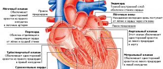
Echocardioscopy is based on the ability of ultrasound waves to be reflected from tissues of different densities and elasticity with different intensities. Tissues prevent the propagation of ultrasound, changing its oscillation period, frequency and wavelength. Changes are captured and recorded by sensors, and then the received information is transmitted to the monitor of the ultrasound machine. This kind of diagnosis makes it possible to assess the size and volume of the cavities of the heart, such as the atria and ventricles, determine the thickness of the walls of the heart, and also detect changes in the valve system and other morphological units of this organ. The answer to the question, cardiac echocardioscopy - what is it, is simple - it is a way to diagnose heart diseases using ultrasound.
Existing types of cardiac ultrasound
Depending on the methodology used, diagnostics are distinguished:
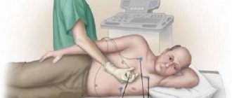
- transthoracic – this is a standard technique that is carried out by applying an ultrasound probe to the patient’s chest and then collecting the results obtained,
- transesophageal - the method involves the introduction of a special sensor into the esophagus - the information content of the results of this diagnosis is especially high, since the esophagus is located in close proximity to the organ being examined,
- intravascular - ultrasound is only possible in case of cardiac catheterization, and it helps to determine whether the patient has a blockage of blood vessels.
Many patients, faced with the need to undergo one of the types of cardiac ultrasound, wonder what ECHO CS is in medicine. This is an informative diagnostic method that makes it possible to diagnose heart disease in a patient without surgical intervention.
There is also a classification of ultrasound examination of the heart depending on the technical methods of its implementation. The first technique, one-dimensional echocardiography, also known as M-examination, is rarely used today on its own - it does not make it possible to visualize cardiac structures, but only displays a record of the heart’s work in the form of a graph on the monitor of the device. Based on the results obtained, the doctor can assess the size of the ventricles, atria, as well as their functional activity.
The second type of ultrasound examination, two-dimensional echocardiography, designated "B", provides information about the organ being examined in the form of a black and white image. This image allows you to evaluate the work of the myocardium in real time - its relaxation and contraction, and see the work of the valve system. Also, passing this examination allows you to obtain information about the size of the heart, the volume of its chambers, the thickness of the heart walls, valve structures - their mobility, as well as the contractility of the ventricles.
The third method, Doppler echocardiography, is considered one of the most modern. The principle of its operation is based on the Doppler effect: ultrasonic waves are reflected from moving objects with different frequencies and lengths. In this case, the moving objects are red blood cells - this technique allows you to observe the flow of blood in the chambers of the heart and large vessels.
Normally, blood flow can only be unidirectional; therefore, forward and reverse movements are recorded on the device’s monitor in contrasting red and blue colors.
Doppler echocardiography also allows you to assess the severity of the reverse movement of blood, measure the diameter of blood vessels and valves, and determine the speed of blood flow - it is used both independently and together with M- or B-methods.
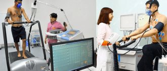
A contrast-enhanced cardiac ultrasound involves injecting a contrast agent into the patient's bloodstream. This allows you to visualize cardiac structures more clearly, and thereby increase the information content of the procedure. Stress cardiography is also used for the examination, which is accompanied by the patient’s physical activity, such as walking on a treadmill or riding an exercise bike. Exercise testing provides a unique opportunity to diagnose the disease at an early stage of its development, since symptoms of various abnormalities may be absent at rest.
Pros and cons of the test
Stress echocardiography, like other instrumental studies, has its pros and cons.
Advantages of the technique:
- the test reliably evaluates heart function during intense exercise;
- The portability and mobility of the equipment allows stress echocardiography to be performed even at the outpatient stage.
Disadvantages of stress echocardiography:
- difficulty in visualizing certain structures (due to movement);
- frequent occurrence of adverse reactions (dizziness, fainting, nausea, arrhythmia, angina attack);
- When using drugs, allergic reactions occur.
If it is impossible to use physical activity, drug or transesophageal stimulation is used. However, these methods are not without drawbacks. They do not fully imitate the necessary conditions, can cause discomfort and have even more contraindications from other organs.
Indications for echocardioscopy
It is recommended to undergo cardiac ultrasound on a regularly scheduled basis for patients suffering from hypertension, congenital or acquired heart defects, or having a hereditary tendency to diseases of the cardiovascular system. This procedure is mandatory for those who have suffered a myocardial infarction (its contractility is assessed) or have angina pectoris. The motivating reason should be dizziness, headaches, shortness of breath, fainting, swelling of the extremities, a feeling of interruptions in the functioning of the heart, pain in the chest.
Ultrasound of the heart helps in diagnosing neoplasms of various types, cardiomyopathy, aneurysm, and allows identifying the presence of fluid accumulations in the pericardium.
The stress type of diagnosis is used if the patient is suspected of coronary heart disease, to assess the patency of blood vessels, determine the degree of risk to the life and health of the patient before performing surgical interventions.
As for transesophageal diagnostics, it is prescribed for suspected:
- malfunction of the prosthetic heart valve,
- infective endocarditis,
- the presence of a thrombus in the atrium,
- aortic aneurysm,
- atrial septal defect.
It is also used when the patient is overweight and therefore unable to conduct a standard examination, as an informative procedure before the patient undergoes cardioversion in order to correct the arrhythmia.
When should it be performed (Echo CG)
The earlier pathologies or diseases of the heart muscle are diagnosed, the greater the chance of a positive prognosis after treatment. An ultrasound should be performed for the following symptoms:
- periodic or frequent pain in the heart;
- rhythm disturbances: arrhythmia, tachycardia;
- dyspnea;
- increased blood pressure;
- signs of heart failure;
- previous myocardial infarction;
- if there is a history of heart disease;
You can undergo this examination not only with the direction of a cardiologist, but also with other doctors: endocrinologist, gynecologist, neurologist, pulmonologist.
Preparation for cardiac ultrasound, examination
No special preparation is required for undergoing standard echocardiography - it is enough to come for the examination on time and, if possible, provide the doctor with the results of a previous ultrasound scan. As for transesophageal cardiography, it is necessary to refrain from eating for three hours before the procedure, and immediately before the procedure, remove dentures and remove the gastric tube, if present.
It is also necessary to remove all jewelry and be naked to the waist so that nothing interferes with the movement of the sensor.
The transesophageal technique requires reducing the patient's gag reflex - for this, a nurse or doctor treats his mouth and throat with lidocaine, and then a mouthpiece is placed in his mouth, through which an endoscope with a sensor is inserted.
When undergoing a transthoracic examination, the patient lies on his left side - this position provides the best possible visualization: the left side of the chest moves closer to the upper part of the heart, and all four heart chambers become visible on the machine’s monitor. The doctor applies a small amount of contact gel to the patient's skin in the area being examined. Immediately after contact between the sensor and the skin, an image of the heart appears on the monitor. The sensor is gradually moved along a standard trajectory - first into the jugular fossa, then into the area of the fifth intercostal space so as to see the apex beat of the heart, and under the xiphoid process of the sternum.
Stress echocardiography involves taking indicators using a standard ultrasound, subsequent physical activity of the patient, obtaining results after a series of exercises and then comparing the first results with the second.
The quality of the results obtained depends on three factors:
- anatomical features of the patient: obesity, emphysema, chest deformities can distort diagnostic results,
- quality of equipment,
- experience of a sonologist.
Transesophageal echocardiography
There are cases in which certain factors prevent transthoracic echocardiography. For example, subcutaneous fat, ribs, muscles, lungs, as well as prosthetic valves, which are acoustic barriers to the path of ultrasonic waves. In such cases, transesophageal echocardiography is used, the second name of which is “transesophageal” (from the Latin “oesophagus” - esophagus). It, like echocardiography through the chest, can be three-dimensional. In this type of examination , the probe is inserted through the esophagus, which is adjacent directly to the left atrium , which makes it possible to better view the small structures of the heart. Such a study is contraindicated in the presence of diseases of the patient’s esophagus (varicose veins of the esophagus, bleeding, inflammatory processes, etc.).
Unlike transthoracic echocardiography, the obligatory preparatory stage for transesophageal echocardiography is the patient fasting for 4-6 hours before the actual procedure. The sensor placed in the esophagus is treated with ultrasound gel and is often in the area for no more than 12 minutes.
Decoding the results obtained
If the diagnosis was carried out as planned and there was no suspicion of disease in the patient, the normal indicators of cardiac echocardioscopy are deciphered. A sonologist analyzes the results obtained, and he also draws up a preliminary conclusion, which includes visualized deviations. Then the conclusion is transferred to the patient’s attending physician, who compares the ultrasound results with laboratory tests, the patient’s complaints, makes the most accurate diagnosis and prescribes appropriate treatment.
Normal parameters
There are normal values for the size and structure of the heart muscle, which make it possible to assess the deviations of the results obtained from the state of a healthy person. The volume of the right and left ventricles at the end of diastole is assessed, which should be 1-2.5 cm and 3.6-5.8 cm, respectively. The volume of the right atrium during ventricular contraction reaches 4 cm, the thickness of the posterior wall of the ventricle at the end of diastole should be from 0.8 to 1.3 cm. The thickness of the interventricular septum (IVS) at the end of diastole is also taken into account - this indicator is at the level of 1 cm. The amplitude of movement of the interventricular septum is on average 0.8 cm, the diameter of the aortic orifice is within 3 cm, the pulmonary artery is 2 cm, and the pulmonary artery trunk is 2.5 cm.
Mandatory thickness measurement is important if hypertension, characterized by myocardial hypertrophy, is suspected.
Attention is also paid to the left ventricular myocardial mass index, the value of which is usually 80 g/m² with an adjustment of 10 g in both directions. The ejection fraction of blood into the bloodstream from the chambers of the heart should be at the level of 65-70%, the stroke volume of the heart should reach a value of 100 ml. During cardiac ECHO, the speed of blood flow in the carotid artery is checked, which is approximately 23 cm/s. Normal films do not show fluid collections in the pericardium, signs of regurgitation, papillary muscle dysfunction, or signs of abnormal growths on the valves.
Deviation rates
Changes in the structure of the heart valves and chambers are assessed relative to normal values, which makes it possible not only to determine whether the patient has abnormalities, but also to assess the extent of their development. Diseases of a vascular nature, such as aortic aneurysm and its dissection, thrombosis, stenosis, are characterized by deformation of the vascular walls - a decrease in their tone, thinning or, conversely, thickening, as well as ruptures.
Ultrasound examination allows you to diagnose:
- ventricular hypertrophy,
- thickening of the muscular walls of the heart,
- ischemic lesions,
- myocarditis,
- aortic defects,
- disorders of the valve apparatus, such as insufficiency of the aortic and mitral valves,
- dilatation and prolapse of the mitral valve,
- aortic and mitral stenoses.
All these conditions are determined by examination results by assessing visible changes in the morphological structures of the heart.
Types of load tests
You can achieve an increase in pressure, stroke volume, and preload in various ways. Physical, pharmacological and electrical stimulation have become widespread. The choice of stress echocardiography technique depends on the condition of the heart, blood vessels, and musculoskeletal system.
For example, if a subject has problems with the lower extremities or an attack of angina occurs during any exercise, it is worth conducting medicinal or electrical stimulation. If, on the contrary, the patient cannot tolerate medications or has GERD, physical stress echocardiography is the method of choice.
Physical test
Exercise echocardiography is the most commonly used. This is explained by the fact that the test is close to the real life conditions of the test subject. There are many types of physical testing.
The most commonly used option is physical exercise on a treadmill or bicycle ergometry. The treadmill test consists of walking on a treadmill, while the angle of elevation and speed of movement are constantly changing.
Bicycle ergometry can be performed in a sitting or lying position. During the study, the subject pedals. EchoCG data is recorded before and after the procedure.
The choice of method depends on the purpose of the study. It is believed that bicycle ergometry performed in a horizontal position allows for better visualization of the ventricular myocardium. At the same time, the treadmill test is a gentle technique, so it is widely used in elderly and weakened people.
Pharmacotest
For exercise intolerance, a pharmacological test should be chosen. It has a small number of side effects and is more easily tolerated by the study subjects. The test is carried out with dobutamine, dipyridamole or adenosine. All these medicinal substances increase blood pressure, pulse, and myocardial oxygen demand. As a result, the most significant pathological processes are identified.
Electrical test
EchoCG with transesophageal electrical stimulation is the most labor-intensive study. A probe is placed through the nose into the esophagus, which generates electrical impulses. Before performing an electrical test, electrocardiography is performed. If paroxysmal or constant rhythm disturbances are recorded on the cardiogram, then it is worth choosing a different technique.
On video there is a master class on transesophageal echocardiography:
What is the difference between echocardioscopy and electrocardiography?
The differences between the two methods of examining the heart lie in their information content, technique, diseases that can become a reason for diagnosis and the form of obtaining results.
Electrocardiography (ECG) is one of the simplest and most well-known methods for studying heart rhythm abnormalities, the result of which is an electrocardiogram. It is provided in the form of a graph on a special paper tape and is carried out using an electric current that is imperceptible to the patient. Based on the results of electrocardiography, it is possible to determine the heart rate and the intervals between systole and diastole. This technique allows you to determine the presence of deviations from the norm, but does not determine the possible cause of their occurrence, which is an undoubted disadvantage.
Echocardiography is a relatively modern and informative diagnostic method that not only determines the presence of abnormalities in a patient, but also identifies the disease that provoked them. The results of the heart examination are visualized on the monitor of the device and can be recorded electronically.
A diagnostician analyzing the images receives all the information about the size of the heart, the volume of the chambers, the structure of the walls, the presence of scars on the tissues, as well as the condition of the valves, pericardium and endocardium. Often, electrocardiography is performed simultaneously with an ultrasound of the heart to obtain more detailed information about the work and anatomical features of the heart muscle. The positive side of ultrasound is its information content, while ease of implementation and accessibility are characteristic of both methods.