Benign and malignant neoplasms of cardiac structures account for less than 2-3% of all primary neoplastic processes in the body.
It is not known for certain what causes such a low incidence, but tumors of this location are infrequent visitors. The lethality of such phenomena has also not been sufficiently studied.
Myxoma is a low-grade neoplasm of the heart that grows from mucoid (mucous) cells of the organ. However, you should not be afraid of this characteristic.
How is the severity of neoplasia assessed?
On three points:
- Proliferative activity. The rate of cell division and structure growth.
- Ability to grow infiltratively. Tumors that are poorly demarcated from surrounding tissues and grow through are considered highly malignant.
- Tendency to metastasize. The formation of third-party lesions in the patient’s body.
Accordingly, based on these three points, low-grade, benign and aggressive neoplasias are distinguished. Myxoma belongs to the first.
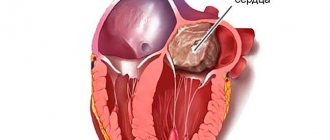
The tumor consists of a stalk, with which it is attached to the heart, and a body of myxoid, mucus-like tissue, covered with a hard shell.
It grows slowly but constantly, partially grows into healthy tissue, but is still demarcated from muscle fibers. In 75% of cases, myxoma is found in the left atrium, and in 20% - in the right. Metastasis is possible, but is not a mandatory feature.
The prognosis with timely intervention is always favorable. Cure without relapse occurs in most cases.
Mechanism of myxoma formation
The mechanism of the process is based on the same factor. Violation of cellular integrity with activation of the regenerative moment and weak immunity.
All injuries received, even minor ones, require recovery. In the case of the heart, we are talking about scarring.
In epithelialization, the main role is played by stem cells. They, like cancerous ones, have “immortality”, are not capable of spontaneous death after a certain number of divisions, and can also differentiate and transform into any others. It is the building material of the body.
When the differentiation process is disrupted, the immune system reacts. It destroys modified structures. With a weak defense system, tumors grow freely.
It becomes clear why myxoma develops after injuries, heart damage and other moments.
Causes
The factors behind the development of the pathological process are not known for certain. Doctors in the post-Soviet space are of the opinion of incidentality.
That is, neoplasia, according to their point of view, is a random process. This indicates that the phenomenon is poorly understood and there is an insufficient amount of empirical material.
Neoplasia develops from mucous cells that remain as vestiges after a person is born. They do not play a functional role and are invisible in the normal state of affairs.
Accordingly, cardiac myxoma begins in the early stages of life. The mechanism of development of the deviation is also unknown.
According to research by specialized experts around the world, the following moments become trigger mechanisms:
Past infectious diseases
The main thing is myocarditis. Inflammation of the heart muscle of septic origin. Accompanied by a pronounced clinical picture, chest pain, pressing sensations, heaviness, rhythm disturbances such as tachycardia, increased body temperature, and others. It is necessary to approach treatment carefully.
The likelihood of developing myxoma has not been determined. Apparently, the sooner therapy is started, the higher the chances of preventing the oncological process.
Staphylococci, pyogenic flora in general, and the waste products of these agents are considered highly carcinogenic, capable of causing neoplasia.
Previous heart surgery
Mostly open. The likelihood of developing a myxoma is also quite low, which is proven by the absence of massive tumor processes among patients with surgical interventions.
If there is a predisposition, the person should be carefully monitored after treatment.
Genetic abnormalities
Congenital pathological processes of varying severity. Usually accompanied by multiple tumors of different locations. These include Carney syndrome.
It is extremely rare and therefore does not have much clinical significance. Most myxomas in such patients begin in childhood. Although the overall prevalence of heart tumors in young people occurs in 10-12% of cases.
Risk factors
In addition to the fundamental factors themselves, predisposing reasons play a role. They increase the risks:
- Belonging to the female gender. According to specialized studies and statistical information, the likelihood of developing myxoma in patients is 3-5 times higher. Men become victims of more aggressive forms of the process. With intense current. Histological evaluation often reveals that the lesion is not a myxoma.
- Older age. The main patient population is people over 45 years of age. Apparently, there is an implicit connection between the development of neoplasia and the level of specific sex hormones. Mainly estrogens. We are also talking about the accumulated burden of somatic diseases associated with cardiac dysfunction. In children, the deviation occurs, but is relatively rare in the general array of clinical cases.
Everything said is just speculation. No one has conducted serious research, since tumors are relatively rare. Cardiac myxoma has a controversial etiology and is poorly understood.
Practical experience suggests good chances for treatment and complete recovery, even at advanced stages (when neoplasia reaches significant sizes, there is evidence of structures of 10-15 cm).
What is myxoma?
Some researchers suggest that this formation develops from a parietal thrombus, which subsequently degenerates into a tumor. But other scientists hold alternative views - they consider myxoma as a true tumor. Proof: particles easily break off from it and are carried along the bloodstream to various places. And there new daughter formations can grow from them.
Upon macroscopic examination, two types of myxomas are distinguished:
- A colorless, translucent tumor with a wide base. It fits tightly to the wall and has a soft consistency.
- Round polyp-like formation on a stalk. This tumor is much denser than the first one.
The surface of the myxoma is smooth. Inside there is a jelly-like mass in which there are few cellular elements. Sometimes formations may become calcified followed by ossification. But this happens with very “old” myxomas. They are supplied with blood through various vessels: capillaries and arterioles. The tumor tissue is quite fragile, which explains its frequent embolization (tearing off of individual sections).
picture: type 2 myxoma in the left atrium
Detected myxomas can have a variety of sizes: from one to several centimeters. It seems, what harm can a small benign formation cause? In general, none. Small tumors do not reveal themselves in any way; they are found by chance during a complete cardiac examination of the patient. But the danger is that sooner or later they grow up. “Old” formations are, of course, easier to diagnose. They are larger in size and exhibit characteristic clinical signs. But, unfortunately, they are the ones that lead to terrible complications.
Why is a tumor dangerous?
Myxoma is malignant, so it needs to be treated as a big problem. It poses a threat to life and health:
- Compression of cardiac structures. Develops as a result of the growth of a neoplasm. With significant volumes, it compresses the tissue and causes artificial ischemia. Blood flow weakens, myocardial contractility decreases. The most likely outcome of a pathology of this kind is a heart attack or stoppage of the muscle organ.
- Another problem is arrhythmia. It manifests itself as a result of a spontaneous increase in the electrical activity of all chambers. This happens as a result of excessive mechanical stimulation. Consequence of compression.
- Myxoma has a tendency to metastasize. It is not known exactly how high, but cases have been recorded. Therefore, it is possible to separate cells from the main tumor and move them with the bloodstream to distant areas of the cardiac structures.
- Stroke. Poor blood circulation in the brain as a result of blocked heart valves. Necrosis of tissues of the central nervous system. The formation of neurological deficits and other “delights” of the condition after the fact.
The likely results will be severe disability and then death of the patient. Prevention of complications is the goal of early therapy. Without treatment, the outcome is always fatal; there can be no spontaneous regression.
Is it possible to live with myxoma?
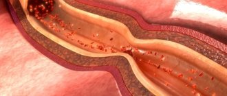
If a myxoma is diagnosed, postponing surgery is life-threatening. The tumor is characterized by intensive growth, which cannot be stopped with medications. Its location in an area of intense blood flow leads to separation of parts and blockage of blood vessels, so the risk of sudden death from thromboembolism remains at any time.
Thromboembolism as a complication of cardiac myxoma
Symptoms
Signs of the oncological process depend on the location of the tumor. Common manifestations for all clinical variants:
- Chest pain. Regular, pressing, aching and burning. The origin of the disorder is not clear until diagnosis. Discomfort increases after physical activity. Depends on the size of the tumor. The larger, the more intense the syndrome.
- Cardiac disorders. According to the type of ventricular fibrillation, sinus tachycardia, and other arrhythmias. Without echocardiography, it is impossible to understand the reasons.
- Breathing disorders. The normal process of gas exchange.
- Nausea.
- Weakness, drowsiness, decreased performance.
- Headache. Vertigo with inability to orientate in space.
In the early stages, when the tumor is small in size, symptoms are completely absent. As it progresses and grows, the first manifestations develop. Among which are the points described above.
Specific signs depend on the localization of the pathogenic structure:
Myxoma of the left atrium makes itself known by circulatory disorders in the systemic circle. The contractility of the chambers decreases, the release of liquid tissue is insufficient for normal nutrition of all organs.
Mortality is high, the probability of sudden death without therapy is 40%.
Myxoma of the right atrium gives symptoms of respiratory failure. Blood flow in the small pulmonary circle is disrupted. Fatality rate is about 50%. Overall mortality without treatment in both cases is close to 100% over a 1-3 year period. Nobody crosses the line.
General symptoms after the development of a neoplasm are as follows:
- Paleness of the skin. Bluish vessels and a mesh appear through the dermal layer.
- Increase in body temperature to subfebrile levels. No more than 38 degrees Celsius. The use of antibiotics and anti-inflammatory drugs has no effect, which indicates a non-septic process.
- Pain in bones, muscles. Of unknown origin. It resembles the influenza variant, but has no connection with infectious phenomena.
- Sudden loss of body weight. Within individual boundaries. Provided that the patient is not on a diet or starving.
- Spasm of peripheral vessels of the extremities (Raynaud's disease).
- Increase in blood pressure. Persistent, stable. The numbers are high. The condition is resistant to therapy and does not respond to antihypertensive drugs. The cause is cardiac, vascular. After the tumor is removed, the symptom gradually disappears.
- Increased venous pressure. Manifested by ascites, enlarged liver, severe abdominal pain. Yellowing of the skin is possible due to the release of burbulin into the bloodstream.
- Pulmonary edema. Productive or dry cough, respiratory distress, hemoptysis.
- Loss of consciousness. Syncope occurs repeatedly with damage to the left atrium and compression of the ventricle.
- Manifestations of pulmonary failure. They subside after changing body position. Such patients cannot lie down due to suffocation.
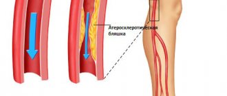
With a long course of the pathological process, thromboembolism develops in half of the patients. Blockage of a vessel by a blood clot.
Depending on the location of the disorder, the target organs are kidneys, liver, intestines, lungs, lower limbs, and brain.
With the development of occlusion, death can occur much earlier. Patients need to be monitored after surgery; the risks persist for at least two weeks.
Forecast and recommendations
Although myxoma grows quite slowly, it can take 2 to 3 years from its onset to probable death (if untreated). After removal of cardiac myxoma, doctors give a positive prognosis. If there are no complications, the patient’s recovery is fairly quick.
Of course, relapses are possible. Experts note insufficient excision of the tumor attachment as the cause of relapse.
Lethal cases have occurred in medical practice. They are considered quite rare. During and after surgery, only 5% of patients with a tumor located in the atrium and 10% of patients with a tumor located inside the ventricle die.
After surgery, patients are advised to follow the following rules:
- sleep 7-8 hours per day;
- avoid stress and conflicts;
- engage in physical therapy;
- avoid chest injury;
- avoid smoking, drinking alcohol;
- adjust the diet;
- follow the doctor’s recommendations;
- undergo sanatorium-resort treatment;
- be in the fresh air;
- avoid contact with infected people;
- undergo regularly scheduled examinations.
People will be able to return to their previous life and go to work after the operation after 2–3 months.
Did you like the article? Save it!
Still have questions? Ask them in the comments! Cardiologist Mariam Harutyunyan will answer them.
Ivan Grekhov
Graduated from the Ural State Medical University with a degree in General Medicine. General practitioner
Diagnostics
It is carried out urgently by a cardiologist. Complaints of pain in the chest are a nonspecific symptom, therefore it is necessary to examine the person using instrumental methods.
Approximate list of events:
- Symptom assessment. It has no pronounced pathognomonicity.
- Anamnesis collection. Family history, other points. A consultation with a geneticist is indicated.
- Measurement of blood pressure, heart rate. There is little or no change in the early stages. Then the manifestations increase.
- Echocardiography. Basic diagnostic method. Allows you to visualize tissue, detect a tumor, and evaluate its signs.
- MRI. With contrast enhancement with gadolinium preparations. The path to better tissue imaging. Allows you to quickly identify the nature of blood flow and proliferative activity.
- Biopsy and histological examination. Used for differential diagnosis of tumors. After confirmation, treatment can begin.
- General blood test, biochemical.
The diagnosis can be made and verified only after taking a tissue sample. A biopsy can be performed during the operation, depending on the treatment tactics chosen by the cardiac surgeon.
Features of intracardiac hemodynamics in pathology
The effect of a tumor on the movement of blood inside the heart depends on its size and location. Any volumetric formation leads to difficulty in the passage of blood:
- left atrial - clogs the left opening between the atrium and the ventricle, preventing outflow from the pulmonary veins;
- right atrial – closes the right orifice and interferes with systemic venous outflow;
- the left ventricular inhibits the release of blood into the arterial network;
- in the right ventricle reduces blood flow to the lungs.
Characteristics of the tumor include rapid growth. Cardiac hemodynamic disturbances appear already from 7 cm.
Treatment
It is carried out urgently immediately after detection of a tumor. Myxoma doesn't give you time to think.
Watchful waiting worsens the prognosis by approximately three times. Death occurs quickly, neoplasia progresses steadily, and a sharp jump in proliferative activity is possible. This is a malignant neoplasm and must be treated as such.
The main method of treatment is open surgery. Myxoma is a delimited tumor; in the early stages and even later, infiltration and growth through tissue are not observed. There is a possibility of radical removal.
Attention:
Radiotherapy and chemotherapy do not play a big role. In addition, myxomas are highly resistant to such treatment methods.
After the operation, observation is indicated for 1-2 weeks. Then regular tomographic screenings every 3 months. In the absence of relapse (which is most likely), the frequency of studies decreases.
After 5 years, MRI is not prescribed at all. The patient is considered healthy.
Correction of disorders that were caused by the tumor can be carried out. Usually everything returns to normal on its own, but when the tumor is large, organic defects form.
Complications during and after surgery
Surgery to remove myxoma of the heart usually takes place without complications, but of course they cannot be excluded. Doctors explain their manifestation with certain nuances:
- poor preparation for the upcoming surgical intervention;
- a sharp attack of arrhythmia;
- localization of the tumor;
- penetration of infection into the heart cavity during surgery.
After the operation, the patient is in the clinic under observation for about 2 days, in the intensive care unit. This is necessary to prevent the occurrence of the following complications:
- ari src=”https://davlenienorm.com/wp-content/uploads/2018/08/miksoma-serdca-9.jpg” class=”aligncenter” width=”695″ height=”208″[/img]
- bleeding;
- heart failure;
- embolism caused by vascular injury during surgery.
Forecast
Depends on many factors.
- Tumor volume. The larger it is, the worse the likely outcome. Neoplasia over 10 cm in diameter compress the heart and cause anatomical disorders.
Treatment is urgent. Without quality help, even if the operation is performed later, a complete recovery of the condition is not expected.
- Age. In younger years, the tumor behaves more aggressively.
- Floor. In men, the pathology is more active.
- Family history. If there were people in the family suffering from myxoma, the disease has a worse prognosis.
- Carney syndrome. It ends in failure in most cases, although the process is not so active.
- Somatic conditions. Diabetes mellitus, atherosclerosis, organ defects, congenital or acquired. They provoke a sharp drop in patient survival.
- Severe hormonal dysfunction is also a negative point.
In general, death occurs within 1-2 years without treatment. Sudden death occurs in 20-30% of situations.
Complete relief with radical intervention occurs in 85% of cases. Partial effect is associated with the occurrence of organic defects. With proper correction, the chances are good.
Prevention
There are no specific preventive measures. You can reduce risks by following a few rules:
- Avoid chest injuries. Especially affecting the heart and surrounding structures.
- Urgently treat all infectious diseases. Myocarditis and other similar processes.
- Maintain hormone concentrations at adequate levels. After the onset of menopause, especially in women, regularly receive replacement therapy with synthetic substances.
Pregnant women are advised to avoid exposure to ionizing radiation. It can cause increased risks for the unborn child.
Eat right, do not drink questionable vitamin and mineral complexes, especially those that are actively advertised. Stress also does not bring anything good.
The best solution would be to undergo genetic testing at the pregnancy planning stage. Also assess the partner’s condition.
In case of violations, monitor the position of the fetus during gestation. The tendency to develop myxoma is not visible, but at the same time the presence of defects can be determined. Especially those incompatible with life.