What is he talking about?
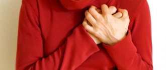
If a patient experiences sounds inside the body, this means that the process of blood flow in the heart vessels is disrupted. There is a widespread belief that systolic murmur occurs in adults.
This means that a pathological process is occurring in the human body, which indicates some kind of illness. In this case, it is necessary to urgently undergo a cardiac examination.
Systolic murmur is defined as its presence between the second heart sound and the first. The sound is recorded on the heart valves or bloodstream.
Division of noise into types
There is a certain gradation of separation of these pathological processes:
- Functional systolic murmur. It refers to innocent manifestation. Does not pose a danger to the human body.
- Systolic murmur of organic type. Such a noise character indicates the presence of a pathological process in the body.
An innocent type of noise may indicate that there are other processes in the human body that are not related to heart disease. They are mild in nature, do not last long, and have a weakly expressed intensity. If a person reduces physical activity, the noise will disappear. Data may vary depending on the patient's posture.
Noise effects of a systolic nature arise due to septal and valvular disorders. Namely, in the human heart there is dysfunction of the partitions between the ventricles and atria. They differ in the nature of their sound. They are hard, tough and stable. A rough systolic murmur is present and its long duration is recorded.
These sound effects extend beyond the boundaries of the heart and are reflected in the axillary and interscapular areas. If a person has subjected his body to exercise, then sound deviations persist after completion. Noises become louder during physical activity. The organic sound effects that are present in the heart are independent of body position. They can be heard equally well in any position of the patient.
Heart murmurs
Heart murmurs
Noises that occur in the cavities of the heart and in the supravalvular section of the ascending aorta or pulmonary trunk when turbulence in the blood flow appears in them. Conditions for the formation of vortex flows and northern latitudes. occur with pathology of the valves and defects of the septa of the heart, expansion of its cavities and sections of the pulmonary trunk or aorta (aneurysm), significant acceleration of blood flow with a decrease in its viscosity (for example, with anemia) or due to hyperkinetics of heart contractions (for example, with thyrotoxicosis). Like other true noises, S. sh. are the result of the summation of sound vibrations of different frequencies and amplitudes, but based on the predominance of a certain frequency, in some cases they can be conditionally characterized as low-, medium-, or high-frequency. In rare cases, high harmonic vibrations are involved in the formation of noise, which are perceived by the ear as musical noise. Although heart sounds, by their physical nature, also belong to noise, in perception they differ from prolonged S. sh. brevity and abruptness of sound.
The main method of identifying S. sh. is Auscultation. To analyze the amplitude-frequency characteristics of S. sh. and their connection with certain phases of systole and diastole of the heart, phonocardiography (Phonocardiography) is used. To identify and evaluate S. sh. Using auscultation, it is recommended to listen to the heart in the patient’s position not only lying on his back, but also, if necessary, in a position on the left and right side, on the stomach, in a sitting and standing position, in the phases of deep inspiration and exhalation, sometimes also before and after physical activity, taking nitroglycerin. Listening is carried out at so-called standard points, i.e. in certain areas of the precordial region ( Fig. 1 ), corresponding to the places of projection of the S. sh. from individual valves: 1) in the area of the apex of the heart - from the bicuspid (mitral) valve (although the bicuspid valve itself is projected higher - at the place of attachment of the third rib to the sternum); 2) in the second intercostal space, at the right edge of the sternum, from the aortic valve; 3) in the second intercostal space, at the left edge of the sternum, from the pulmonary valve; 4) at the right edge of the lower third of the sternum - from the tricuspid valve; 5) in the fourth intercostal space at the left edge of the sternum (fifth point, or Botkin-Erb point) - to listen to the diastolic murmur of aortic insufficiency, and in some cases, murmurs associated with lesions of the mitral valve. When identifying S. sh. Auscultation is not limited to standard points, determining the best places to listen to the noise, its conduction to the vessels of the neck or other vessels. During a phonocardiographic study, the microphone is placed at standard auscultation points.
By origin S. sh. are usually divided into organic, associated with defects or aneurysm of the heart or vessels extending from it, and functional, caused, for example, by accelerating blood flow, reducing its viscosity, incl. and so-called innocent heart murmurs, often detected in healthy people, especially in children and young people.
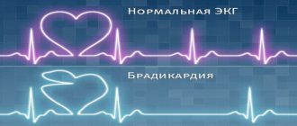
In relation to the phases of the cardiac cycle ( Fig. 2, 3 ), heart murmurs are divided into systolic, i.e. heard between the 1st and 2nd heart sounds (in the systole phase), and diastolic, heard in the diastolic pause between the 2nd and 1st heart sounds. S. sounds that occur during systole and continue after the second heart sound are called systolic-diastolic murmurs. Depending on what part of systole or diastole the noise occupies - initial, middle, final (late), it is designated as proto-, meso- and telesystolic (or proto-, meso- and telediastolic), respectively, and if it is heard from the beginning until the end of the pause - as pan-, or holosystolic (respectively, pan-, or holo-diastolic). Telediastolic murmur is more often called presystolic.
Listened S. sh. can be short and long, increasing, decreasing, increasing-decreasing (on a phonocardiogram - diamond-shaped, spindle-shaped), etc. In terms of pitch and timbre, the noise can be rough, gentle, blowing, scraping, sawing, rumbling, musical. The loudness of the murmur depends on many conditions, so in itself it cannot be a characteristic of the severity of a defect or other heart lesion. In thin people with a thin chest wall, as well as in children, murmurs are louder than in obese people. Pulmonary emphysema reduces the sonority of noises due to the air gap separating the heart from the anterior chest wall. In heart failure, the intensity of many murmurs caused by organic valve disease decreases, and sometimes these murmurs even disappear. Volume of diastolic S. sh. decreases with tachycardia (due to shortening of diastole); these noises usually disappear when the heart rate exceeds 100 per minute. Due to respiratory fluctuations in cardiac output, the intensity of S. sh. may vary according to the phases of breathing; Intracardiac murmurs, both valvular and extravalvular, sharply weaken when straining after a deep breath. During physical exertion, mental excitement, fever, when the speed of blood circulation increases, the strength of S. sh. usually increases; in this case, it is often possible to listen to noises that are not detectable under normal conditions. S. sh. caused by valve defects, are best heard in a horizontal position of the patient, but in general they are less dependent on changes in body position than functional noises.
Diagnostic value of heart murmurs . Murmurs that occur with heart defects have characteristics of origin and sound that reflect the nature of the pathology. Depending on the type of defect, the murmur is formed in certain phases of the cardiac cycle, so determining the phase of the cardiac cycle in which the murmur is heard has diagnostic significance.
Systole-diastolic murmurs occur when there is a septal defect in the heart or a shunt between large vessels. The most common cause of this noise is a patent ductus arteriosus. The noise begins immediately after the first tone ( Fig. 3, f ) with a slight deviation from it. As the pressure in the aorta increases, the volume of the noise increases. The pressure gradient between the aorta and the pulmonary trunk reaches a maximum at the end of systole, and at the same time the maximum sound of the murmur is noted. It ends in the middle or at the beginning of diastole. The noise is often accompanied by shaking. Gromkiy S. sh. heard over the entire region of the heart, but has maximum intensity in the second left intercostal space, from where it radiates to the third intercostal space; When the patient is lying down, the noise intensifies. An aneurysm of the sinus of Valsalva, when it ruptures into the right ventricle, is accompanied by a systolic-diastolic murmur, which is heard to the left of the sternum. The diastolic component of the murmur is louder than the systolic component.
Systolic murmurs are heard most often, because formed in a variety of heart diseases, including myocarditis, cardiosclerosis, cardiomyopathy, as well as anemia and hyperkinetic syndrome; they make up the largest portion of “innocent” noises in practically healthy individuals and, in addition, can be important auscultatory symptoms of the most common valvular heart defects - atrioventricular valve insufficiency or stenosis of the aorta or pulmonary trunk (see Acquired heart defects (Acquired heart defects)) , as well as septal defects of the heart.
In mitral regurgitation, systolic murmur occurs due to regurgitation of blood from the left ventricle into the left atrium. It begins immediately after the weakened first sound and decreases towards mid-systole ( Fig. 3, a ), but often continues until the beginning of the second sound, i.e. happens pansystolic. The noise may have the same intensity throughout the entire systole, but may intensify towards the end of it, often having a blowing character. Damage to the papillary muscles in acute myocardial infarction (myocardial infarction), traumatic separation of the papillary muscles leads to the formation of acute mitral regurgitation; in this case, a rough and prolonged systolic murmur is heard, which begins with separation from the first tone, has a diamond shape, and is sometimes carried out to the vessels of the neck and into the interscapular space. Telesystolic (late) murmur is often heard in patients with atherosclerotic cardiosclerosis, with sclerotic degeneration of the papillary muscles, as well as with mitral valve prolapse.
The systolic murmur of tricuspid regurgitation is better heard at the base of the sternum, often intensifies at the height of inspiration and often has varying intensity, in contrast to the stable murmur of mitral regurgitation. Inconsistency of noise is more typical for relative tricuspid insufficiency. As the patient's condition improves, the sound of organic failure does not change or even intensifies due to increased contractility of the right ventricular myocardium, while the sound of relative failure becomes quieter or disappears.
A systolic murmur in the second intercostal space to the right of the sternum is characteristic of aortic stenosis (ejection murmur). Most often, it has a diamond-shaped or spindle-shaped shape, begins with some separation from the first sound ( Fig. 3, b ), ends before the beginning of the second sound, and often radiates to the apex of the heart and carotid arteries. The systolic murmur of aortic stenosis is usually accompanied by tremors of the chest wall determined by palpation (“cat purring”). The same systolic ejection murmur, but in the second left intercostal space, is heard with stenosis of the mouth of the pulmonary trunk ( Fig. 3, d ). Sometimes it is best heard in the third or fourth intercostal space, to the left of the sternum.
Systolic murmur with a ventricular septal defect is loud, prolonged, sharp and even rough, accompanied by tremors, the epicenter of which is in the third or fourth intercostal space, at the left edge of the sternum. The murmur usually covers the first sound and occupies the entire systole ( Fig. 3, c ); sometimes it is recorded as increasing and decreasing. In a horizontal position, the noise is louder, often heard at a distance, and is carried into the interscapular space. After the introduction of mezatone, it becomes more intense; sublingual administration of nitroglycerin reduces its intensity.
With an atrial septal defect, the systolic murmur begins immediately after the first sound, it is not intense, blowing, and is accompanied by a bifurcation of the first sound. The intensity of the noise increases with physical activity, but does not reach the degree that occurs with ventricular septal defects.
Diastolic murmurs are almost always associated with organic heart pathology. Most often, diastolic murmurs are caused by insufficiency of the aortic valve or pulmonary trunk or stenosis of the atrioventricular orifices.
The diastolic murmur of aortic insufficiency begins at the very beginning of diastole, during the period of the second sound, which is often replaced or covered by this noise. The noise is high-frequency, gentle, usually blowing. The duration of the murmur depends on the severity of aortic insufficiency. With minor aortic insufficiency, the diastolic murmur is short (protodiastolic; Fig. 3, e ), it is difficult to detect, only in the absence of extraneous noise. With more severe aortic insufficiency, it occupies 1/2-2/3 of diastole and is characterized by decreasing intensity; with severe aortic defects, the murmur often occupies the entire diastole. Many researchers believe; that in severe aortic insufficiency the murmur is usually more intense. It is heard at the auscultation point of the aortic valve and at the fifth point, and is better heard with the patient lying on his stomach with calm, shallow breathing.
A fading diastolic murmur in the second (less often in the third) intercostal space to the left of the sternum is a sign of pulmonary valve insufficiency. With relative pulmonary valve insufficiency resulting from hypertension of the pulmonary circulation (pulmonary circulation hypertension), a quiet blowing diastolic murmur called Still's murmur is heard. It begins immediately after the second sound and is heard in a limited area in the second intercostal space on the left; in cases of bifurcation of the second tone, the noise begins from its second component.
The diastolic murmur of mitral stenosis occurs immediately after the second component of the bifurcated II tone, has presystolic amplification, often musical, and is accompanied by chest trembling. The murmur is usually low-frequency, better heard at the apex of the heart when the patient is horizontally positioned on the left side, and intensifies after physical activity. It can occupy the entire diastole; at the same time, it first decreases, and then, from approximately the middle of diastole, continues with a constant amplitude until presystolic increase ( Fig. 3, e ) associated with atrial systole. This is explained by the fact that during the phase of rapid filling of the left ventricle, blood rushes at high speed through a narrow opening, creating a noise that subsides as the flow rate decreases and intensifies again with its additional acceleration associated with atrial systole. With atrial fibrillation, there is no presystolic increase in noise. Along with diastolic murmur, an increase in the first tone and a bifurcation of the second tone are detected, which creates the so-called melody of mitral stenosis.
Diastolic murmur with presystolic amplification is determined with myxoma of the left atrium (see Heart), it is variable, its appearance and intensity depend on the position of the body and the speed of blood flow.
With severe organic aortic insufficiency, functional stenosis of the left atrioventricular orifice occurs due to the fact that the blood stream regurgitating into the left ventricle lifts the mitral valve leaflet. In this case, a diastolic murmur with presystolic amplification (Flint murmur) is heard at the apex of the heart. It is not as long and loud as with organic mitral stenosis, and is not accompanied by an increase in the first sound and the sound of the mitral valve opening.
Often, in children with mitral valve insufficiency and a ventricular septal defect with significant dilatation of the atria or ventricles, a fusiform diastolic murmur is detected, separated by an interval from the second sound and lasting no more than 0.2 s (Coombs noise); it is heard near the apex of the heart only in the presence of a third sound.
Diagnostic differentiation of cardiac murmurs is carried out primarily with extracardiac (paracardial) murmurs. The greatest similarity with S. sh. have pericardial friction rub, cardiopulmonary and pleuropericardial murmurs. Pericardial friction noise is detected in pericarditis, myocardial infarction, usually in the form of short scratching sounds during systole or diastole or in both phases. Cardiopulmonary (cardiopulmonary) murmurs occur in those parts of the lungs that are in contact with the heart. Changes in the size and position of the heart during systole cause rapid movement of air in the adjacent areas of the lungs, which generates high-frequency noise. Cardiopulmonary murmurs are often heard in persons with a reduced anteroposterior chest size, in young people with excited cardiac activity (the so-called hyperkinetic type of cardiac activity), as well as in cases of severe cardiac hypertrophy. They are usually heard during systole, and when breathing is held during exhalation, they usually disappear. Pleuropericardial murmurs, i.e. friction noises that occur with dry pleurisy in the areas of contact between the pleura and the pericardium intensify during inspiration. When differentiating the nature of murmurs, it is also necessary to remember about the various vascular murmurs.
Significant difficulties sometimes arise when differentiating organic and functional S. sh. To distinguish organic and “innocent” noises, various techniques (physical activity, change of body position) and pharmacological tests were proposed. Taking nitroglycerin causes an increase in the systolic murmur of aortic stenosis, a weakening of the murmur of mitral regurgitation and an increase in the systolic murmur of tricuspid regurgitation. Functional pulmonary systolic murmur is heard in the second intercostal space, at the edge of the sternum. The murmur usually has a spindle-shaped shape and occupies the first half of systole, always blowing, is better heard when the patient is lying down, and intensifies under the influence of stress and with fever. Exhalation improves the audibility of this noise. An “innocent” aortic systolic murmur occurs due to systolic vibration of the distended aortic root. It occupies mid-systole, is heard in the second intercostal space, and is usually carried out to the apex of the heart at the right edge of the sternum.
Diagnosis of the nature of the North. more accurate for dynamic noise estimation. As heart function improves, organic murmurs usually become more distinct. We can also assume that murmurs heard in the diastole phase and murmurs that occupy the entire systole are never “innocent”. If there is doubt about the nature of the noise, a comprehensive examination of the patient and dynamic observation is necessary.
Rice. 3. Scheme of phonocardiographic image of cardiac murmurs (shaded areas) for some heart defects in relation to the first and second heart sounds: a - decreasing protosystolic murmur with mitral insufficiency; b — diamond-shaped mesosystolic murmur with aortic stenosis; c — holosystolic murmur with ventricular septal defect; d — fusiform holosystolic murmur with pulmonary stenosis; e — presystolic (due to mitral stenosis) and protodiastolic (due to aortic insufficiency) murmurs; e - systolic-diastolic murmur with open ductus arteriosus.
Rice. 2. Schematic representation of an electrocardiogram (ECG) and phonocardiogram (PCG) of the I and II heart sounds with the distribution of the phases of systole (between the I and II sounds) and diastole (between the II and I sounds) into time intervals (separated by a dotted line), which indicate audible During these intervals, heart murmurs: a - presystolic; b - protosystolic; c — mesosystolic; g - telesystolic; d - protodiastolic; e - mesodiastolic; g - holosystolic; h - hunger-diastolic.
Rice. 1. Scheme of the projection of the heart valves onto the anterior chest wall (A - projection of the aortic valve; L - projection of the pulmonary valve; M - projection of the mitral valve; T - projection of the tricuspid valve) and the main points for listening to heart murmurs: 1 - apex of the heart (murmurs are heard from the mitral valve); 2 - second intercostal space at the right edge of the sternum (aortic valve); 3 - second intercostal space at the left edge of the sternum (pulmonary valve); 4 - body of the sternum above the xiphoid process (tricuspid valve); 5 - Botkin-Erb point - fourth intercostal space to the left of the sternum (diastolic murmur of aortic insufficiency and murmur of the mitral valve are performed); Roman numerals indicate the ribs.
Source: Medical Encyclopedia on Gufo.me
Meanings in other dictionaries
- CARDIAC NOISE - CARDIAC NOISE - a sound phenomenon caused by obstacles to blood flow due to heart defects, changes in its muscles, disturbances in the composition of the blood, for example. for anemia, etc. Big encyclopedic dictionary
- Heart murmurs are sound phenomena accompanying the work of the heart, which can occur in pauses between heart sounds (See Heart sounds) and, in comparison, represent longer aperiodic sound vibrations. Determined by auscultation (See Great Soviet Encyclopedia
- Blog
- Jerzy Lec
- Contacts
- Terms of use
© 2005—2020 Gufo.me
What factors influence the occurrence of noise?
What are the causes of systolic murmur? There are several main ones. These include:
- Aortic stenosis. It can be either congenital or acquired. This disease occurs due to narrowing of the aorta. With this pathology, the walls of the valve become fused. This position makes it difficult for blood to flow inside the heart. Aortic stenosis can be considered the most common heart defect in adults. The consequence of this pathology can be aortic insufficiency, as well as mitral disease. The aortic system is designed in such a way that calcification is produced. In this regard, the pathological process intensifies. It is also worth mentioning that with aortic stenosis, the load on the left ventricle increases. At the same time, the brain and heart experience insufficient blood supply.
- Aortic insufficiency. This pathology also contributes to the occurrence of systolic murmur. With this pathological process, the aortic valve does not close completely. Infectious endocarditis causes aortic insufficiency. The impetus for the development of this disease is rheumatism. Lupus erythematosus, syphilis and atherosclerosis can also provoke aortic insufficiency. But injuries and congenital defects rarely lead to the occurrence of this disease. A systolic murmur in the aorta indicates that the valve has aortic insufficiency. The reason for this may be expansion of the ring or aorta.
- Washing of the acute course is also the reason why systolic murmurs appear in the heart. This pathology is associated with the rapid movement of liquids and gases in the hollow regions of the heart during their contraction. They are moving in the opposite direction. As a rule, this diagnosis is made when the functioning of the dividing partitions is impaired.
- Stenosis. This pathological process is also the cause of systolic murmurs. In this case, a narrowing of the right ventricle, namely its tract, is diagnosed. This pathological process occurs in 10% of cases of murmurs. In this situation, they are accompanied by systolic tremors. The vessels of the neck are especially susceptible to irradiation.
- Tricuspid valve stenosis. With this pathology, the tricuspid valve narrows. As a rule, rheumatic fever leads to this disease. Patients experience symptoms such as cold skin, fatigue, and discomfort in the neck and abdomen.
Treatment and diagnosis
If murmurs are detected, you should first consult a cardiologist who will conduct a diagnosis and identify the root cause of the deviation. Do not neglect your doctor's recommendations. Health and future life directly depend on the timeliness of actions taken. Of course, each of the subtypes of such manifestations has its own characteristics, however, heart murmurs cannot be attributed to a natural phenomenon.
To detect noise, a specific analysis scheme is used:
- First, determine the phase of the heart in which it is heard (systole or diastole).
- Next, its strength is determined (one of the degrees of loudness).
- The next step is to determine the relationship to heart sounds, that is, it can deform heart sounds, merge with them or be heard separately from the tones.
- Then its shape is determined: decreasing, increasing, diamond-shaped, ribbon-shaped.
- Consistently listening to the entire area of the heart, the doctor determines the place where the murmur is more clearly audible. Checking the irradiation of a deviation consists of determining its location.
- The penultimate stage of diagnosis is to determine the influence of respiratory phases.
- After this, the doctor determines the dynamics of the noise over time: it can be a day, a week, a month, etc.
For differential diagnosis, the moment of occurrence of systolic murmurs and their duration are determined using laboratory tests.
As a rule, the following tests are prescribed:
- radiography, which allows you to determine thickening of the walls of the heart, hypertrophy or enlarged chambers of the heart;
- ECG - determines the level of overload in various areas;
- EchoCG - used to detect organic changes;
- catheterization
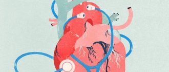
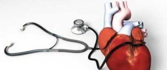
Listening to the work of the heart using a phonendoscope is one of the main methods for diagnosing diseases of the cardiovascular system. A competent specialist can easily differentiate suspicious signs from normal manifestations.
Doctors believe that it is especially important to evaluate the systolic murmur at the apex of the heart, since this indicator helps to identify certain pathologies. Consultation with a cardiologist will help the patient learn more about heart murmurs.
Systolic heart murmur can be organic or functional.
The heart is the main organ of the cardiovascular system. This is a muscle pump that maintains constant blood movement in the vessels and blood supply to all tissues of the body.
Thanks to the contractions of the organ, venous blood returns from the cells to the lung tissue for oxygenation, and arterial blood constantly transports oxygen and nutrients. Even a short-term failure of the heart muscle can lead to the death of the patient. Organs that are heavily dependent on blood supply are primarily damaged, including the brain and kidneys.
From an anatomical point of view, the heart is divided into four sections - two atria and two ventricles.
The left atrium and left ventricle contain arterial blood, and the right atrium and right ventricle contain venous blood. During contraction of the heart muscle, blood from the right side enters the lung tissue, and blood from the left side is thrown into the aorta and enters the arteries of the body. In this case, the organ enters a phase of activity during contraction (systole) and returns to a short resting phase between contractions (diastole) to fill the chambers of the heart before a new contraction.
Since the work of the cardiovascular system is accompanied by various noises, cardiac auscultation is an effective first examination. The doctor applies the head of the phonendoscope to certain points on the front surface of the patient's chest to listen to sounds and evaluate the work of the heart. Certain noises are caused by the moment of myocardial contraction, the collapse of the internal valves of the organ, the reflux of blood and other conditions. Conventionally, murmurs are divided into systolic and diastolic.
In addition to noise, it is important for the doctor to consider heart sounds. There are 4 tones that arise in different phases of the organ’s operation. The first two sounds are associated with myocardial contractile activity and valves, so they are best audible. To assess the functioning of different parts of the heart and blood vessels, the doctor can apply the head of the phonendoscope to different areas, including the intercostal spaces and the substernal area.
Possible reasons
According to classification, most noises are divided into functional and organic. Functional murmurs, which include systolic murmur at the apex of the heart, are not necessarily a sign of pathology and often occur in healthy people, while organic murmurs indicate a certain structural pathology of the heart.
It is believed that apical noise during myocardial contraction occurs due to a change in the nature of blood movement through the vessels.
Causes of “innocent” noises:
- High physical activity.
- Pregnancy.
- Fever.
- Insufficient number of red blood cells (the blood is thinner, causing turbulent flow).
- Excessive hormonal activity of the thyroid gland (hyperthyroidism).
- The period of rapid growth of organs and tissues (childhood and adolescence).
Thus, harmless heart murmurs at the apex of the organ occur during rapid blood flow and other completely normal conditions.
More information about the causes of heart murmurs in children can be found in the video:
Possible causes of pathological noises:
- The presence of an open foramen ovale between the atria. This leads to mixing of blood and disruption of the pumping function of the organ.
- Violation of the anatomy and functions of the heart valves. Most congenital anomalies affect valve closure. In patients with valve stenosis, there is a disturbance in the movement of blood through the parts of the heart.
- Valve calcification is a hardening of the anatomical structure that makes it difficult for the heart to function.
- – an infectious disease characterized by viral or bacterial damage to the inner lining of the heart and valves. The infection can spread to the organ from other anatomical areas. If such a disease is not treated in time, structural pathology may occur.
- Rheumatic fever is an autoimmune disease in which the body's defense systems attack healthy tissue. Rheumatic heart disease can occur due to improper treatment of infectious diseases.
Risk factors for heart disease:
- Family history of heart disease and abnormalities.
- Disorders of pregnancy.
- Taking medications that affect the condition of the organ.
Often, heart murmurs are the only noticeable manifestations of pathology.
Additional signs
Pathological systolic murmur at the apex of the heart can be accompanied by a wide variety of symptoms, since such a sign indicates various pathologies of the heart. Often, patients with the abnormality have no symptoms for a long time.
Possible signs:
- Swelling of the neck and limbs.
- Breathing problems.
- Chronic cough.
- Enlarged liver.
- Swollen neck veins.
- Loss of appetite.
- Heavy sweating.
- Chest pain.
- and weakness.
If you notice any of the above symptoms, you should consult a doctor.
Why does noise appear in children?
Why might a child have a heart murmur? There are many reasons. The most common ones will be listed below. So, heart murmurs may occur in a child due to the following pathologies:
- Violation of the interatrial septum. In this case, we are talking about the absence of fabric in it. This position leads to the discharge of blood. The volume of blood discharged depends on the size of the defect and the compliance of the ventricles.
- Abnormal state of venous return of the lungs of the child's body. There are cases of improper formation of pulmonary veins. The essence of this is that the pulmonary veins do not communicate with the atrium on the right. They can grow together with the veins of the systemic circle.
- Aortic coarctation. In this case, we are talking about narrowing of the thoracic aorta. The child is diagnosed with a heart defect. The segmental lumen of the aorta is smaller in size than it should be. This pathology is treated through surgery. If medical care is not provided, as you get older, the narrowing of the aorta will increase.
- Pathology of the interventricular septum. This defect also leads to the occurrence of systolic heart murmurs. This pathology can be isolated. That is, it can develop on its own or be combined with other cardiac dysfunctions.
- Congenital heart defects in children. An open arterial defect can also cause the presence of systolic murmurs in a child. There is a vessel in the structure of the cardiac system. It is the connecting element between the pulmonary artery and the descending aorta. The function of this organ is to allow the baby to take its first breath after birth. Then, after a short amount of time, the vessel closes. There are cases where this process fails. Then the process of shunting blood from the systemic circulation to the small circulation continues. This is the defect in the functioning of the body. In the case where the breakthrough allows a small blood flow through it, this does not particularly affect the child’s health. But if there is a large blood flow, then the baby may experience complications. Namely, there may be an overload in the work of the heart. In this situation, certain symptoms appear in the body, for example, shortness of breath. It also matters what cardiac straits are present in the baby’s body. If their flow is large, then it is possible that the condition of the newborn will be extremely serious. In this situation, in addition to systolic murmurs, the heart itself increases in size. The child is prescribed urgent surgical intervention.
Congenital heart defects in children
It is worth saying a few words about newborn babies. Immediately after birth, a complete examination of the body is carried out. This includes listening to the heart rate. This is done in order to exclude or detect any pathological processes in the body.
With such an examination, there is a possibility of detecting any noise. But they shouldn't always be a cause for concern. This is due to the fact that noises are quite common in newborn babies. The fact is that the child’s body adapts to the external environment. The cardiac system is reconfigured, so different noises are possible. Further examination through methods such as x-ray and electrocardiogram will show whether any abnormality is present or not.
The presence of congenital noises in the baby’s body is determined during the first three years of life. Murmurs in newborn babies may indicate that the heart was not fully formed during development before birth for various reasons. In this regard, after birth the baby develops noises. They talk about congenital defects of the cardiac system. In cases where pathologies have a high risk for the child’s health, doctors decide on a surgical method of treating a particular pathology.
Noise features: systolic murmur at the apex of the heart and in other parts of it
It is worth knowing that the characteristics of noise may vary depending on their location. For example, there is a systolic murmur at the aortic apex.
- Mitral valve pathology and associated acute insufficiency. In this position, the noise is short-lived. Its manifestation occurs early. If this type of noise is detected, then the patient is diagnosed with the following pathologies: hypokinesis, chord rupture, bacterial endocarditis, etc.
- Systolic murmur on the left sternal border.
- Chronic mitral valve insufficiency. This type of noise is characterized by the fact that they occupy the entire duration of ventricular contraction. The size of the valve defect is proportional to the volume of blood returned and the nature of the murmur. This noise is better heard if a person is in a horizontal position. As the heart defect progresses, the patient experiences vibration in the chest. There is also a systolic murmur at the base of the heart. Vibration is felt during systole.
- Mitral insufficiency of a relative nature. This pathological process is treatable with proper treatment and compliance with recommendations.
- Systolic murmur in anemia.
- Pathological disorders of the papillary muscles. This pathology refers to myocardial infarction, as well as ischemic disorders in the heart. This type of systolic murmur is variable. It is diagnosed at the end of systole or in the middle. There is a short systolic murmur.
Noise classifications
Functional noises are classified as follows:
- with mitral insufficiency, heard above the apex of the heart;
- above the aorta when it enlarges;
- arising from aortic valve insufficiency;
- above the pulmonary artery during its expansion;
- during nervous excitement or physical exertion, accompanied by tachycardia and ringing tones;
- appearing with fever;
- arising from thyrotoxicosis or severe anemia.
By its nature, the noise is distinguishable from a heartbeat, and treatment depends on its volume, frequency and strength. There are six volume levels:
- Barely visible.
- Disappearing at times.
- Constant noise, more sonorous and without trembling of the walls.
- Loud, accompanied by vibrations of the walls (can be distinguished by placing your palm).
- Loud, which can be heard in any area of the chest.
- The loudest one can be easily heard, for example, from the shoulder.
Volume is affected by body position and breathing. For example, when you inhale, the noise increases, as the reversal of blood to the heart muscle increases; When standing, the sound will be much quieter.
The appearance of heart murmurs during pregnancy in women
When a woman is pregnant, processes such as systolic murmurs cannot be ruled out in her heart. The most common cause of their occurrence is the load on the girl’s body. As a rule, heart murmurs appear in the third trimester.
If they are detected in a woman, the patient is placed under more careful monitoring. At the medical institution where she is registered, her blood pressure is constantly measured, her kidney function is checked, and other measures are taken to monitor her condition. If a woman is constantly under observation and follows all the recommendations that doctors give her, then bearing a child will be in a good mood without any consequences.
Causes
Systolic murmurs can occur in children already in the first year of life, which, as a rule, is a sign of restructuring of the circulatory system.
Quite often, similar symptoms are diagnosed in children 11-18 years old. The reasons for the occurrence of noises in adolescence include the rapid growth of the child’s entire body and the restructuring of the endocrine system. The heart muscle does not keep up with growth, and therefore certain sounds appear, which are temporary phenomena and stop as the work of the child’s body stabilizes.
Common phenomena include the occurrence of noise in girls during puberty and the onset of menstruation. Frequent and heavy bleeding may be accompanied by anemia and heart murmurs. In such cases, parents need to take measures to normalize the menstrual cycle after consultation with a pediatric gynecologist.
An excess of thyroid hormones can also cause a heart murmur.
If they are diagnosed in adolescents, doctors first of all refer for an examination of the thyroid gland in order to identify the true causes of the disorders.
Insufficient or overweight in adolescent children affects the functioning of the heart muscle, which is why proper nutrition is so important during the period of active growth of the body.
However, vegetative-vascular dystonia is the most common cause of murmurs. Additional symptoms include headaches, permanent weakness, and fainting.
If such deviations occur in adults over 30 years of age, which is quite a rare occurrence, then I associate them with an organic narrowing of the carotid artery.
How are diagnostic procedures carried out to detect heart murmurs?
First of all, doctors are faced with the task of determining whether there is a heart murmur or not. The patient undergoes an examination such as auscultation. During it, the person must first be in a horizontal position and then in a vertical position. Listening is also performed after physical exercise in a position on the left side while inhaling and exhaling. These measures are necessary to accurately determine noise. Since they can have a different nature of occurrence, an important point is their accurate diagnosis.
For example, in case of mitral valve pathology, it is necessary to listen to the apex of the heart. But in case of tricuspid valve defects, it is better to examine the lower edge of the sternum.
An important point in this matter is the exclusion of other noises that may be present in the human body. For example, with a disease such as pericarditis, murmurs may also occur.
Diagnostic methods
If you suspect a heart or vascular disease, you should consult a physician or cardiologist. During the appointment, the doctor will ask the patient about complaints, review medical history to identify risk factors, and perform a physical examination.
Listening to the heart, as well as a general examination, helps to identify signs and complications of diseases. To clarify the patient’s condition, the doctor prescribes instrumental and laboratory tests.
Prescribed diagnostic procedures:
- – a method for assessing the bioelectrical activity of the heart. The resulting cardiogram helps to identify organ dysfunction.
- – a visual examination of the heart to determine the efficiency of the organ. Ultrasonic equipment is used to carry out the test.
- Stress test – conducting electrocardiography during physical activity to detect hidden diseases.
- Computed tomography and magnetic resonance imaging are high-precision scanning methods that provide high-resolution images of organs.
- Blood test for hormones, electrolytes, formed components, plasma biochemistry and markers of heart disease.
After diagnosis, the doctor can select a specific treatment.
Diagnostic options
In order to diagnose noise effects in the human body, special technological means are used, namely: PCG, ECG, radiography, echocardiography. X-ray of the heart is done in three projections.
There are patients for whom the above methods may be contraindicated, since they have other pathological processes in the body. In this case, the person is prescribed invasive examination methods. These include probing and contrast methods.
Samples
Also, to accurately diagnose the patient’s condition, namely, to measure the intensity of noise, various tests are used. The following methods are used:
- Loading the patient with physical exercises. Isometric, isotonic, carpal dynamometry.
- Listen to the patient's breathing. It is determined whether the noise increases when the patient exhales.
- Extrasystole.
- Changing the posture of the person being examined. Namely, raising the legs when a person is standing, squatting, etc.
- Holding your breath. This examination is called the Valsalva maneuver.
Basic recommendations
It is worth saying that it is necessary to carry out timely diagnostics to identify murmurs in a person’s heart. An important point is to establish the cause of their occurrence. It should be remembered that systolic murmur may mean that a serious pathological process is occurring in the human body. In this case, identifying the type of noise at an early stage will help to take all necessary measures to treat the patient. However, they also may not have any serious deviations behind them and will pass after a certain time.
It is necessary for the doctor to carefully diagnose the noise and determine the cause of its appearance in the body. It is also worth remembering that they accompany a person at different age periods. These manifestations of the body should not be taken lightly. It is necessary to complete diagnostic activities. For example, if a noise is detected in a woman who is pregnant, then monitoring her condition is mandatory.
Causes
To understand what causes heart murmurs exist, you must first look at their classification. So, systolic murmur in the heart happens:
- inorganic;
- functional;
- organic.
The latter is associated with morphological changes in the heart muscle and valves. It is divided into ejection and regurgitation murmurs, pulmonary aortic narrowing or pulmonary arrhythmia, and valvular abnormalities, respectively.
In the first case, the noise is quite strong and sharp, heard in the second intercostal space on the right and spreads towards the right clavicle. A systolic oscillation is felt at the site where it is heard and on the carotid artery. The time of occurrence is determined by the first sound and intensifies towards the median systole. With a sharp narrowing, the peak of the noise occurs in the second part of systole due to the slow expulsion of blood.