Central venous pressure (abbreviated as CVP) is an integral level that characterizes indicators in the right atrium and, partially, in the pulmonary circulation at the moment of greatest relaxation of the muscular organ. That is, diastole. It is measured in mm, but not in the column of mercury, but in the water column.
An assessment of this level is not always necessary. There are several indications. All of them, one way or another, relate to urgent, critical conditions. For example, acute heart failure, cardiogenic shock and others.
In this case, the task of diagnosis is not only to determine the severity of the disorder, but also to evaluate the effectiveness of the infusion of drugs (infusion therapy).
CVP is a kind of marker, an indicator of myocardial contractility, pumping function, and hemodynamic quality. Any deviations indicate dangerous violations. In such a situation, the condition that caused the jumps in numbers is corrected.
What is this indicator and what is its norm?
Two important hemodynamic factors depend on the level of central venous pressure:
- the amount of venous blood returned to the heart;
- filling of this organ at the time of diastole (relaxation interval between contractions).
The value of central venous pressure is determined in case of circulatory disorders accompanied by a critical condition of a person in order to establish a diagnosis.
Determining venous pressure is equally important for the correct conduct of infusion therapy (intravenous administration of drugs by drip or catheterization), as it helps to monitor and control the patient’s condition.
Theoretically, the normal value of venous return is close to 0 mmHg. Art. But such values are difficult to capture or measure with sensors. Therefore, it is usually measured in millimeters or centimeters of water column.
The normal average central venous pressure is:
- in children - from 60 to 120 mm of water. st;
- in adults - from 30 to 120 mm of water. Art.
Sharp deviations in numbers indicate the presence of serious pathologies that are dangerous to human health and life, which require emergency assistance through medical intervention.
What does central venous pressure depend on and what is it?
Blood circulation usually depends on the condition of the heart and blood vessels. The heart is the central point of blood circulation.
The right parts of the heart receive blood from the veins, with the help of the lungs they transform it into arterial blood and transfer it to the left parts, which drive oxygenated blood throughout the body.
This process depends on many factors and must be constant and constant in order to ensure life activity.
Central venous pressure is the amount of blood entering the right atrium. Thanks to it, you can determine the state of blood circulation.
How is the measurement carried out?
In order to measure the CVP value, you can use several methods:
- measuring technique with a ruler and dropper;
- Medifix system;
- direct manometry method using a Swan-Ganz catheter;
- alternative non-invasive methods are visual determination, for example, checking for bulging veins in the neck or arms in different body positions.
However, the most common method for determining central venous pressure, characterized by accuracy and ease of use, is the Waldman apparatus (phlebotonometer).
This is a tall stand with a graduation scale, which is located along a long glass tube with an adapter, filled with saline solution.
The algorithm for measuring central venous pressure using a phlebotonometer is as follows:
- All air bubbles are first removed from the system by opening the tap and flushing the system with saline solution. The entry of bubbles into the patient’s body is unacceptable, as it can not only lead to errors in measurements, but also provoke the occurrence of an air embolism.
- The device is positioned so that the zero division is parallel to the right atrium.
- To carry out the procedure, the patient takes a lying position. The height of the sternum above the surface of the bed is then measured. The resulting value is divided by three and correlated with the zero level, which is located 2/3 above the bed.
- The phlebotonometer is connected to the infusion system using an adapter and a connecting tube that has a clamp.
- This tube is then filled with saline and secured with a clamp to prevent possible leaks.
- Next, the device is connected to the jugular or subclavian vein.
- The fixing clamp is removed so that the solution begins to flow.
- A couple of minutes after the solution arrives, the phlebometer shows its level on the scale, which is used to determine the central venous pressure.
Improved methods allow monitoring using electrical sensors, the readings and diagrams of which are displayed on the device screen.
Central venous pressure, norm, measurement, therapy
Update date: 09/07/2019
Measuring central venous pressure is very important for diagnosing many serious diseases of the respiratory and cardiovascular systems.
In order to find out its level, a catheter is inserted into the right atrium through a peripheral vein.
This invasive procedure, which requires special equipment for patient preparation and highly qualified physicians, has a specific list of indications, contraindications and side effects.
Letters from our readers
What does central venous pressure depend on and what is it?
Blood circulation usually depends on the condition of the heart and blood vessels. The heart is the central point of blood circulation.
The right parts of the heart receive blood from the veins, with the help of the lungs they transform it into arterial blood and transfer it to the left parts, which drive oxygenated blood throughout the body.
This process depends on many factors and must be constant and constant in order to ensure life activity.
Central venous pressure is the amount of blood entering the right atrium. Thanks to it, you can determine the state of blood circulation.
In order to measure central venous pressure, a Waldmann apparatus is used in medicine. This method guarantees high accuracy. It is a glass tube with a division scale, which is filled with saline solution.
Before starting, the patient is given general anesthesia. The procedure consists of inserting a catheter into the subclavian or jugular vein through the superior vena cava into the right atrium, and the catheter is connected to a device. This method very accurately records the CVP numbers; it is used in cases of severe, life-threatening conditions.
To indirectly measure central venous pressure, a conventional catheter is used, which is attached to a manometer. Indicators may not be accurate.
Mandatory indications for the procedure are:
- artificial ventilation;
- before the start of heart surgery;
- severe blood loss or loss of fluid (profuse vomiting, diarrhea).
The procedure is contraindicated only in cases where other manipulations through the central venous catheter currently take precedence over measuring CVP.
Side effects can occur when the doctor uses a catheter to damage the vessels or walls of the heart. This leads to acute cardiac arrhythmia, hemorrhage, and possible cardiac arrest.
Normal CVP value
The normal value of central venous pressure is 70-150 mm H2O or 20 mm Hg for a healthy adult. The minimum permissible CVP is 5-10 mm water column, the maximum permissible is 100-120 mm water column.
The norm for an adult varies depending on gender, age, body weight and muscle mass, physical activity, and time of day.
For example, during a coughing attack it can increase to maximum normal readings, and when holding your breath or pushing during childbirth, it drops.
The normal value of central venous pressure in a newborn baby and children under one year of age is 2.7-6.7 cm of water column, the norm of central venous pressure in children older than one year is approximately 3.0-10.0 cm of water column.
The physiologically normal value during pregnancy is 5-8 cm of water column.
Causes of pathological increase and decrease in central venous pressure
It should be noted that the change in venous pressure is a relative indicator; it must be interpreted based on a variety of paraclinical studies, concomitant diseases and possible errors.
If the central venous pressure drops below the permissible value, this indicates a serious pathology of the cardiovascular system. Pathological conditions associated with increased levels of central venous pressure:
- cardiac tamponade;
- pulmonary embolism or pulmonary stenosis;
- restrictive pericarditis;
- mitral or tricuspid valve stenosis;
- acute myocardial infarction;
- acute insufficiency of contractility of the right ventricle and other heart diseases.
The main reasons for increased central venous pressure:
- vasospasm;
- decreased contractile function of the heart muscle (necrosis or inflammation of the myocardium, fluid or blood pressing on the myocardium);
- pericardial pathology (restrictive pericarditis);
- pathology of the central nervous system and specifically the vasomotor center (trauma, stroke).
The main pathogenetic causes of decreased central venous pressure:
- decreased blood volume (bleeding);
- vasodilation and vascular atony;
- loss of fluid (vomiting, diarrhea, diuretic overdose).
A decrease in central venous pressure indicates a serious damage to vascular tone, which occurs during shock (hypovolemic, toxic, septic, traumatic), when the sympathetic nervous system is decompensated.
Symptoms of changes in central venous pressure
When venous pressure decreases, the patient will have a characteristic appearance. There may be a lack of consciousness or impaired consciousness; the patient may complain of dry mouth, weakness, dizziness, darkening of the eyes. With increased central venous pressure, the patient complains of pain in the chest space, shortness of breath, and cyanosis of the skin is observed.
The state of blood circulation can be objectively assessed by the pulsation of the jugular veins. Normally, in a supine position with the torso slightly elevated at 45°, pulsation is not observed if the central venous pressure is low. The pulsation will not be visible even when the patient is moved to a full horizontal position.
With an increase in central venous pressure, pulsation will be clearly visible. The veins will be swollen and full of blood; upon palpation in the right hypochondrium, an increase in the size of the liver is determined. There may be ascites, fluid in the pleural cavity.
With increased pressure in the right atrium, blood through the inferior and superior vena cava can return to the systemic circulation, which leads to the development of peripheral edema up to anasarca, increased and insufficiency of liver function.
A decrease in venous pressure leads to hypotension, which increases the risk of developing cerebral ischemia.
Therapy for changes in central venous pressure
Treatment of pathological conditions that cause changes in the norm of central venous pressure requires immediate consultation with a doctor and supportive therapy.
To normalize blood pressure and diagnose the cause of elevated central venous pressure, infusion therapy is often used. At the same time, the pressure itself is constantly measured.
If, after administration of physiological fluid through the central catheter, venous pressure rises by more than 5 cm of water, infusion therapy is suspended. This sign indicates a pathology of myocardial contractile function.
If, after the administration of saline solution, the central venous pressure rises by approximately 2 cm of water column or less, this indicates a decrease in circulating blood volume (CBV).
To correctly assess the patient’s general condition, in addition to central venous pressure, many indicators are measured: systolic and mean pressure, volume of urine excreted per hour, blood parameters (hemoglobin, hematocrit), pulse oximetry.
During infusion therapy, it is necessary to prescribe treatment for the disease that led to the pathology. For example, acute heart failure is treated with digitalis, diuretics for high blood pressure, steroids and vasoconstrictors for low blood pressure, and narcotic analgesics.
For hypovolemia, blood transfusions and crystalloid solutions are prescribed.
For what pathologies is monitoring prescribed?
Indications for CVP monitoring are serious disturbances of hemodynamic processes provoked by the following pathologies:
- acute circulatory failure;
- massive blood transfusion syndrome due to severe blood loss;
- chronic heart failure requiring invasive monitoring of the response during infusion therapy;
- the threat of developing shock conditions due to severe sepsis;
- suspicion of the development of cardiac tamponade;
- monitoring the condition during and after surgery in the abdominal area.
The CVP indicator helps to correctly assess the BCC (circulating blood volume) and the ability of the myocardium to contract.
And also control of its level allows you to avoid the occurrence of water intoxication due to the introduction of too large a volume of liquid during infusion activities.
Indications for monitoring
There are not many reasons for measuring central venous pressure. These are always life-threatening conditions. Among them.
- Formation of acute heart failure. Without adequate therapy, the deviation in almost 95% of cases ends in the death of the patient within a matter of hours or days. The response to the therapy is needed immediately; the high speed of data acquisition is precisely what ensures control of the central venous pressure. Another indication for assessing the level is also partially touched upon here.
- Study of the safety and effectiveness of intravenous drugs for heart failure. Dysfunction of cardiac structures requires careful infusion of pharmaceuticals, because there is a change in hemodynamics and fluid volume. It is not known how the muscular organ will react even to such a minor intervention. In case of severe insufficiency, measurement of central venous pressure is indicated as part of the therapy and to determine its safety and effectiveness.
- Assessment of the state of the cardiovascular system, pumping ability of the myocardium after surgical interventions. It is not always required, usually if there is a tendency to cardiac dysfunction, a diagnosis of heart failure has already been made, or after surgical treatment of blood vessels and abdominal structures. The task is to prevent collapse and death of the patient from spontaneous changes.
- Massive blood loss. In such a situation, even the transfusion itself carries enormous danger. Sharp jumps in central venous pressure can lead to the opposite effect: a decrease in myocardial contractility, critical heart failure, or even stoppage of organ function (asystole). Pressure control is used as a monitoring method.
- Pericardial tamponade or suspicion of this condition. The essence is the accumulation of fluid or blood in a special membrane that encloses the heart itself. If the pressure in this bag exceeds the levels in the cardiac structures, the patient will stop and die. Careful monitoring can prevent such an outcome.
- Finally, it makes sense to carry out measurements during the acute phase of blood poisoning, sepsis. To avoid the development of a shock state or timely recognition of it.
Based on objective data, specialists adjust therapy and infusion volumes.
Symptoms and possible causes of changes in normal levels
Visually, changes in the level of central venous pressure can be assessed by the appearance or absence of pulsation in the external jugular vein, which is located approximately in the middle of the neck.
Click on the picture to enlarge
In addition, signs of changes in venous pressure appear with the following changes in body position:
- vertical position - at a normal level of pressure, the veins will not stand out or pulsate;
- horizontal position, but the upper part of the body is elevated at an angle of 45 degrees (reclining) - pulsation and distinct manifestation of the veins indicate increased venous pressure;
- completely horizontal position of the body - if the veins are not visible and there is no pulsation, this indicates low pressure.
A clear symptom of increased central venous pressure will be swollen veins in the neck, even when the body is in an upright position. High blood pressure is also indicated by pulsation of the veins in the neck during palpation of the liver.
The level of central venous pressure depends on the following factors:
- Circulating blood volume (CBV) - when blood volume decreases, a similar decrease in pressure occurs in the vena cava and right atrium. For example, with severe blood loss or dehydration, low or sharply negative CVP values are observed.
- The condition of the myocardium - deterioration of its contractile function can lead to stagnation of blood in the right side of the heart and the development of acute heart failure, as a result, the central venous pressure increases.
- The condition of the lung tissue and breathing - when you exhale, venous pressure increases, and when you inhale, it decreases. Pathologies of the respiratory system and lungs lead to the development of stagnation in the organs, resulting in an increase in indicators.
Accordingly, the reasons for changes in normal venous pressure in one direction or another are various pathologies and conditions that require urgent hospitalization of the patient.
Promotion
The reasons for the increased level of central venous pressure are directly related to a sharp decrease in the contractile activity of the heart muscle or the presence of cardiovascular diseases.
Pathologies and conditions that provoke an increase in central venous pressure:
- Myocardial infarction.
- Inflammatory damage to the myocardium (myocarditis).
- Cardiogenic shock.
- Decompensated heart failure.
- Severe TBI (traumatic brain injury).
- Development of tamponade and hemopericardium due to trauma, cardiac rupture or dissection of an aortic aneurysm.
- Inflammation of the pericardium (constrictive pericarditis).
- Interruptions of heart rhythm (angina pectoris, arrhythmia, tachycardia), hypotension, hypertension.
- Pathologies of the tricuspid valve.
- Shock condition caused by exo and endotoxins of bacteria or viruses.
- Arterial pulmonary hypertension, which leads to acute right ventricular failure.
- Pulmonary embolism.
- Artificial ventilation.
- Pneumothorax due to trauma, injury or disease of the lungs.
Demotion
The main reasons for a decrease in the central venous pressure level:
- Significant blood loss (at least 10–15% of blood volume).
- Excessive vomiting and diarrhea, contributing to dehydration.
- Decreased blood volume (hypovolemia) due to septic, spinal, hemocoagulative or anaphylactic shock.
- Severe pain or fear, even anaphylaxis.
- Uncontrolled use of diuretic or diuretic drugs.
- Exposure to vasodilators (drugs that dilate peripheral blood vessels).
Pressure parameters in the central veins are usually interpreted in conjunction with clinical data and indications of additional studies prescribed by the doctor.
In some cases, the development of heart failure or compensatory vascular spasm, accompanied by bleeding, can lead to an increase in the level of central venous pressure.
Central Venous Pressure: Norm + Causes of Abnormalities
The blood pressure on the walls of the vena cava and the right atrium of the heart is called central venous pressure.
The norm or its (CVP) deviations significantly affect the volume of venous return to the heart and the filling of the right atrium during diastole (the phase of myocardial relaxation after contraction). It is not possible to measure the central venous pressure at home or in a clinic. Blood pressure in the central vena cava is measured only in hospital settings
CVP is an important criterion in the diagnosis of pathological changes in the cardiovascular and pulmonary systems. This indicator is also monitored to calculate infusion therapy during certain surgical operations and in patients on a ventilator.
Anesthesiology instructions stipulate that the doctor must stop infusion therapy if the patient's CVP exceeds the normal value.
Urgent indications
CVP is an important indicator for assessing central hemodynamics.
The main reasons for performing urgent central venous pressure monitoring are:
- Acute heart failure.
- Suspicion of cardiac tamponade or pulmonary embolism.
- Conditions of the patient in which there is a need for blood transfusion.
- Treatment of sepsis.
- Monitoring the adequacy of perfusion when using a heart-lung machine.
- Artificial, including mechanical ventilation.
- Spontaneous breathing.
- Determining and monitoring the body's fluid needs.
By the way, the CVP indicator is more reliable in patients with mild heart pathologies.
On a note. For patients with severe diseases of the cardiovascular system and lungs, additional diagnostics are indicated using a Ppcw examination - catheterization of the pulmonary artery (indicated in a light lilac shade in the figure above) and measuring the wedge pressure in the pulmonary capillaries.
There are no contraindications to measuring central venous pressure. Its monitoring is not performed if other resuscitation or therapeutic interventions need to be performed to preserve life and health, and for this all 3 points of central venous access are needed (pictured below).
However, in practice this situation does not actually occur.
Reference values and deviations
The normal value of central venous pressure is 60–120 mm H2O. Subject to bed rest and muscle rest, its value remains virtually unchanged.
However, if a person leads a normal lifestyle, then the morning pressure in the central vena cava is 60–70 mm water column. in the evening it rises to 100–120 mm water column. The CVP indicator in pregnant women by the end of gestation, starting from the 30th week, physiologically increases and its morning value can normally fluctuate between 50 and 80 mm water column.
Possible catheter insertion points for CVP measurement
A decrease in CVP increases the volume of venous return, and an increase in it decreases venous inflow to the heart. When the value drops below 60 mm water column. the patient requires an infusion of fluid or blood.
Stable values above 120 mm water column. indicate cardiac tamponade, stenosis or thromboembolism of the pulmonary artery, pericarditis of restrictive origin, increased mass of the walls of the left ventricle.
For your information. An increase in intrapleural pressure, which is accompanied by tension in the abdominal muscles, for example, during coughing attacks, causes a sharp but short-term jump in central venous pressure to 120–130 mm water column. But a deep breath and subsequent breath-holding causes a drop in central venous pressure down to negative values.
A low central venous pressure is characteristic of reduced vascular tone, and is typical for conditions after spinal cord injuries, in diseases leading to persistent damage to the functions of the sympathetic nervous system, during sepsis.
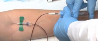
Measurement procedure
The volume of circulating blood, the condition of the heart muscle and pulmonary parenchyma, the patency of the heart valve apparatus and the adequacy of breathing are the parameters that affect central venous pressure. How to measure its value?
For this purpose, a disposable central venous catheter, a blood transfusion system, isotonic Na saline solution (400 ml) and a Waldman phlebotonometer are used. The price of such a procedure, performed as planned, starts at 12,000 rubles.
For those who are interested in how exactly pressure is measured in the central vena cava, we recommend watching a short final video. The visual is accompanied by an audio track in Ukrainian, but to get an idea of the procedure, you can watch the video even without sound.
Source: https://Cardio-help.ru/davlenie/centralnoe-venoznoe-davlenie-norma-179
Decoding indicators
Determining the level of pressure in the central veins allows you to assess the severity of the patient’s condition and promptly recognize dangerous pathologies. To do this, you need to know not only the CVP norm, but also the reasons that can affect the parameters.
Decoding of indicators:
| Level | Values in cm water. Art. |
| Short | Less than 6 |
| Normal | From 6 to 12 |
| High | Above 12 |
Due to physiology, in the third trimester of pregnancy (30-42 weeks), increased values of central venous pressure are allowed, while normal values vary within 5-8 cm of water. Art.
Changes in blood volume, vascular tone and cardiac activity can appear separately or in combination with each other.
Therefore, to assess the severity of a patient’s condition with heart failure, a decrease in blood volume, or hemodynamic disorders, an “express index” is often used - the ratio between CVP, heart rate and blood pressure.
Express index indicators:
| Patient's condition | EI value |
| Normal for a healthy person | 60-75 |
| Hypovolemia (decreased blood volume) in combination with heart failure | 90-140 |
| Isolated hypovolemia | 20-25 |
| Cardiac weakness with normovolemia (normal blood volume) | 150-190 |
| Heart failure combined with hypervolemia (excess fluid in the body) | 200-300 |
Since normal CVP values vary within wide limits, monitoring its level over time is considered the most significant in identifying pathologies.
To do this, tests are carried out with the introduction of different volumes of infusion solution. That is, a small amount of fluid is administered while simultaneously checking for changes in pressure levels in the central veins.
The body's response to the administration of the solution may indicate different conditions, for example:
- an increase in the parameter by more than 5 cm of water. Art. - indicates a violation of myocardial contractility, the administration of the solution is stopped;
- increase in indicator by 2 cm of water. Art. and less - indicates a decrease in blood volume, the solution is continued to be administered.
In this case, the test is carried out in several stages, since a lot of other values are simultaneously assessed - blood pressure readings, daily urine output, saturation and others.
Only a specialist’s correct interpretation of the central venous pressure level over time, taking into account other indicators, will allow the correct selection of treatment methods.
CVP (central venous pressure): measurement algorithm, norm
Central venous pressure (CVP) in medicine is the level of pressure in the right atrium. This indicator is very important for the correct diagnosis of cardiovascular diseases and pulmonary pathologies.
In the article we will take a detailed look at the cases in which CVP determination is required, learn about measurement methods and what can provoke its increase or decrease.
What is this indicator and what is its norm?
Two important hemodynamic factors depend on the level of central venous pressure:
- the amount of venous blood returned to the heart;
- filling of this organ at the time of diastole (relaxation interval between contractions).
The value of central venous pressure is determined in case of circulatory disorders accompanied by a critical condition of a person in order to establish a diagnosis.
Determining venous pressure is equally important for the correct conduct of infusion therapy (intravenous administration of drugs by drip or catheterization), as it helps to monitor and control the patient’s condition.
Theoretically, the normal value of venous return is close to 0 mmHg. Art. But such values are difficult to capture or measure with sensors. Therefore, it is usually measured in millimeters or centimeters of water column.
The normal average central venous pressure is:
- in children - from 60 to 120 mm of water. st;
- in adults - from 30 to 120 mm of water. Art.
Sharp deviations in numbers indicate the presence of serious pathologies that are dangerous to human health and life, which require emergency assistance through medical intervention.
How is the measurement carried out?
In order to measure the CVP value, you can use several methods:
- measuring technique with a ruler and dropper;
- Medifix system;
- direct manometry method using a Swan-Ganz catheter;
- alternative non-invasive methods are visual determination, for example, checking for bulging veins in the neck or arms in different body positions.
However, the most common method for determining central venous pressure, characterized by accuracy and ease of use, is the Waldman apparatus (phlebotonometer).
This is a tall stand with a graduation scale, which is located along a long glass tube with an adapter, filled with saline solution.
The algorithm for measuring central venous pressure using a phlebotonometer is as follows:
- All air bubbles are first removed from the system by opening the tap and flushing the system with saline solution. The entry of bubbles into the patient’s body is unacceptable, as it can not only lead to errors in measurements, but also provoke the occurrence of an air embolism.
- The device is positioned so that the zero division is parallel to the right atrium.
- To carry out the procedure, the patient takes a lying position. The height of the sternum above the surface of the bed is then measured. The resulting value is divided by three and correlated with the zero level, which is located 2/3 above the bed.
- The phlebotonometer is connected to the infusion system using an adapter and a connecting tube that has a clamp.
- This tube is then filled with saline and secured with a clamp to prevent possible leaks.
- Next, the device is connected to the jugular or subclavian vein.
- The fixing clamp is removed so that the solution begins to flow.
- A couple of minutes after the solution arrives, the phlebometer shows its level on the scale, which is used to determine the central venous pressure.
Improved methods allow monitoring using electrical sensors, the readings and diagrams of which are displayed on the device screen.
For what pathologies is monitoring prescribed?
Indications for CVP monitoring are serious disturbances of hemodynamic processes provoked by the following pathologies:
- acute circulatory failure;
- massive blood transfusion syndrome due to severe blood loss;
- chronic heart failure requiring invasive monitoring of the response during infusion therapy;
- the threat of developing shock conditions due to severe sepsis;
- suspicion of the development of cardiac tamponade;
- monitoring the condition during and after surgery in the abdominal area.
The CVP indicator helps to correctly assess the BCC (circulating blood volume) and the ability of the myocardium to contract.
And also control of its level allows you to avoid the occurrence of water intoxication due to the introduction of too large a volume of liquid during infusion activities.
Symptoms and possible causes of changes in normal levels
Visually, changes in the level of central venous pressure can be assessed by the appearance or absence of pulsation in the external jugular vein, which is located approximately in the middle of the neck.
https://www.youtube.com/watch?v=v9UVcwGbsmI\u0026list=PL12OVkpesEy4F7yQoZa_vk5MtapnUBrm1
Click on the picture to enlarge
In addition, signs of changes in venous pressure appear with the following changes in body position:
- vertical position - at a normal level of pressure, the veins will not stand out or pulsate;
- horizontal position, but the upper part of the body is elevated at an angle of 45 degrees (reclining) - pulsation and distinct manifestation of the veins indicate increased venous pressure;
- completely horizontal position of the body - if the veins are not visible and there is no pulsation, this indicates low pressure.
A clear symptom of increased central venous pressure will be swollen veins in the neck, even when the body is in an upright position. High blood pressure is also indicated by pulsation of the veins in the neck during palpation of the liver.
The level of central venous pressure depends on the following factors:
- Circulating blood volume (CBV) - when blood volume decreases, a similar decrease in pressure occurs in the vena cava and right atrium. For example, with severe blood loss or dehydration, low or sharply negative CVP values are observed.
- The condition of the myocardium - deterioration of its contractile function can lead to stagnation of blood in the right side of the heart and the development of acute heart failure, as a result, the central venous pressure increases.
- The condition of the lung tissue and breathing - when you exhale, venous pressure increases, and when you inhale, it decreases. Pathologies of the respiratory system and lungs lead to the development of stagnation in the organs, resulting in an increase in indicators.
Accordingly, the reasons for changes in normal venous pressure in one direction or another are various pathologies and conditions that require urgent hospitalization of the patient.
Promotion
The reasons for the increased level of central venous pressure are directly related to a sharp decrease in the contractile activity of the heart muscle or the presence of cardiovascular diseases.
Pathologies and conditions that provoke an increase in central venous pressure:
- Myocardial infarction.
- Inflammatory damage to the myocardium (myocarditis).
- Cardiogenic shock.
- Decompensated heart failure.
- Severe TBI (traumatic brain injury).
- Development of tamponade and hemopericardium due to trauma, cardiac rupture or dissection of an aortic aneurysm.
- Inflammation of the pericardium (constrictive pericarditis).
- Interruptions of heart rhythm (angina pectoris, arrhythmia, tachycardia), hypotension, hypertension.
- Pathologies of the tricuspid valve.
- Shock condition caused by exo and endotoxins of bacteria or viruses.
- Arterial pulmonary hypertension, which leads to acute right ventricular failure.
- Pulmonary embolism.
- Artificial ventilation.
- Pneumothorax due to trauma, injury or disease of the lungs.
Demotion
The main reasons for a decrease in the central venous pressure level:
- Significant blood loss (at least 10–15% of blood volume).
- Excessive vomiting and diarrhea, contributing to dehydration.
- Decreased blood volume (hypovolemia) due to septic, spinal, hemocoagulative or anaphylactic shock.
- Severe pain or fear, even anaphylaxis.
- Uncontrolled use of diuretic or diuretic drugs.
- Exposure to vasodilators (drugs that dilate peripheral blood vessels).
Pressure parameters in the central veins are usually interpreted in conjunction with clinical data and indications of additional studies prescribed by the doctor.
In some cases, the development of heart failure or compensatory vascular spasm, accompanied by bleeding, can lead to an increase in the level of central venous pressure.
Decoding indicators
Determining the level of pressure in the central veins allows you to assess the severity of the patient’s condition and promptly recognize dangerous pathologies. To do this, you need to know not only the CVP norm, but also the reasons that can affect the parameters.
https://www.youtube.com/watch?v=QVG6FpZHRio\u0026list=PL12OVkpesEy4F7yQoZa_vk5MtapnUBrm1
Decoding of indicators:
| Level | Values in cm water. Art. |
| Short | Less than 6 |
| Normal | From 6 to 12 |
| High | Above 12 |
Due to physiology, in the third trimester of pregnancy (30-42 weeks), increased values of central venous pressure are allowed, while normal values vary within 5-8 cm of water. Art.
Changes in blood volume, vascular tone and cardiac activity can appear separately or in combination with each other.
Therefore, to assess the severity of a patient’s condition with heart failure, a decrease in blood volume, or hemodynamic disorders, an “express index” is often used - the ratio between CVP, heart rate and blood pressure.
Express index indicators:
| Patient's condition | EI value |
| Normal for a healthy person | 60-75 |
| Hypovolemia (decreased blood volume) in combination with heart failure | 90-140 |
| Isolated hypovolemia | 20-25 |
| Cardiac weakness with normovolemia (normal blood volume) | 150-190 |
| Heart failure combined with hypervolemia (excess fluid in the body) | 200-300 |
Since normal CVP values vary within wide limits, monitoring its level over time is considered the most significant in identifying pathologies.
To do this, tests are carried out with the introduction of different volumes of infusion solution. That is, a small amount of fluid is administered while simultaneously checking for changes in pressure levels in the central veins.
The body's response to the administration of the solution may indicate different conditions, for example:
- an increase in the parameter by more than 5 cm of water. Art. - indicates a violation of myocardial contractility, the administration of the solution is stopped;
- increase in indicator by 2 cm of water. Art. and less - indicates a decrease in blood volume, the solution is continued to be administered.
In this case, the test is carried out in several stages, since a lot of other values are simultaneously assessed - blood pressure readings, daily urine output, saturation and others.
Only a specialist’s correct interpretation of the central venous pressure level over time, taking into account other indicators, will allow the correct selection of treatment methods.
How is treatment and normalization carried out?
Treatment of a patient with an increase or decrease in pressure in the veins and right atrium depends directly on the factor that provoked this condition:
- In case of cardiovascular failure, restorative therapy of the contractility of the heart muscle is carried out.
- For hypovolemia, administration of blood substitutes or crystalloids is prescribed.
- Shock conditions - eliminate the cause of shock with subsequent restoration of organ function.
- Cardiac tamponade - a puncture is performed to remove excess fluid.
To normalize and prevent central venous pressure, venotonics are prescribed, which have the following effects:
- strengthen the walls of blood vessels, making them less permeable;
- have a positive effect on the tone of the veins, increasing their elasticity;
- actively counteract inflammatory processes.
Possible complications during measurement
Overhydration (infusion of an excessive amount of solution) of the body can provoke not only an increase in the level of central venous pressure, but also lead to organ damage, even death.
The following complications are most often observed with invasive methods for determining central venous pressure:
- pneumothorax;
- artery damage;
- infection.
And there are also cases with more rare, but quite serious consequences of the following plan:
- hydrothorax;
- air (or catheter) embolism;
- thrombosis;
- damage to nerve receptors;
- puncture by the end of the catheter of the superior vena cava or the right atrium with the subsequent appearance of hydromediastinum or hydropericarditis;
- perforation of the endotracheal tube cuff.
It is worth noting that if all the rules of technique for performing the method are observed, the possibility of complications occurring is minimized.
Forecast
Since changes in CVP usually occur against the background of severe pathological conditions, the prognosis depends on the cause that provoked them:
- When the level of pressure in the veins decreases due to blood loss, the use of infusion therapy with blood replacement solutions leads to complete recovery.
- With CVP hypotension against the background of sepsis and septic shock, 50-75% of patients are cured.
- In case of CVP hypertension against the background of acute heart failure, the restoration of the patient’s health completely depends on the results of treatment of the underlying pathology.
Dmitrieva Yulia (Sych) – In 2014, she graduated with honors from Saratov State Medical University named after V. I. Razumovsky. Currently working as a cardiologist at the 8th City Clinical Hospital in the 1st clinic.
Source: https://infoserdce.com/davlenie/cvd/
How is treatment and normalization carried out?
Treatment of a patient with an increase or decrease in pressure in the veins and right atrium depends directly on the factor that provoked this condition:
- In case of cardiovascular failure, restorative therapy of the contractility of the heart muscle is carried out.
- For hypovolemia, administration of blood substitutes or crystalloids is prescribed.
- Shock conditions - eliminate the cause of shock with subsequent restoration of organ function.
- Cardiac tamponade - a puncture is performed to remove excess fluid.
To normalize and prevent central venous pressure, venotonics are prescribed, which have the following effects:
- strengthen the walls of blood vessels, making them less permeable;
- have a positive effect on the tone of the veins, increasing their elasticity;
- actively counteract inflammatory processes.
Treatment methods
Treatment depends on the specific condition. So, against the background of a decrease in the volume of circulating fluid or connective tissue, intravenous solutions are administered, and if necessary, blood transfusions are prescribed.
Cardiogenic shock and heart failure are corrected symptomatically. Then the disorder is corrected (after the patient has been removed from the emergency condition).
Pericardial tamponade requires puncture, drainage of the cavity, pumping out fluid, exudate or blood.
It is important to monitor CVP levels throughout the initial treatment, reducing or increasing the dose of the drug if necessary.
Possible complications during measurement
Overhydration (infusion of an excessive amount of solution) of the body can provoke not only an increase in the level of central venous pressure, but also lead to organ damage, even death.
The following complications are most often observed with invasive methods for determining central venous pressure:
- pneumothorax;
- artery damage;
- infection.
And there are also cases with more rare, but quite serious consequences of the following plan:
- hydrothorax;
- air (or catheter) embolism;
- thrombosis;
- damage to nerve receptors;
- puncture by the end of the catheter of the superior vena cava or the right atrium with the subsequent appearance of hydromediastinum or hydropericarditis;
- perforation of the endotracheal tube cuff.
It is worth noting that if all the rules of technique for performing the method are observed, the possibility of complications occurring is minimized.
Forecast
Since changes in CVP usually occur against the background of severe pathological conditions, the prognosis depends on the cause that provoked them:
- When the level of pressure in the veins decreases due to blood loss, the use of infusion therapy with blood replacement solutions leads to complete recovery.
- With CVP hypotension against the background of sepsis and septic shock, 50-75% of patients are cured.
- In case of CVP hypertension against the background of acute heart failure, the restoration of the patient’s health completely depends on the results of treatment of the underlying pathology.
Author of the article: Yulia Dmitrieva (Sych) - In 2014, she graduated with honors from Saratov State Medical University named after V. I. Razumovsky. Currently working as a cardiologist at the 8th City Clinical Hospital in the 1st clinic.