Main arteries in the human body
Arterial vessels carry blood from the heart to organs and tissues, supplying them with oxygen. The only exception is the pulmonary trunk, which carries unenriched blood to the lungs for the oxygenation process (O2 saturation). It is worth noting that in some cases, for example, with congenital heart defects (tetralogy of Fallot, patent ductus arteriosus, ventricular septal defects, etc.), the arteries may contain both arterial and venous blood. Arteries are translated as “carrying air,” which is exactly how the Greek healer, physician Erasistratus, imagined them. He believed that only veins distill blood. Arterial vessels pulsate in time with heart contractions. This can be easily felt by placing a finger on the wrist; a rhythmic pulsation of the radial artery is detected. The most important arteries are the following:
- aorta. It emerges from the left ventricle and is the largest vessel, its diameter can reach three centimeters;
- coronary - nourish the heart muscle;
- brachiocephalic trunk;
- general sleepiness;
- subclavian;
- axillary;
- humeral, ulnar, radial and four arterial palmar arches - supply blood to the upper limbs;
- ileal;
- femoral, popliteal, small and large tibial and plantar - enrich the lower extremities with blood.
From all large arteries branch smaller ones in diameter, and from the latter the smallest are arterioles, which end in microvasculature vessels - capillaries. Their main function is to carry blood with nutrients and oxygen from the heart to the internal organs under a certain pressure, which normally does not fall below 100/60 mm Hg. Art. (mercury column), and does not rise above 139/89 mmHg. Art. The wall of each arterial vessel consists of three layers:
- internal, represented by endothelial cells;
- middle - from muscle structures and elastic elastic tissue;
- external, consisting of a connective tissue base - adventitia.
Arteries are divided into muscular (precapillaries, arterioles), elastic (aorta) and mixed (medium-sized vessels) types.
When the integrity of all three layers of arteries is damaged, arterial bleeding occurs.
How to recognize capillary bleeding
The integrity of small blood vessels may be compromised at the epithelial surface. In this case, the bleeding is called external. Such damage is dangerous only if there is a violation of hemostasis - coagulation.
Attention ! Prolonged slow bleeding from internal organs negatively affects health. Whatever its nature - a trickle or evenly, accumulation in the body cavity can provoke the development of peritonitis or a purulent-inflammatory process.
One type of internal bleeding is parenchymal, when small ducts in several internal organs are damaged simultaneously.
Signs of capillary bleeding:
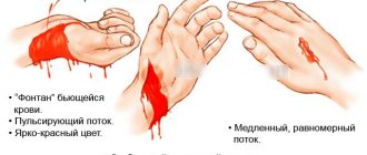
- slow expiration;
- bright scarlet or brightly saturated pink color - due to increased oxygen saturation;
- no pulsation.
Spasms can be felt if there are large vessels - veins or arteries - near the damage.
The following symptoms will help you distinguish one type of bleeding from another. With arterial blood, scarlet blood beats under pressure, and with venous blood, it is abundant, dark in color, the flow is slow, but blood loss is pronounced. Both conditions require immediate assistance.
It also happens that the wound is shallow, profuse bleeding is not noticeable, and the color is dark. Such a clinical picture cannot occur with capillary bleeding. A small vein is damaged, and even with normal hemostasis the body does not always cope with such damage.
Types of bleeding
Depending on the damaged vessel
Bleeding can be classified in several ways. Depending on the damaged vessel, there are:
- capillary. The least dangerous type, with an adequately functioning coagulation system (responsible for the release of biologically active substances - coagulation factors, thromboplastin fibrinogen and others that help stop the bleeding that has begun), the stop can occur on its own within five to ten minutes. Blood is released in small portions and is deep red in color (symptom of “blood dew”). They are dangerous only because a secondary bacterial infection may occur with the development of a purulent process;
- venous. They occur when veins are damaged and are a more dangerous condition. They are characterized by the release of dark-colored blood containing metabolic products and carbon dioxide at a slow pace. Venous vessels are located in more superficial layers, and therefore are injured much more often;
- arterial. An extremely dangerous species that occurs when exposed to a strong traumatic agent. Their seriousness is also justified by their rather deep location. In some cases, this type of bleeding stops on its own, since arterial vessels have the ability to spasm due to the presence of a muscular membrane in them;
- parenchymatous. They occur when the spongy substance of bone structures and the following internal organs are injured - lungs, liver, kidneys, spleen, pancreas. The entire wound surface is bleeding. Damaged vessels are not able to go deep into the tissue and cannot be compressed or contracted by it. Bleeding is very profuse and often poses a direct threat to the life of the affected person; it is extremely difficult to stop it;
- mixed. Occurs with penetrating wounds of the abdominal or thoracic cavity. At the same time, venous and arterial vessels and various internal organs are damaged.
In the direction of blood flow
- external, in which blood flows through the natural openings of the body or from a wound into the external environment. Example, bleeding from the upper or lower extremities, uterus (metrorrhagia), intestines (melena);
- internal. Blood rushes into various tissues of the body, hollow organs and cavities - the ventricles of the brain, interfascial spaces, articular, pleural, abdominal and pericardial. They can be quite massive with changes in the patient’s vital signs or minor, in quantities that can only be determined by specialized methods (feces for occult blood, etc.).
By origin
- traumatic. They are formed as a result of the impact of a traumatic agent on tissues and organs (a blow with a knife, ax, saber, screwdriver, etc.), which exceeds their strength characteristics and leads to damage to the artery or other vessels;
- pathological. They occur in the presence of various diseases - vasculitis (an autoimmune pathology that damages arteries and veins), diabetes mellitus, hemophilia A and B types, etc. For bleeding to occur, a minor provoking effect is required.
According to the degree of blood loss (severity)
- lungs - up to 500 ml or 10 - 15% of the bcc (circulating blood volume);
- medium - up to one liter, 20% bcc;
- heavy - up to one and a half liters, 30% bcc;
- massive - more than one and a half liters or 30% bcc;
- fatal - more than three liters, 50 - 60% of the bcc.
The most dangerous bleeding is from the aorta and common carotid arteries; death without prompt medical assistance is guaranteed within a few seconds.
Complications due to arterial bleeding
If help is not provided in a timely manner, the person may bleed and die. Due to the rapid loss of blood, the body does not have time to activate protective mechanisms, which leads to the fact that the heart does not receive enough blood and, as a result, blood circulation stops.
If a tourniquet is applied, but further assistance is not provided by doctors, the limb dies. If blood does not flow in the required quantity for 8 or more hours, then gangrene begins to develop, leading to tissue necrosis. In this case, saving the patient’s life is only possible through amputation, which is carried out well above the onset of death.
What can lead to arterial bleeding?
Among the main causes of arterial bleeding are:
- mechanical injuries received during road traffic accidents, plane crashes and other types of accidents; stab injuries at home or at work;
- vascular pathology. Among them: vasculitis and other autoimmune diseases that occur with vascular damage (systemic lupus erythematosus, etc.); malignant neoplasms (hemangiosarcoma and others); suppurative processes affecting the walls of the arteries; atherosclerosis; viral diseases (for example, influenza);
- blood and liver diseases - hemophilia, von Willebrand disease - Diana (violation of three parts of hemostasis - the process of stopping bleeding), cirrhosis, etc.;
- systemic pathologies - diabetes mellitus, sepsis (massive blood poisoning with various infectious agents), deficiency of vitamins, especially C and PP.
Why is it the most dangerous type of bleeding?
Arterial blood is the most important supplier of oxygen for all internal organs. Severe circulatory deficiency leads to an ischemic condition in which organs do not receive the required amount of O2. A structure such as the intestines, for example, can remain without it for tens of minutes without serious consequences, but in the brain, significant changes are formed after just six minutes of oxygen starvation.
There is an important concept - collapse, a condition in which hemorrhagic shock develops due to a sharp drop in blood pressure and circulating blood volume.
It can lead to sudden cardiac arrest. Also, when large-caliber arteries are damaged, centralization of blood circulation is formed. In this condition, blood flows from the upper and lower extremities to vital organs - the heart, lungs, and brain. This condition is a protective mechanism for emergency preservation of life support.
Signs of venous bleeding
Symptoms of internal bleeding
The internal, or deep, veins are located among the muscle fibers and carry more than 60% of the blood supply back to the heart.
Trauma threatens:
- local disorders in the limbs;
- fatal.
The main signs of venous bleeding, which may or may not be visible to visual inspection, are:
- Dark blood color, high flow rate. The main difference from arterial is the absence of pulsation, “fountain”.
- General health indicators decrease, especially blood pressure. Collapse is coming.
- With palpation and physical impact on the superficial veins, the rate and nature of blood loss do not change.
- The main location is the inside of the arms and legs. The veins of the thigh and shoulder are usually affected.
Diagnosis of arterial bleeding
Diagnosis of external types of bleeding, of course, does not present any difficulties. To detect internal bleeding, the following laboratory tests and instrumental types of research are used:
- general blood analysis. Allows you to detect a decrease in hemoglobin, red blood cells and hematocrit (the ratio of red cells to plasma);
- fibroesophagogastroduodenoscopy (FEGDS) and colonoscopy if bleeding from the gastrointestinal tract is suspected. Also, during this procedure, it is possible to stop it using the endoscopic method using electrical cauterization;
- ultrasound examination - to detect blood accumulation in the abdominal cavity;
- plain radiography of the chest organs - if there is a suspicion of blood in the pleural cavity;
- computed tomography and magnetic resonance imaging are the most reliable ways to determine internal types of bleeding.
Ways to stop bleeding
For this purpose, the following methods can be used:
- pressing a bleeding artery with a finger. Hold it in this position for at least ten minutes. It is worth noting that with this method it will be possible to stop only minor bleeding; for deeper damage this will only be a temporary measure;
- pressure bandage. It is a simple method to implement - gauze is placed on the wound, a tight roller is placed on top of it, and then this structure is wrapped in several layers of bandage. If the bleeding originates from the vessels of the neck, then the roller is tied through a raised arm on the opposite side;
- applying a tourniquet or twist from improvised means above the bleeding site;
- bending the injured limb is the most unreliable method, but is the only one in a number of situations.
The victim must bend the bleeding limb as much as possible and fix it to the body, holding it in this way until qualified medical assistance arrives.
The above methods are first aid and can only be used to stop external bleeding. To finally stop arterial bleeding in specialized hospitals, surgeons and vascular surgeons use various biological, medicinal, mechanical and physical methods. The latest are:
- electrocoagulation - cauterization of blood vessels with high-frequency current;
- Irrigation of bleeding tissue with hot isotonic sodium chloride solution.
Mechanical ones include:
- ligation of bleeding vessels;
- suturing damaged arteries;
- wound tamponade;
- ligation of blood vessels.
Medications include the following hemostatic (stopping bleeding) agents for general or local use:
- 3% hydrogen peroxide solution;
- 5% aminocaproic acid;
- 12.5% sodium ethamsylate (Dicinone);
- 1% solution of Vikasol (onset of action after 8 - 12 hours);
- bandages, applicators with Celox granules - a hemostatic agent based on Chitosan (a natural polysaccharide present in the shell of various mollusks).
Biological:
- tamponade with your own tissue (for example, in case of internal bleeding, the greater omentum can be used for this purpose);
- blood transfusion;
- collagen hemostatic sponge.
If arterial bleeding occurs in a patient suffering from hemophilia, then immediately resort to transfusion of fresh frozen plasma with the necessary clotting factors or cryoprecipitate, a drug used to stop bleeding in this disease.
Bleeding from superficial veins
Superficial veins located on the lower leg
Even with a complete intersection of any of the saphenous veins, the general blood circulation will not be disrupted. But the bleeding that occurs during this process can be fatal. The most dangerous damage to the following veins is:
- Located on the inside of the wrist.
- A large vein running down the leg and thigh.
- Venous plexuses present on the foot (dorsal side).
- Veins located on the outside of the forearm and shoulder.
Signs
Let's look at how this type of bleeding is characterized:
- If small saphenous veins have been damaged, then in this case the bleeding may stop on its own some time after the injury.
- Most bleeding occurs from the lower end of the vein, since the direction of blood flow is from top to bottom.
- Increased bleeding can occur due to alcohol intoxication, blood diseases, and high blood pressure.
Important! If the veins of the shoulder or thigh are crossed, it is very rarely possible to stop such bleeding on your own. In this case, you should immediately seek professional help.
Video: first aid for venous bleeding
How to give first aid
Stopping bleeding when superficial veins rupture using a tourniquet
Now let's look at how to stop venous bleeding:
- The easiest way to stop is to clamp the vein. This is done just below the rupture site.
Advice! If the above option does not bring results, then try squeezing the vein above the wound.
- The wound is washed with an antiseptic solution such as hydrogen peroxide, after which it should be covered with a gauze bandage covering the skin both above and below the wound. For greater effectiveness, a gauze roll is first placed on the wound, after which it is bandaged.
- In order to completely eliminate the bleeding, it is necessary to suture the skin wound, which is done exclusively by a specialist.
First aid for external bleeding
Remember, your actions will determine whether the victim survives.
First of all, you should call an ambulance. Then:
- It is necessary to quickly decide on a method to stop the bleeding. Depending on the location, you can apply a tourniquet, try to press the vessel with your finger, or apply a pressure bandage.
- Try to pinch the artery with your fingers; you need to press it against the bone, not against the soft tissue.
- Apply a tourniquet above the bleeding site. It must be present in every medical machine first aid kit.
- If this is not available, you need to find a piece of wide fabric and, by twisting it on a longitudinal object (pen, pencil, branch), place it slightly above the wound site.
- If it was not possible to complete the previous steps, then you can pack (cover) the bleeding area with this tissue, apply as much pressure as possible and wait for the ambulance to arrive.
Injury to neck vessels
In case of arterial bleeding from the neck area, it is necessary to understand which vessel is damaged - the carotid artery (located on both sides of the neck) or the subclavian artery (slightly below the collarbones). In the first option, the artery must be immediately pressed towards the spine (towards the seventh cervical vertebra). In the second, the vessel is clamped in the area of the supraclavicular fossa, following the first rib. Without removing your finger, wait for the ambulance to arrive.
The cervical arteries must be compressed below the wound site.
A tourniquet can be applied to the neck area in three ways:
- The tourniquet is applied to a roll of fabric or bandage, which is used to pack the wound, and the other side of the tourniquet is wrapped around the arm placed behind the head. As a result, one side of the neck will not be pinched, and blood will flow freely to the brain.
- It is applied similarly to the first method, but the second end is passed through the armpit of the injured person.
- Initially, a Kramer wire splint is placed on the undamaged area of the neck, and a tourniquet is applied over it.
Vascular injury of the lower extremities
There are three main arteries in this area - the femoral, popliteal and dorsalis pedis. Stopping external bleeding from this area depends on the location of the injury. Help in this case will be provided by applying a tourniquet or finger pressure:
- when bleeding from the thigh, press the femoral artery as tightly as possible with your fingers gathered into a fist above the site of bleeding, to the femur or pubic bones;
- if bleeding develops in the lower leg area, the artery is fixed in the popliteal fossa;
- in case of bleeding from the arteries of the dorsum of the foot, the area of the ankle joint is pressed.
Then they begin to apply a hemostatic tourniquet slightly above the site of bleeding, and if the arteries of the foot are damaged, it is applied to the underlying bone.
Arterial bleeding from the lower extremities can be stopped by bending the leg.
- Place a roll of bandage or fabric in the area of the popliteal fossa, bend the limb at the knee joint as much as possible and bandage it to the body.
- Place the roller in the groin fold, bend the leg at the hip joint and secure it to the body with a belt or bandage.
Vascular injury of the upper extremities
In this case, the subclavian, brachial, radial, ulnar and palmar arterial arches may be damaged:
- in case of arterial bleeding from the armpit, the arms, bent at the elbow joint, are placed behind the back and tied. Thus, the subclavian artery is pressed against the first rib or collarbone.
This method should absolutely not be used for fractures of the bones of the upper extremities;
- The brachial artery is pressed with four fingers to the inner surface of the humerus. Bleeding can also be stopped by placing a cushion in the armpit, and the arm bent at the elbow must be bandaged to the chest;
- if the radial or ulnar artery is damaged, the arteries should be pressed against the corresponding bones and a hemostatic tourniquet should be installed above the bleeding site;
- for wounds of the hand and forearm, bleeding can be stopped by flexing the limb. To do this, you need to install a bolster in the elbow bend, bend your arm as much as possible at the elbow joint, and fix the forearm to the shoulder. It is also possible to compress the blood vessels in the area of the wrist joint.
In case of bleeding from small arteries or injury to the abdomen, chest, head, back, a tight pressure bandage is applied. The wound should be covered with several layers of sterile gauze and bandaged tightly. You should not use cotton wool for arterial or other types of bleeding, as often its small particles remain in the wound, which attract a secondary bacterial infection, which ends in the formation of suppurative processes.
Bleeding from the neck veins
The main veins in the human body are the internal and external jugular veins. Damage to the latter is more common, but problems with the internal vein are fraught with more serious consequences.
Signs
Despite the fact that the clinical manifestations of bleeding from the neck veins do not differ from other venous bleeding, the consequences and rules of first aid in this case are different.
Important! The main difference in providing first aid for cervical venous bleeding is that in this case it is forbidden to apply tight bandages.
Correctly stopping neck bleeding
The danger of damaging the neck veins is as follows:
- Severe blood loss may occur.
- An air embolism may occur.
- There is significant bleeding at the upper end of the vein.
- Possibility of cerebral circulatory disorders and swelling.
Features of first aid
In this case, it is important to know the correct way to stop bleeding. Let's take a closer look at it:
- Through the skin, you need to firmly press down the ends of the bleeding vessels with your fingers.
- Apply a tampon with peroxide to the wound, pressing it firmly.
Important! It is necessary to contact a specialist as soon as possible, since the internal jugular vein should be sutured as quickly as possible.
Location of the jugular vein
The characteristics and methods of stopping venous bleeding were discussed above. The main thing when any of the above types of bleeding occurs is to remain calm. In order to minimize the consequences of bleeding, it is necessary to provide first aid correctly and in a timely manner and contact a specialist as soon as possible.
← How to stop bleeding from the mouth after tooth extraction?
Emergency first aid for pulmonary hemorrhage →
We recommend studying similar materials:
- 1. Autoimmune hemolytic anemia in children: what is missing and how it manifests itself
- 2. How to choose a diet based on your blood type: losing weight together
- 3. Causes and dangers of increased basophil levels in children
- 4. The level of basophils in adults has decreased: how to treat basophilia
- 5. Reasons for an increase or decrease in neutrophils in a blood test in children?
- 6. Functions and possible causes of pathologies of segmented neutrophils
- 7. Norms for the content of neutrophils in the blood and what functions they perform
Rules for treating wounds and applying various dressings
To protect the wound site from bacterial flora and help stop bleeding, it is necessary to apply a bandage. A material suitable for this purpose is gauze. It has high hygroscopicity - the ability to absorb moisture. When applying a bandage, it is important to follow the following rules:
- first, it is necessary to lay or sit the victim, since even with minor bleeding there is a threat of loss of consciousness;
- It is impossible to wash the wound surface with water, as it can cause infection;
- if possible, treat your hands with alcohol or alcohol-containing solutions before applying a bandage;
- if there are various objects, fragments, or earth in the wound, it is worth removing them if they are located on the surface;
- the dressing material that will be used to close the wound defect must also be absolutely sterile;
- if you don’t have one, a scarf or piece of fabric will do;
- the skin at the wound site should be treated with iodine or another antiseptic. You need to wipe in the direction away from the wound;
- After laying the gauze bandage, proceed to the last stage - bandaging. Do it in a circular motion, from left to right.
This method is suitable for temporarily stopping minor bleeding from the lower or upper extremities. For better hemostasis, it is worth raising the limb upward, above the level of the heart. This will reduce blood flow to the area.
First aid for internal bleeding
If you suspect internal bleeding, you should immediately call an ambulance. Then place an ice pack on the victim’s stomach, which helps to narrow the blood vessels and helps to slightly stop the bleeding. You need to be prepared for vomiting. If present, turn the victim onto his left side to prevent vomit from entering the respiratory tract. It is very important to monitor the victim's breathing, heart rate and blood pressure. Report all data to the emergency medical team. Such patients are transported in a semi-sitting position.
Places of vascular compression during various arterial bleedings
Depending on the location of the wound and the affected arterial vessel, proceed to the following manipulations:
- if the external maxillary artery is injured, you need to press on the lower jaw at the edge of the masticatory muscle;
- in case of damage to the temporal artery, the area located at the temporal bone in front of the upper part of the ear is pressed;
- the popliteal arterial vessel is compressed on the back of the leg to the tibia;
- femur to the pubic or thigh bones, as forcefully as possible and preferably with a fist;
- subclavian towards the first rib;
- carotid to the seventh cervical vertebra (it is the most prominent in this area);
- brachial artery to the brachial hand in the medial area (inner head of the biceps);
- radius and ulna to the corresponding bones.
It is necessary to hold damaged vessels in this position for at least 10 minutes.
This time will be enough to stop minor bleeding.
How can you stop bleeding?
Several techniques are used to stop bleeding. It is worth choosing one of them depending on the location of the damaged vessel, its size, and the intensity of the hemorrhage.
These are the techniques:
- Finger compression of the vessel,
- Application of a tourniquet,
- Wound tamponade.
Pressure points of arteries
The first and last methods of stopping are suitable if the carotid, maxillary or temporal arteries are damaged, that is, those vessels on which it is impossible to apply a tourniquet. Arterial bleeding from wounds of the extremities can most effectively be stopped using a tourniquet.
How to apply a tourniquet correctly?
This method helps stop arterial bleeding for a long time. In case of serious bleeding, it is applied immediately, without using other methods, such as finger pressure, maximum flexion, or packing the wound. It is used only for external types of bleeding. If you don’t have a tourniquet, you can use a piece of fabric, a tie, or a wide belt from your waist.
Wire, rope or other narrow objects are not suitable as a tourniquet, as they contribute to excessive pressure on the vessels and rapid tissue death.
When applying a tourniquet, it is important to use the following rules:
- it is applied as close as possible to the bleeding wound;
- they fix it on top of several layers of bandage or clothing, and it should be visible to others and not covered by anything;
- tighten the tourniquet until the arterial bleeding stops completely. But it is worth remembering that excessive tightening can injure nerve trunks and increase pain, and weak tightening can increase bleeding;
- be sure to put a note under it indicating the exact time of application (hours and minutes), but since they are often lost during various transportation of the victim, it is better to leave a note on the skin and forehead of the victim;
- transport the patient to a nearby hospital or medical center.
In the summer, the tourniquet should be applied for no more than one hour, and in the winter, up to thirty minutes.
Prolonged use of a tourniquet can lead to necrosis (death) of the underlying tissues. If arterial bleeding continues after this period, the tourniquet can be loosened for up to ten minutes, then reapplied. This time is enough to enrich the tissues with such an important element as oxygen. The criterion for the correct placement of the tourniquet is the absence of pulsation of the artery in the underlying section.
Applying a tourniquet for venous bleeding
If there is massive blood loss, a tourniquet should be used immediately.
When using a tourniquet, you should adhere to the basic rules for applying it:
- It is applied below the location of the wound/trauma.
- It is very important to record the time when the tourniquet was applied.
- It is absolutely prohibited to use it for more than 90-120 minutes.
- It is recommended to regularly loosen the tourniquet, squeezing the damaged area with your fingers as it loosens.
How to act in case of large blood losses?
Arterial bleeding, for example, from the carotid or femoral arteries, pose a direct threat to the life of the victim. The victim’s reaction to such acute blood loss will depend on age, the presence of concomitant pathology (heart failure, diabetes mellitus, etc.). It leads to significant changes and damage to the internal organs, which can progress even after the bleeding has completely stopped. Such blood loss leads to small output syndrome, i.e. the heart pushes a much smaller volume of blood into the systemic circulation; stimulation of endocrine and sympathetic activity (the release of various hormones and biologically active substances occurs); disruption of peripheral blood flow and centralization of blood circulation; hypoxia (lack of oxygen) of various tissues and organs; changes in the kidneys and disruption of coagulation processes.
First aid for injuries to large vessels includes the following:
- apply pressure to the damaged artery;
- you need to unbutton tight clothes and give a flow of fresh air;
- the victim must be laid on his back, with his legs placed on an elevation so that the blood is concentrated as much as possible in the central part of the body;
- If there is no heartbeat, you should immediately begin chest compressions.
Further drug treatment will include correction of hypovolemic disorders by prescribing:
- dextran preparations (Polyglyukin, Reopoliglyukin);
- gelatin solution (Gelatinol);
- hydroxyethyl starch (Infukol, Refortan, Stabizol);
- saline solutions, such as Ringer's.
Blood products used:
- fresh frozen plasma;
- red blood cell mass;
- albumen.
In cases where there is no increase in blood pressure numbers or initially low values, enter:
- adrenalin;
- dopamine;
- dobutamine;
- norepinephrine.
Dangers of capillary bleeding
It is not immediately possible to determine internal blood loss from your own sensations. Signs of damage to small vessels supplying internal organs:
- tachycardia;
- a sharp decrease in blood pressure;
- severe weakness;
- dyspnea;
- semi-fainting state.
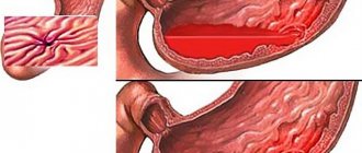
When the mucous membrane of the stomach or intestines bleeds, melena - black stool - may indicate pathology. In other cases, the patient is often treated already at the stage of peritonitis.
Considering how capillary bleeding is characterized, external damage is easier to identify. People who faint even with a small scratch immediately treat the problem area. But there are also “heroes” who do not pay attention to minor injuries and do not realize that they, too, can pose a danger.
With capillary bleeding that covers a large area, signs of oxygen deficiency appear - temporary weakness and impaired impulse conduction occur. If hemostasis is impaired, then it is impossible to cope with the worsening condition without outside help.
Important! An open wound is a gateway to infection. Through it, pathogenic microorganisms can enter the body, which will cause a purulent-inflammatory process and even sepsis. Therefore, it is not enough to stop the bleeding from a broken knee - you need to reduce the risk of infection.