Hypovolemia is a pathological condition of the body that occurs with significant loss of fluid and electrolytes. Accordingly, hypovolemic shock must necessarily be associated with a decrease in water-salt balance.
Dehydration is possible as a result of loss of interstitial fluid or blood plasma with significant blood loss, massive burns, diarrhea, and uncontrollable vomiting. Feverish conditions and prolonged periods without water in hot climates are also accompanied by dehydration.
Children are most sensitive to fluid loss. In them, hypovolemic shock occurs quickly with dyspeptic and infectious diarrhea, in a hot room. As first aid, victims should be given something to drink.
The importance of fluid in human physiology
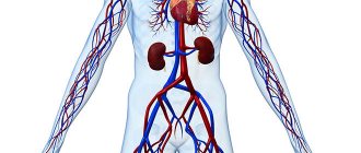
Water is part of the entire complex of fluids that wash organs and tissues. It is the main component of blood, lymph, cerebrospinal and interstitial fluid, secretions of the salivary glands, gastric and other juices produced by internal organs, tears, and urine.
Liquid creates a universal internal environment for the existence of cells. Through it the following is carried out:
- nutrition and removal of toxins;
- “orders” are delivered from the nervous and endocrine centers;
- the necessary brain structures are excited.
The role of liquid in ensuring metabolic processes is great. All biochemical reactions take place only in dissolved form. By changing the amount of water in the body in accordance with physiological needs, the required concentration of salts, electrolytes, acids and alkalis, biological substances, everything that ensures the internal environment or homeostasis is maintained.
The preservation of homeostasis indicators is guaranteed by natural tissue barriers (skin, mucous membranes of organs and blood vessels). Equilibrium can change under the influence of regulatory systems, but within very narrow limits.
Therefore, any disturbances in the composition of liquid media can be used to judge the pathology that has arisen. A decrease in fluid causes significant changes in homeostasis: some substances are lost along with water, others sharply increase in concentration. Pathophysiological disorders may concern:
- cellular composition of blood;
- alkaline balance;
- concentration of dissolved substances.
The functional capacity of systems depends on the norms of water distribution in the body.
Changed conditions cause many diseases.
In humans, it is convenient to judge the volume of fluid by the indicator of circulating blood. It is calculated in the laboratory. A decrease of 25% in healthy people is well compensated and does not cause any significant changes in homeostasis. 90% of the blood is in the vascular bed, the rest is deposited in the spleen and bones. If necessary, it is thrown out of storage and replenishes losses.
Large losses lead to varying degrees of hypovolemia, and in the absence of compensation and assistance - to a hypovolemic shock state.
Introduction
Hypovolemic shock is a life-threatening condition in which rapid loss of fluid from the body leads to severe disruption of the functioning of many organs due to their inadequate blood supply.
Loss of fluid leads to a decrease in circulating blood volume, a drop in blood pressure and a deterioration in perfusion (blood supply) of all organs. For a patient with hypovolemic shock to survive, they need immediate medical attention. If the blood supply to vital organs is not improved as soon as possible, irreversible changes in the tissues appear and the patient dies.
What happens during hypovolemic shock?
With timely and correct treatment, most patients can quickly improve blood supply to all organs. The prognosis for patients depends on the causes of the condition.
All patients with shock require treatment in intensive care units (resuscitation), so their treatment is carried out by anesthesiologists.
What causes hypovolemic shock?
The most common causes of hypovolemic shock are uncompensated losses:
- blood in case of massive acute bleeding, external or internal, caused by trauma, surgery, accumulation in different parts of the body during fractures, against the background of hemophilia;
- plasma - in the case of widespread burn surfaces, effusion into the peritoneal cavity during peritonitis, intestinal obstruction, pancreatitis, ascites;
- isotonic liquid - with frequent vomiting, prolonged diarrhea (for example, in the case of cholera, salmonellosis, gastroenteritis), with sweat and high fever caused by infectious diseases with severe intoxication.
With cholera, a large amount of fluid is lost with vomiting and diarrhea
A special place is occupied by the option of deposition (redistribution) of free blood volume in peripheral capillaries. This is typical for combined injuries and some infections. In such cases, the severity of the patient's condition is due to mixed types of shock (hypovolemic + traumatic + toxic) and damaging factors.
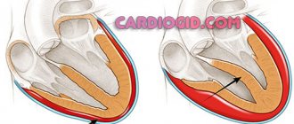
What is happening in the victim's body?
The pathogenesis of the shock state during hypovolemia begins with the body’s attempts to independently stop fluid loss and compensate for the deficiency:
- a reserve volume of blood enters the general channel from the depot;
- arterial vessels narrowing towards the periphery (arms and legs) in order to retain the required amount of blood for the brain, heart and lungs.
When the compensation mechanism is exhausted, spasm of peripheral vessels is replaced by complete paralysis, loss of tone and dilation. Because of this, most of the blood goes to the extremities and there is an insufficient supply of oxygen to vital organs, homeostasis is grossly disrupted, and metabolism suffers.
It is customary to distinguish 3 stages (phases) of shock development:
- Deficient - leading to the occurrence of acute fluid deficiency, a decrease in blood volume, which leads to a drop in venous pressure in the central veins, a decrease in blood flow to the heart. Fluid from the interstitial space passes into the capillaries.
- Stimulation of the sympathoadrenal system - receptors that control blood pressure signal to the brain and cause an increase in the synthesis of catecholamines (adrenaline, norepinephrine) by the adrenal glands. They increase the tone of the vascular wall, promote spasm in the periphery, increase the heart rate and increase stroke volume. Actions are aimed at supporting arterial and venous pressure for blood circulation in vital organs by reducing blood flow to the skin, muscles, kidneys, and digestive system. With prompt treatment, complete restoration of blood circulation is possible. If the period favorable for emergency interventions is missed, then a full-scale picture of shock develops.
- Actually hypovolemic shock - the volume of circulating blood continues to fall, the flow to the heart, lungs and brain sharply decreases. Signs of oxygen deficiency in all organs and changes in metabolism appear. The skin, muscles and kidneys are the first to suffer from the loss of compensatory protection, followed by organs located in the abdominal cavity, then life-supporting organs.
The mechanisms of shock development and the consequences for the body are described in detail in this video:
Reasons and mechanism of formation
The pathological hypovolemic process is based on severe dehydration.
Leads to it:
- Vomiting, characteristic of withdrawal syndrome after binge drinking.
- Diarrhea, with the help of which the body cleanses itself of alcohol toxins.
- Liver diseases characteristic of alcoholism (toxic hepatitis and cirrhosis). In this case, non-functional leakage of plasma into the intercellular space occurs due to a decrease in the oncotic pressure of the blood.
- Insufficient intake of fluid and protein into the body.
Hypovolemia is preceded by increased venous and hydrostatic pressure in small arteries, reflecting the negative influence of factors predisposing to a painful condition. Secondarily, the permeability of the vascular wall increases. Sweating of blood through arteries and veins leads to its general deficiency.
The body tries to compensate by:
- Producing the required amount of plasma.
- Slowing venous return.
- Spasms of small vessels.
The protective reaction allows you to maintain the activity of the brain and heart in a relatively normal state for some time and avoid circulatory complications.
Clinical manifestations of hypovolemic shock
The clinic of hypovolemic shock is determined by:
- total volume of fluid loss;
- rate of blood loss during hemorrhagic shock;
- the body’s ability to compensate (related to age, the presence of chronic diseases, training).
Athletes and people who live for a long time in hot climates and high altitude conditions are resistant to loss of blood and other fluids.
Based on the symptoms, one can judge the amount of blood loss, and vice versa; doctors use a classification to assess the patient’s condition depending on the volume of circulating blood (CBV). They are shown in the table.
| Degree of loss of bcc in % | Hemodynamic signs | Features of symptoms |
| up to 15 | When getting out of bed, the heart rate increases by 20 or more per minute | not detected in a supine position |
| 20–25 | blood pressure decreases, but the upper pressure is not lower than 100 mm Hg. Art., pulse within 100 – 110 per minute | in a supine position, blood pressure is normal |
| 30–40 | upper pressure below 100 mm Hg. Art., thread-like pulse more often than 100 | pale skin, cold hands and feet, decreased urine output |
| more than 40 | blood pressure is sharply reduced, the pulse in the peripheral arteries is not detected | the skin is pale with a marbled tint, cold to the touch, consciousness is impaired to the point of coma |
Common patient complaints:
- dizziness,
- drowsiness,
- shortness of breath.
The state of consciousness can vary from lethargy to periods of excitement. Breathing is shallow. In the absence of emergency medical assistance, the patient falls into a coma. Death occurs from cardiac arrest.
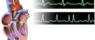
Clinical picture of hypovolemia
- cardiopalmus;
If the heart rate is more than 100 per minute, urgent measures are required to stop the bleeding and provide emergency medical care.
- decreased blood pressure;
- cold, sweaty, pale bluish or marbled skin;
- decrease in central venous pressure;
- decrease in body temperature;
- decrease or absence of urine - oligoanuria;
- dry mouth and severe thirst;
- dilated pupils;
- disturbance of consciousness - at first there is excitement and anxiety, which is then replaced by lethargy, up to the development of coma;
- dyspnea.
When pressing on the nail, a slow restoration of blood flow is observed, which indicates a violation of microcirculation.
Diagnostics
In diagnosis, it is important to determine the type of fluid loss. If there is or information about bleeding, vomiting, diarrhea, or a large burn surface, the symptoms themselves indicate the root cause of the pathological disorders. The doctor experiences significant difficulties if the bleeding is internal with an unclear cause.
The patient should be taken to the hospital as quickly as possible. Here you must take:
- blood tests;
- the group and Rh factor are determined;
- OCC;
- urine is examined for specific gravity (an indicator of concentration), protein and red blood cells.
X-rays are taken to identify hidden fractures.
If blood in the abdominal cavity is suspected, laparoscopy is necessary.
During treatment, the electrolyte composition and alkaline balance are examined. These indicators are important for choosing solutions of the desired concentration and composition.
Hemorrhagic shock is considered a type of hypovolemic shock. It is practically important to determine the amount of blood loss. There are different ways to do this.
Calculation of the shock index by dividing the pulse rate by the upper pressure: if normally this coefficient is about 0.54, then with shock it increases.
To establish blood loss during fractures in an adult, average values are used depending on the type:
- femur fracture - 1 l;
- shin bones - about 750 ml;
- shoulder - up to 500 ml;
- pelvic bones - up to 3 liters.
When examining the chest organs, radiologists approximately determine the amount of blood that has spilled into the pleural cavities:
- if you can clearly see the liquid level - up to 0.5 l;
- when the fields of lung tissue are darkened - up to 2l.
When examining a patient with suspected internal bleeding into the abdominal cavity, the surgeon focuses on the symptom of fluid leakage. This means that there is at least a liter of liquid in the cavity.
Providing first aid for HS
It is impossible to cope with hypovolemic shock without the help of doctors. Even if it is caused by dehydration, it will not be possible to quickly restore blood volume by feeding the patient; he requires intravenous infusions. Therefore, the first action that others should take when symptoms of shock appear is to call an ambulance .
Emergency aid algorithm before doctors arrive:
- If there is bleeding, place the patient so that the injury is 30 cm above the heart. If shock is caused by other reasons, ensure blood flow to the heart: place the patient on his back, with a cushion of things under his feet. If you suspect a spinal injury (a sign is a lack of sensation in the limbs), changing your body position is prohibited.
- Turn your head to the side so that the patient does not choke if vomiting begins. If he is unconscious, check for breathing. If it is weak or noisy, determine if the airway is clear. To do this, clean the oral cavity and remove the sunken tongue with your fingers.
- Clean the surface of the wound. If foreign objects get deep into the tissues, touching them is prohibited. Try to stop the bleeding:
— If the cause of shock is an injured limb, apply a tourniquet or twist above the wound. Record the time, write it on a piece of paper and slip it under the tourniquet. Simply informing the patient about the time of application of the tourniquet is not enough. By the time he is taken to the hospital, he may already be unconscious.
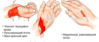
— For venous bleeding (signs of dark, evenly flowing blood), a tight bandage is sufficient. It is better if it is antiseptic. When bandaging, try to bring the edges of the wound together.
— If it is impossible to apply a bandage or tourniquet, stop the bleeding using a gauze swab, or, if it is not available, with any cloth or even a plastic bag. A bandage is applied in several layers to the wound and pressed with your hand for 20 minutes. You cannot remove the tampon during this time, even for a couple of seconds. If it is soaked in blood, new layers of bandage are added.
- Cover the patient, reassure him if possible, and do not leave him until the ambulance arrives.
- If there is external bleeding or suspected internal bleeding, you should not give the patient anything to drink, much less feed him. This way you will reduce the likelihood of asphyxia.
Expert opinion
Shalaeva Svetlana Sergeevna
endocrinologist, highest category, 18 years of experience
Note! All that is required of those around you is the correct implementation of the above emergency care algorithm. If you are not a doctor, a patient in hypovolemic shock should not be given any medications, IVs, or pain-relieving injections.
Treatment
The main goal of treatment is:
- restoration of blood supply to the heart, brain and lung tissue, elimination of their oxygen deficiency (hypoxia);
- combating acid-base imbalance;
- replacement of lost electrolytes and vitamins;
- normalization of blood supply to the kidneys and daily diuresis;
- symptomatic support for the functioning of the heart and brain.
Mild symptoms of hypovolemia can be eliminated by slowly drinking plain water, or better yet, lightly salted water. At high temperatures, profuse sweating, and diarrhea, doctors recommend drinking more tea, juices, compote, and herbal decoctions. Avoid coffee, alcohol, and carbonated drinks that affect vascular tone and the surface of the stomach.
Sterile disposable catheters allow you to quickly set up a transfusion system
The emergency aid algorithm includes the initial actions of people around who can assist the victim.
We advise you to read: Causes of low red blood cells
- Therapeutic measures for hypovolemic shock should begin with the fight against bleeding if the victim has a wound: application of a tourniquet, tight bandaging, immobilization of the damaged area of the body (do not forget to record the time of application of the tourniquet).
- It is necessary to call an ambulance, and until it arrives, ensure the person’s peace and immobility. If unconscious, it is better to turn him on his side.
- Infusion therapy (intravenous administration of fluid) begins at the inpatient stage, the ambulance doctor installs an intravenous system and administers a saline solution containing a minimum of sodium. Small doses of glycosides are indicated to support cardiac activity.
- Hospitalization is carried out, depending on the reason, in the intensive care unit of a surgical hospital or the intensive care ward of an infectious diseases hospital.
- Due to the need for transfusion of a large volume of fluid, a catheter is placed in the subclavian vein.
- While the blood type of the victim is unknown, blood substitutes such as Poliglyukin or Reopoliglyukin are quickly administered by drip. The preparations are solutions of dextrans.
- In case of large blood loss, a jet infusion of up to 0.5 liters of single-group blood, plasma, Protein or Albumin solutions is indicated.
- To relieve peripheral vascular spasm, glucocorticoids are administered intravenously in large dosages.
- Breathing with an oxygen-air mixture through nasal catheters is indicated.
Planned therapy
Planned measures include:
- correction of metabolic acidosis using sodium bicarbonate solutions (up to 400 ml per day);
- Panangin (a drug with potassium and magnesium) is added to the infused solutions.
Blood substitutes are able to maintain the biological and physical properties of blood, reduce viscosity, improve blood circulation in the vascular network, and maintain blood flow in the kidneys
The effectiveness of activities is judged by:
- sufficient stabilization of blood pressure levels;
- control of urine output (diuresis).
The discharge of 50–60 ml of urine per hour through the urinary catheter is considered normal. If the fluid loss deficit is considered to be replenished, and not enough urine is excreted, stimulation with Mannitol is necessary (daily slow drip of no more than 1 liter).
Measuring central venous pressure and increasing it to 120 mmH2O. Art. allows you to verify the achieved stabilization.
Shock. Etiology. Pathogenesis. Classification
The term "shock", meaning a blow, shock, shock in English and French, was accidentally introduced in 1743 by a now unknown translator into English of a book by Louis XV's army consultant Le Dran to describe the condition of patients after a gunshot injury. Until now, this term has been widely used to describe the emotional state of a person when exposed to unexpected, extremely strong mental factors, without implying specific damage to organs or physiological disorders. In clinical medicine, shock means a critical condition, which is characterized by a sharp decrease in organ perfusion, hypoxia and metabolic disorders. This syndrome is manifested by arterial hypotension, acidosis and rapidly progressive deterioration in the functions of vital body systems. Without adequate treatment, shock quickly leads to death.
Acute short-term hemodynamic disturbances can be a transient episode when there is a violation of vascular tone, reflexively caused by sudden pain, fear, the sight of blood, stuffiness or overheating, as well as cardiac arrhythmia or orthostatic hypotension due to anemia or hypotension. This episode is called collapse and in most cases resolves on its own without treatment. Due to a transient decrease in blood supply to the brain, syncope - a short-term loss of consciousness, which is often preceded by neuro-vegetative symptoms: muscle weakness, sweating, dizziness, nausea, darkening of the eyes and tinnitus. Characterized by pallor, low blood pressure, bradycardia or tachycardia. The same thing can develop in healthy people at high ambient temperatures, since heat stress leads to a significant dilation of skin vessels and a decrease in diastolic blood pressure. Longer hemodynamic disorders always pose a danger to the body.
Causes of shock
Shock occurs when the body is exposed to super-strong irritants and can develop as a result of various diseases, injuries and pathological conditions. Depending on the cause, hemorrhagic, traumatic, burn, cardiogenic, septic, anaphylactic, blood transfusion, neurogenic and other types of shock are distinguished. There may also be mixed forms of shock caused by a combination of several reasons. Taking into account the pathogenesis of changes occurring in the body and requiring certain specific therapeutic measures, four main types of shock are distinguished
Hypovolemic shock occurs with a significant decrease in blood volume as a result of massive bleeding or dehydration and is manifested by a sharp decrease in venous return of blood to the heart and severe peripheral vasoconstriction.
Cardiogenic shock occurs when there is a sharp decrease in cardiac output due to impaired myocardial contractility or acute morphological changes in the heart valves and interventricular septum. It develops with normal bcc and is manifested by overflow of the venous bed and pulmonary circulation.
Redistribution shock is manifested by vasodilation, a decrease in total peripheral resistance, venous return of blood to the heart and an increase in the permeability of the capillary wall.
Extracardiac obstructive shock occurs due to the sudden occurrence of an obstruction to blood flow. Cardiac output drops sharply despite normal blood volume, myocardial contractility and vascular tone.
Pathogenesis of shock
Shock is based on generalized perfusion disturbances, leading to hypoxia of organs and tissues and disorders of cellular metabolism ( Fig. 15. 2. ). Systemic circulatory disorders are a consequence of decreased cardiac output (CO) and changes in vascular resistance.
Primary physiological disorders that reduce effective tissue perfusion are hypovolemia, heart failure, impaired vascular tone, and obstruction of large vessels. With the acute development of these conditions, a “mediator storm” develops in the body with the activation of neuro-humoral systems, the release into the systemic circulation of large quantities of hormones and pro-inflammatory cytokines, affecting vascular tone, vascular wall permeability and CO. In this case, the perfusion of organs and tissues is sharply disrupted. Acute severe hemodynamic disorders, regardless of the reasons that caused them, lead to the same type of pathological picture. Serious disturbances of central hemodynamics, capillary circulation and critical disruption of tissue perfusion with tissue hypoxia, cell damage and organ dysfunction develop.
Hemodynamic disorders
Low CO is an early feature of many types of shock, except for redistribution shock, in which in the initial stages the cardiac output may even be increased. CO depends on the strength and frequency of myocardial contractions, venous blood return (preload) and peripheral vascular resistance (afterload). The main reasons for a decrease in CO during shock are hypovolemia, deterioration in the pumping function of the heart and increased arteriolar tone. The physiological characteristics of various types of shock are presented in table. 15.2 .
In response to a decrease in blood pressure, the activation of adaptive systems increases. First, reflex activation of the sympathetic nervous system occurs, and then the synthesis of catecholamines in the adrenal glands increases. The content of norepinephrine in plasma increases 5-10 times, and the level of adrenaline increases 50-100 times. This enhances the contractile function of the myocardium, increases cardiac activity and causes a selective narrowing of the peripheral and visceral venous and arterial beds. Subsequent activation of the renin-angiotensin mechanism leads to even more pronounced vasoconstriction and the release of aldosterone, which retains salt and water. The release of antidiuretic hormone reduces urine volume and increases its concentration.
In shock, peripheral vasospasm develops unevenly and is especially pronounced in the skin, abdominal organs and kidneys, where the most pronounced decrease in blood flow occurs. Pale and cool skin observed during examination and pallor of the intestine with weakened pulse in the mesenteric vessels visible during surgery are clear signs of peripheral vasospasm.
Constriction of the blood vessels of the heart and brain occurs to a much lesser extent compared to other zones, and these organs are provided with blood longer than others due to a sharp limitation of the blood supply to other organs and tissues. The metabolic rates of the heart and brain are high, and their reserves of energy substrates are extremely low, so these organs do not tolerate prolonged ischemia. Neuroendocrine compensation of a patient in shock is primarily aimed at providing the immediate needs of vital organs - the brain and heart. Sufficient blood flow in these organs is maintained by additional autoregulatory mechanisms as long as blood pressure exceeds 70 mmHg. Art.
Centralization of blood circulation is a biologically appropriate compensatory reaction. In the initial period, it saves the patient’s life. It is important to remember that initial shock reactions are adaptation reactions of the body aimed at survival in critical conditions, but beyond a certain limit, they begin to be pathological in nature, leading to irreversible damage to tissues and organs. Centralization of blood circulation, which persists for several hours, along with protection of the brain and heart, is fraught with a mortal danger, although more distant. This danger lies in the deterioration of microcirculation, hypoxia and metabolic disorders in organs and tissues.
Correction of central hemodynamic disturbances during shock includes intensive infusion therapy aimed at increasing blood volume, the use of drugs that affect vascular tone and myocardial contractility. Only in case of cardiogenic shock is massive infusion therapy contraindicated.
Disorders of microcirculation and tissue perfusion
The microvasculature (arterioles, capillaries and venules) is the most important link in the circulatory system in the pathophysiology of shock. It is at this level that nutrients and oxygen are delivered to organs and tissues, and metabolic products are also removed.
The developing spasm of arterioles and precapillary sphincters during shock leads to a significant decrease in the number of functioning capillaries and a slowdown in the speed of blood flow in the perfused capillaries, ischemia and tissue hypoxia. Further deterioration of tissue perfusion may be associated with secondary capillary pathology. The accumulation of hydrogen ions, lactate and other products of anaerobic metabolism leads to a decrease in the tone of arterioles and precapillary sphincters and an even greater decrease in systemic blood pressure. In this case, the venules remain narrowed. Under these conditions, the capillaries become overfilled with blood, and albumin and the liquid part of the blood intensively leave the vascular bed through pores in the walls of the capillaries (“capillary leak syndrome”). Thickening of blood in the microcirculatory bed leads to an increase in blood viscosity, while the adhesion of activated leukocytes to endothelial cells increases, red blood cells and other formed elements of blood stick together and form large aggregates, peculiar plugs, which further worsen microcirculation until the development of sludge syndrome.
Vessels blocked by the accumulation of blood cells are switched off from the bloodstream. The so-called “pathological deposition” develops, which further reduces the bcc and its oxygen capacity and reduces the venous return of blood to the heart and, as a result, causes a drop in CO and a further deterioration in tissue perfusion. Acidosis, in addition, reduces the sensitivity of blood vessels to catecholamines, preventing their vasoconstrictor effect and leads to atony of the venules. Thus, a vicious circle is closed. A change in the ratio of the tone of the precapillary sphincters and venules is considered a decisive factor in the development of the irreversible phase of shock.
An inevitable consequence of slowing capillary blood flow is the development of hypercoagulation syndrome. This leads to disseminated intravascular thrombus formation, which not only increases capillary circulation disorders, but also causes the development of focal necrosis and multiple organ failure.
Ischemic damage to vital tissues consistently leads to secondary damage that maintains and aggravates the shock state. The resulting vicious circle can lead to a fatal outcome.
Clinical manifestations of impaired tissue perfusion are cold, moist, pale cyanotic or marbled skin, prolongation of capillary refill time over 2 seconds, temperature gradient over 3 °C, oliguria (urination less than 25 ml/hour). To determine the capillary refill time, squeeze the tip of the nail plate or the pad of the toe or hand for 2 seconds and measure the time during which the pale area regains its pink color. In healthy people this happens immediately. If microcirculation deteriorates, blanching lasts for a long time. Such microcirculation disorders are nonspecific and are a constant component of any type of shock, and the degree of their severity determines the severity and prognosis of shock. The principles of treatment of microcirculation disorders are also not specific and practically do not differ for all types of shock: elimination of vasoconstriction, hemodilution, anticoagulant therapy, disaggregant therapy.
Metabolic disorders
Under conditions of reduced perfusion of the capillary bed, adequate delivery of nutrients to tissues is not ensured, which leads to metabolic disorders, dysfunction of cell membranes and cell damage. Carbohydrate, protein, and fat metabolism are disrupted, and the utilization of normal energy sources—glucose and fatty acids—is sharply inhibited. In this case, pronounced catabolism of muscle protein occurs.
The most important metabolic disorders in shock are the destruction of glycogen, a decrease in dephosphorylation of glucose in the cytoplasm, a decrease in energy production in mitochondria, disruption of the sodium-potassium pump of the cell membrane with the development of hyperkalemia, which can cause atrial fibrillation and cardiac arrest.
The increase in plasma levels of adrenaline, cortisol, glucagon that develops during shock and the suppression of insulin secretion affect the metabolism in the cell by changes in the use of substrates and protein synthesis. These effects include increased metabolic rate, increased glycogenolysis and gluconeogenesis. A decrease in tissue glucose utilization is almost always accompanied by hyperglycemia. In turn, hyperglycemia can lead to a decrease in oxygen transport, disruption of water-electrolyte homeostasis and glycosylation of protein molecules with a decrease in their functional activity. Significant additional damaging effects of stress hyperglycemia during shock contribute to the deepening of organ dysfunction and require timely correction while maintaining normoglycemia.
Against the background of increasing hypoxia, oxidation processes in tissues are disrupted, their metabolism proceeds along the anaerobic pathway. At the same time, acidic metabolic products are formed in significant quantities, and metabolic acidosis develops. The criterion for metabolic dysfunction is a blood pH level below 7.3, a base deficiency exceeding 5.0 mEq/L and an increase in the concentration of lactic acid in the blood above 2 mEq/L.
An important role in the pathogenesis of shock belongs to the disturbance of calcium metabolism, which intensively penetrates into the cytoplasm of cells. Elevated intracellular calcium levels increase the inflammatory response, leading to intense synthesis of potent mediators of the systemic inflammatory response (SIR). Inflammatory mediators play a significant role in the clinical manifestations and progression of shock, as well as in the development of subsequent complications. Increased production and systemic distribution of these mediators can lead to irreversible cell damage and high mortality. The use of calcium channel blockers improves survival in patients with various types of shock.
The action of pro-inflammatory cytokines is accompanied by the release of lysosomal enzymes and free peroxide radicals, which cause further damage - “sick cell syndrome”. Hyperglycemia and an increase in the concentration of soluble products of glycolysis, lipolysis and proteolysis lead to the development of hyperosmolarity of the interstitial fluid, which causes the transition of intracellular fluid into the interstitial space, dehydration of cells and further deterioration of their functioning. Thus, cell membrane dysfunction may represent a common pathophysiological pathway for various causes of shock. And although the exact mechanisms of cell membrane dysfunction are unclear, the best way to eliminate metabolic disorders and prevent the irreversibility of shock is the rapid restoration of blood volume.
Inflammatory mediators produced during cellular damage contribute to further disruption of perfusion, which further damages cells within the microvasculature. Thus, a vicious circle is completed - impaired perfusion leads to cell damage with the development of systemic inflammatory response syndrome, which in turn further worsens tissue perfusion and cell metabolism. When these excessive systemic responses persist for a long time, become autonomous and cannot be reversed, multiple organ failure syndrome develops.
In the development of these changes, the leading role belongs to tumor necrosis factor (TNF), interlekins (IL-1, IL-6, IL-8), platelet activating factor (PAF), leukotrienes (B4, C4, D4, E4), thromboxane A2, prostaglandins (E2, E12), prostacyclin, interferon gamma. The simultaneous and multidirectional action of etiological factors and activated mediators during shock leads to endothelial damage, disruption of vascular tone, vascular permeability and organ dysfunction.
Persistence or progression of shock may result from either ongoing perfusion defects, cellular damage, or a combination of both. Since oxygen is the most labile vital substrate, its inadequate delivery by the circulatory system forms the basis of the pathogenesis of shock, and timely restoration of tissue perfusion and oxygenation often completely stops the progression of shock.
Thus, the pathogenesis of shock is based on deep and progressive disorders of hemodynamics, oxygen transport, humoral regulation and metabolism. The interrelation of these disorders can lead to the formation of a vicious circle with complete depletion of the body’s adaptive capabilities. Preventing the development of this vicious circle and restoring the body's autoregulatory mechanisms is the main task of intensive care for patients with shock.
Stages of shock
Shock is a dynamic process that begins with the action of the aggression factor, which leads to systemic circulatory disorders, and, as the disorders progress, ends with irreversible damage to organs and death of the patient. The effectiveness of compensatory mechanisms, the degree of clinical manifestations and the reversibility of the resulting changes make it possible to distinguish a number of successive stages in the development of shock.
Preshock stage
Shock is usually preceded by a moderate decrease in systolic blood pressure, not exceeding 20 mm Hg. Art. from normal (or 40 mm Hg if the patient has arterial hypertension), which stimulates the baroreceptors of the carotid sinus and aortic arch and activates the compensatory mechanisms of the circulatory system. Tissue perfusion is not significantly affected and cellular metabolism remains aerobic. If the influence of the aggression factor ceases, then compensatory mechanisms can restore homeostasis without any therapeutic measures.
Early (reversible) stage of shock
This stage of shock is characterized by a decrease in systolic blood pressure below 90 mmHg. Art. , severe tachycardia, shortness of breath, oliguria and cold clammy skin. At this stage, compensatory mechanisms are independently unable to maintain adequate CO and satisfy the oxygen needs of organs and tissues. Metabolism becomes anaerobic, tissue acidosis develops, and signs of organ dysfunction appear. An important criterion for this phase of shock is the reversibility of the resulting changes in hemodynamics, metabolism and organ functions and a fairly rapid regression of developed disorders under the influence of adequate therapy.
Intermediate (progressive) stage of shock
This is a life-threatening critical situation with a systolic blood pressure level below 80 mmHg. Art. and pronounced but reversible organ dysfunction with immediate intensive treatment. This requires artificial pulmonary ventilation (ALV) and the use of adrenergic drugs to correct hemodynamic disorders and eliminate organ hypoxia. Prolonged deep hypotension leads to generalized cellular hypoxia and critical disruption of biochemical processes, which quickly become irreversible. It is on the effectiveness of therapy during the first so-called “golden hour” that the patient’s life depends.
Refractory (irreversible) stage of shock
This stage is characterized by severe disorders of central and peripheral hemodynamics, cell death and multiple organ failure. Intensive therapy is ineffective, even if the etiological causes are eliminated and blood pressure temporarily increases. Progressive multiorgan dysfunction usually leads to irreversible organ damage and death.
Diagnostic testing and monitoring for shock
Shock does not leave time for an orderly collection of information and clarification of the diagnosis before starting treatment. Systolic blood pressure during shock is most often below 80 mmHg. Art. , but shock is sometimes diagnosed at higher systolic blood pressure if there are clinical signs of a sharp deterioration in organ perfusion: cold skin covered with clammy sweat, mental status changes from confusion to coma, oligo- or anuria, and insufficient skin capillary refill. Rapid breathing during shock usually indicates hypoxia, metabolic acidosis and hyperthermia, and hypoventilation indicates depression of the respiratory center or increased intracranial pressure.
Diagnostic tests for shock also include a clinical blood test, determination of electrolytes, creatinine, blood clotting parameters, blood group and Rh factor, arterial blood gases, electrocardiography, echocardiography, and chest radiography. Only carefully collected and correctly interpreted data helps make the right decisions.
Monitoring is a system for monitoring the vital functions of the body, capable of quickly notifying about the occurrence of threatening situations. This allows you to start treatment on time and prevent the development of complications. To monitor the effectiveness of shock treatment, monitoring of hemodynamic parameters, heart, lung and kidney activity is indicated. The number of controlled parameters must be reasonable. Monitoring for shock must necessarily include recording of the following indicators:
- Blood pressure, using intra-arterial measurement if necessary;
- heart rate (HR);
- intensity and depth of breathing;
- central venous pressure (CVP);
- pulmonary artery wedge pressure (PAWP) in severe shock and unknown cause of shock;
- diuresis;
- blood gases and plasma electrolytes.
To approximate the severity of shock, you can calculate the Algover-Burri index, or, as it is also called, the shock index - the ratio of the pulse rate per minute to the value of systolic blood pressure. And the higher this indicator, the greater the danger to the patient’s life. The inability to monitor any of the listed indicators makes the correct choice of therapy difficult and increases the risk of developing iatrogenic complications.
Central venous pressure
Low central venous pressure is an indirect criterion of absolute or indirect hypovolemia, and its rise above 12 cm of water. Art. indicates heart failure. Measuring central venous pressure and assessing its response to a low fluid load helps to select a fluid therapy regimen and determine the appropriateness of inotropic support. Initially, the patient is given a test dose of liquid over 10 minutes: 200 ml at an initial CVP below 8 cm aq. Art. ; 100 ml – with a central venous pressure within 8-10 cm aq. Art. ; 50 ml – with a central venous pressure above 10 cm aq. Art. The reaction is assessed based on the rule “5 and 2 cm aq. Art. ": if the central venous pressure increases by more than 5 cm, the infusion is stopped and the question of the advisability of inotropic support is decided, since such an increase indicates a breakdown of the Frank-Starling contractility regulation mechanism and indicates heart failure. If the increase in central venous pressure is less than 2 cm water. Art. – this indicates hypovolemia and is an indication for further intensive fluid resuscitation without the need for inotropic therapy. Increase in central venous pressure in the range of 2 and 5 cm aq. Art. requires further infusion therapy under the control of hemodynamic parameters.
It must be emphasized that CVP is an unreliable indicator of left ventricular function, since it depends primarily on the condition of the right ventricle, which may differ from the condition of the left. More objective and broader information about the condition of the heart and lungs is provided by monitoring hemodynamics in the pulmonary circulation. Without its use, the hemodynamic profile of a patient with shock is incorrectly assessed in more than a third of cases. The main indication for catheterization of the pulmonary artery in shock is an increase in central venous pressure during infusion therapy. The response to the introduction of a small volume of fluid when monitoring hemodynamics in the pulmonary circulation is assessed according to the rule “7 and 3 mm Hg. Art. "
Hemodynamic monitoring in the pulmonary circulation
Invasive monitoring of blood circulation in the pulmonary circulation is performed using a catheter installed in the pulmonary artery. For this purpose, a catheter with a floating balloon at the end (Swan-Gans) is usually used, which allows you to measure a number of parameters:
- pressure in the right atrium, right ventricle, pulmonary artery and pulmonary artery, which reflects the filling pressure of the left ventricle;
- SV by thermodilution method;
- partial pressure of oxygen and oxygen saturation of hemoglobin in mixed venous blood.
Determination of these parameters significantly expands the possibilities of monitoring and assessing the effectiveness of hemodynamic therapy. The resulting indicators allow:
- differentiate cardiogenic and non-cardiogenic pulmonary edema, identify pulmonary embolism and rupture of the mitral valve leaflets;
- assess blood volume and the state of the cardiovascular system in cases where empirical treatment is ineffective or is associated with an increased risk;
- adjust the volume and rate of fluid infusion, doses of inotropic and vasodilator drugs, and the value of positive end-expiratory pressure during mechanical ventilation.
Decreased oxygen saturation of mixed venous blood is always an early indicator of inadequate cardiac output.
Diuresis
A decrease in diuresis is the first objective sign of a decrease in blood volume. Patients with shock must have a permanent urinary catheter installed to monitor the volume and rate of urination. When carrying out infusion therapy, diuresis should be at least 50 ml/hour. During alcohol intoxication, shock can occur without oliguria, since ethanol inhibits the secretion of antidiuretic hormone.