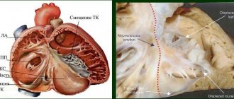
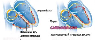
The heart is the most important human organ that does not stop for a minute. Its contraction is ensured thanks to a complex system of nerve impulses transmitted first from the sinus node in the right atrium, spreading to the atrioventricular node and the entire area of the septum. This process is considered normal and is called sinus rhythm. Sometimes, due to various reasons, patients experience an atrial rhythm on the ECG. In the article we will look at what this means and what the possible reasons for such a violation may be.
Development mechanism
Any cardiac conduction route that does not follow the path described above is called an ectopic rhythm. In this case, an electrical impulse that occurs not in the sinus node, but outside it, excites the heart before the signal is transmitted from the normal pacemaker, that is, the sinus node. In simple words, atrial rhythm is a condition in which the heart muscle contracts faster due to the advance of a healthy impulse by a pathological (minor) one.
Often such a violation occurs due to various blockades due to congenital or acquired causes. In this case, a separate part of the heart cannot be excited due to blocking the propagation of the nerve impulse. When activation occurs, an additional contraction is noted, which disrupts the sequence of heart and atrium impulses. This causes the so-called atrial rhythm.
According to some scientists, the pathological condition can develop as a result of hypoxia or inflammatory myocardial diseases. Cases of ectopic rhythm occurring after influenza, sore throat and other infectious diseases have been reported.
Important! Regardless of the cause of cardiac conduction disturbances, it is necessary to promptly identify the pathological condition and take measures to treat it.
What it is
The transmission of an electrical impulse, which sequentially excites all parts of the heart, begins from the right atrium. The conduction system distributes impulses to distant ventricular tissues. The heart begins to contract, and satisfactory blood flow into the arteries is observed. When rhythm and conduction disturbances occur, various diseases of the cardiovascular system develop. A variety of factors can cause the sinus node to lose its ability to produce the energy that is necessary to propagate the impulse to the most distant parts of the heart.
The norm is the formation of periodic excitation in the area of the sinoatrial node with subsequent spread to the atria and ventricles. When processes that affect the transmission of cardiac contraction change, replacement contractions are formed. In this case, impulses develop beyond the natural, physiological boundaries intended for their formation.
How does it show up on an ECG?
Pathological conduction of the heart can be determined using an electrocardiogram. This instrumental diagnostic technique allows you to identify the localization of rhythm disturbances and the cause of the deviation. Using an ECG, a specialist can determine one of the types of ectopia:
- left atrial rhythm - in this case, deviations such as a P wave of two parts are noted, when the first of them has a dome-shaped appearance, and the second looks like a high and narrow peak. In addition, PI can be smoothed, and PV 1 and 2 are positive, PV 5 and 6 are negative;
- right atrial - in the area of the third lead a negative P wave is observed, while in the first and second leads it is positive. This is characteristic of a mid-lateral right atrial rhythm. The lower type of this disorder is characterized by a negative P wave in the second and third leads, as well as a smoothed one, and aVF in the chest leads - 5 and 6;
- lower atrial rhythm - on the electrocardiogram it looks like a shortening of the PQ interval, a negative P wave in the first, third and aVF leads.
Electrocardiography allows for accurate diagnosis
Based on this, we can say that a specialist determines the conductivity of the heart by assessing the P wave, which, with an ectopic rhythm, is characterized by pathological amplitude and polarity. An experienced doctor should diagnose the condition, since it can be quite difficult to determine the pathology on an ECG due to the vagueness of the signs. Holter monitoring is often used to make an accurate diagnosis.
Signs on the electrocardiogram
An ECG is an accessible, simple and fairly informative way to obtain data on various heart rhythm disorders. What does the doctor evaluate on the cardiogram?
- The state of the P wave, reflecting the process of depolarization (appearance of an electrical impulse) in the atria.
- The PQ region demonstrates the features of the excitation wave traveling from the atria to the ventricles.
- The Q wave marks the initial stage of ventricular excitation.
- The R element displays the maximum level of ventricular depolarization.
- The S tooth indicates the final stage of propagation of the electrical signal.
- The QRS complex is called the ventricular complex; it shows all stages of the development of excitation in these sections.
- The T element registers the phase of decline in electrical activity (repolarization).
Using the available information, the specialist determines the characteristics of the heart rhythm (frequency and periodicity of contractions), the source of impulse generation, and the location of the electrical axis of the heart (EOS).
The presence of atrial rhythm is indicated by the following signs on the ECG:
- negative P wave with unchanged ventricular complexes;
- the right atrial rhythm is reflected by the deformation of the P wave and its amplitude in additional leads V1-V4, the left atrial rhythm - in leads V5-V6;
- teeth and intervals have increased duration.
EOS displays electrical parameters of cardiac activity. The position of the heart as an organ with a three-dimensional volumetric structure can be represented in a virtual coordinate system. To do this, the data obtained by the electrodes during the ECG is projected onto a coordinate grid to calculate the direction and angle of the electrical axis. These parameters correspond to the localization of the excitation source.
Normally, it has a vertical (from +70 to +90 degrees), horizontal (from 0 to +30 degrees), intermediate (from +30 to + 70 degrees) position. A deviation of the EOS to the right (over +90 degrees) indicates the development of an ectopic abnormal right atrial rhythm; a deviation to the left (up to -30 degrees and beyond) is an indicator of a left atrial rhythm.
What diseases can cause rhythm disturbances?
Atrial rhythm on the ECG can appear in patients regardless of age, gender and region of residence. Often the pathology is temporary and due to certain physiological reasons. In such cases, the duration of conduction disturbance lasts no more than several hours or days.
The situation is different with the development of certain diseases that can provoke an ectopic rhythm. These include myocardial inflammatory processes, ischemic disorders, and sclerotic changes. Let's look at the most common of them.
Myocarditis
Myocarditis is an inflammatory process of the myocardium. The causes of the pathology are damage to the heart muscle by viruses, bacteria or toxins. Often the disease develops due to an allergic reaction or an autoimmune disorder. Doctors note that myocarditis can act as an independent pathology or be provoked by other diseases. The course of inflammation can be chronic or acute. The latter form often develops into cardiomyopathy.
Myocarditis is a common cause of disruption of normal rhythm
Common symptoms of myocarditis are atrial rhythm, chronic fatigue, dizziness, rapid heartbeat, chest pain, and more. If the pathology is diagnosed in a timely manner and the necessary treatment is carried out, the prognosis for the patient is quite favorable.
Cardiomyopathy
A group of diseases that combine pathological changes in myocardial tissue are called cardiomyopathies. As a result of these disorders, disruptions in the functioning of the heart occur. The disease develops both under the influence of cardiac and non-cardiac factors. That is, there are a lot of reasons that can provoke cardiomyopathy. The disorder is primary or secondary in nature and is almost always accompanied by an extracardiac rhythm.
Rheumatism
Rheumatism is a disease accompanied by an inflammatory process of connective tissues and heart muscle. It mainly affects children under 15 years of age. The main cause of inflammation is an attack of the body by streptococcal infection, which provokes diseases such as tonsillitis, tonsillitis, pharyngitis, etc. Rheumatism occurs as a complication, leading to impaired contractility of the heart, increased temperature, joint and heart pain and the development of other symptoms in the patient.
Heart defects
Heart defects are congenital or acquired. The concept of “defect” implies a deviation in the structure or structure of an organ, as a result of which electrical conductivity or blood flow in it is disrupted. In addition to the congenital or acquired type, doctors classify the disease into combined or isolated, as well as the presence of a symptom such as cyanosis (bluish tint of the skin) or its absence.
Sick sinus syndrome
A dangerous condition that carries the risk of sudden cardiac arrest. The causes of this disorder are congenital or acquired. These include coronary heart disease, cardiomyopathy, trauma to the heart muscle, cancer in this area, defects, toxic damage to the organ and much more.
Sick sinus syndrome provokes a disturbance in the electrical conductivity of the heart
A person suffers from arrhythmia, decreased heart rate, weakness, headaches, paresis, decreased vision, hearing, and memory. Without the necessary treatment, the pathology is often accompanied by death.
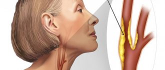
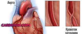
Cardiac ischemia
IHD is a very common disease, which is accompanied by many disturbances in the functioning of a vital organ. Pathology is provoked by many factors, the most common of which are smoking, anatomical aging of the body, genetic predisposition, diabetes mellitus, hypertension, etc. The atrial rhythm is shown on the cardiogram of many people with this deviation. In addition, signs such as shortness of breath, headache, chest discomfort, and chronic fatigue are noted.
Vegetovascular dystonia
VSD is a set of functional disorders that are caused by a violation of the regulation of vascular tone of the autonomic nervous system. In this case, an ectopic rhythm is formed, the patient experiences periodic or constant disturbances in heart rate, excessive sweating, frequent headaches, coldness in the extremities, lightheadedness or fainting.
Important! Vegetative-vascular dystonia often provokes a persistent increase in blood pressure and nervousness, significantly reducing the patient’s quality of life.
Description of the pathology
When the sinus node is weakened or stops working, and this happens either on an ongoing basis or from time to time, ectopic rhythms arise (or they are also called replacement rhythms).
Their frequency is less than that of sinus rhythm. Ectopic atrial rhythm can be considered non-sinus. The further away its source is, the less frequent its pulses will be. What is the reason for changes in heart function?
Other reasons
In addition to various diseases that lead to the development of ectopic heart rhythm, other causes can be identified. These include:
- persistent increase in blood pressure;
- smoking and drinking alcoholic beverages;
- carbon monoxide intoxication;
- taking certain medications;
- frequent stress;
- hormonal disorders;
- regular physical activity;
- professional sports.
Arrhythmia often occurs during heavy physical exertion.
These and other changes in the body can lead to intra-atrial conduction, which causes the development of many unpleasant symptoms.
Therapy for ventricular extrasystole
Treatment of untimely contractions of the ventricles consists of getting rid of the cause that provoked them, as well as stopping attacks of severe ventricular arrhythmia, if any.
Treatment of the functional form of extrasystole
If ventricular extrasystole is functional in nature, then you can get rid of it in the following ways:
- quit bad habits;
- take medications to relieve nervous tension (valerian, sedatives or tranquilizers, depending on the severity of anxiety);
- adjust your diet (give up coffee, strong tea, energy drinks);
- observe a sleep and rest schedule, engage in physical therapy.
Treatment of organic form
Treatment of the organic form of type 4 disease involves taking antiarrhythmic drugs that help get rid of attacks of ventricular arrhythmia. The doctor prescribes Sotalol, Amiodarone or other similar medications.
Features of the patient's symptoms
Often, cardiac conduction disturbances occur without visible symptoms, but more often the following clinical picture is observed:
We also recommend reading: Sinus rhythm on an ECG - what does it mean?
- attacks of heartbeat sensations. The patient seems to feel tremors in the chest;
- the average number of heartbeats increases;
- depending on the pathology that provoked the extracardiac rhythm, a slowdown in organ contractions may be diagnosed;
- sometimes the heart freezes for a few seconds. In this case, the patient may feel panic, fear, and a feeling of excitement;
- sweat production often increases;
- headaches and dizziness occur;
- due to shortness of breath, breathing is impaired;
- discomfort and pain, burning or tingling sensation are observed in the chest area;
- the skin of the face acquires a bluish tint, cyanosis may spread to the fingertips.
In addition, patients complain of the appearance of a veil before the eyes, difficulty breathing, and bouts of fever. In severe situations, nausea occurs, often accompanied by vomiting and abdominal pain. There is a malfunction in the functioning of the digestive system, which entails belching, heartburn, hiccups, flatulence, and bowel dysfunction. A common sign of the disease is pre-syncope or fainting.
Tachycardia is a common symptom of atrial rhythm
People with this pathology are characterized by an increase in heart rate. Signs of atrial tachycardia include palpitations, shortness of breath, panic, increased sweating, and flushing of the face. Attacks occur mainly at night. Their duration is usually short, lasting from several minutes to several hours. At the same time, the patient experiences panic and fear for his life.
Classification and severity
In cardiology, there are several classifications of extrasystoles of the lower cardiac chambers. Depending on quantitative and morphological criteria, the following forms of ventricle gradation are divided (see table).
| Class | Lown classification | Classification (gradation) according to Ryan |
| 0 | No rhythm disturbances observed | |
| 1 | Very rare, isolated (up to 30 per minute) | |
| 2 | Rare, single (more than 30 per minute) | |
| 3 | Polytopic | |
| 4A | Doubles | Monomorphic (come from one focus), paired |
| 4B | 3 or more ventricular impulses during atrial rest | Polymorphic (come from different foci), paired |
| 5 | Early PVCs (registered at 0.8 T waves) | 3 or more ventricular impulses during atrial rest |
There is also a Myerburg classification (Robert J. Myerburg is an American cardiologist, author of books on medicine).
- By frequency:
- very rare;
- rare;
- infrequent;
- moderately rare;
- frequent;
- very frequent.
- According to the characteristics of rhythm disturbance:
- single, monomorphic;
- single, polymorphic;
- pairs;
- stable;
- unstable.
Based on the results of daily ECG monitoring according to Holter, 6 classes of ventricular extrasystole were identified:
- class 0 - no ventricular extrasystoles;
- Class 1 – during any hour of monitoring, less than 30 single monomorphic (monotopic) ventricular extrasystoles are recorded;
- Class 2 - during any hour of monitoring, more than 30 frequent single monomorphic (monotopic) ventricular extrasystoles are recorded;
- Class 3 – polymorphic (polyfocal) ventricular extrasystoles are recorded;
- class 4a – monomorphic paired (2 at a time) ventricular extrasystoles are recorded;
- Class 4b – polymorphic paired ventricular extrasystoles are recorded.
- Class 5 – volley (group) polymorphic ventricular extrasystoles are recorded (3-5 in a row for 30 seconds), as well as episodes of paroxysmal ventricular tachycardia.
Class 1 ventricular extrasystoles do not manifest themselves clinically and are not accompanied by hemodynamic disturbances, therefore they are classified as functional. Ventricular extrasystoles of classes 2-5 are associated with an increased risk of developing ventricular fibrillation and sudden coronary death.
According to the prognostic classification of ventricular arrhythmias, there are:
- Benign ventricular arrhythmias – characterized by the absence of signs of organic heart damage and objective signs of left ventricular myocardial dysfunction; the risk of sudden cardiac death is minimal;
- ventricular arrhythmias of a potentially malignant course - characterized by the presence of ventricular extrasystoles against the background of organic heart lesions, a decrease in ejection fraction up to 30%; are accompanied by an increased risk of sudden cardiac death;
- ventricular arrhythmias of a malignant course - characterized by the presence of ventricular extrasystoles against the background of severe organic lesions of the heart; are accompanied by the highest risk of sudden cardiac death.
Atrial rhythm in children
The regulation of the autonomic nervous system and the cardiac conduction system in newborns differs from that in adults. They do not function fully due to insufficient formation. This provokes the development of atrial rhythm in infants and preschool children. Normally, such a rhythm is independently transformed into a sinus rhythm; the condition does not require additional treatment methods.
Cardiac conduction disorders in children often develop with minor abnormalities of the organ. For example, with mitral valve prolapse or accessory chord. But this does not mean that you should not pay attention to this symptom, because often a deviation can indicate serious heart defects, infectious myocardial lesions, hypoxia, intoxication and other conditions.
The risk group includes children who have suffered an intrauterine infection or prolonged intoxication with alcohol or nicotine, as well as during severe pregnancy and childbirth. Such patients should undergo a thorough examination after birth to ensure timely detection of various diseases and prevent the development of severe complications.
Symptoms and manifestations
We have learned that non-sinus rhythms depend on the underlying disease and its causes. This means that there are no specific symptoms. Let's look at some signs that indicate that it is time to see a doctor yourself or together with your child if his condition worsens.
Single ventricular premature contractions are recorded in half of healthy young people during monitoring for 24 hours (Holter ECG monitoring). They do not affect your well-being. Symptoms of ventricular extrasystole appear when premature contractions begin to have a noticeable effect on the normal rhythm of the heart.
Types of ectopic disorders
Ectopic arrhythmias include various disorders that develop in the area of the ventricles and atria. According to research, it turned out that often the right atrial rhythm, which does not manifest itself on the electrocardiogram, does not come from the sinus node. That is, an electrical impulse is provoked in neighboring areas of the organ.
The most dangerous type of disorder is considered to be atrial fibrillation.
Types of atrial rhythm:
- extrasystole - occurs quite often (about 60% of people), this condition consists of untimely contraction of the heart muscle or its individual parts;
- paroxysmal tachycardia is a separate type of atrial rhythm, accompanied by periodic attacks of accelerated heartbeat. In this case, the number of strokes can reach 220 per minute. Paroxysms replace the normal heart rhythm and have a sudden onset and end. Electrical impulses are generated in the atria, atrioventricular node or ventricles. The duration of the attack varies;
- ectopic accelerated rhythms - attacks of accelerated heartbeat, in which the heart rate increases to 130 beats. In this case, electrical impulses are generated in the atria, ventricles or atrioventricular junction. During an ECG, a non-sinus pacemaker is recorded in the P–QRS–T complex;
- atrial fibrillation or atrial fibrillation - frequent excitation of the atria or individual muscle fibers of a chaotic nature. In some cases, heart rate reaches 600 beats per minute. The prolonged course of such an attack significantly increases the risk of blood clots and the development of ischemic stroke. If left untreated, acute heart failure occurs.
Important! Therapy for any type of ectopic rhythm should be carried out immediately, regardless of the severity and symptoms of the patient.
How to distinguish from sinus
Atrial rhythms occur when the functioning of the sinus nodes is inhibited. They are distinguished by their slow, substitutive nature. There is a decrease in heart contraction, acceleration of heart activity, and the development of pathological activity in the atria. Often the heart rate exceeds the heart rate, and a left atrial or right atrial contraction is formed.
To distinguish atrial rhythm from sinus rhythm, electrocardiography is required. The doctor examines the nature of changes in contractions, pays attention to the heart rate, the duration of the intervals, how correctly the ventricles contract, and whether deformed, negative teeth are formed.
With an atrial rhythm, atrial flutter can accelerate to 400 beats per minute, while with a sinus rhythm, it remains uniform. In the ECG picture, in the first case, saw-like teeth will be observed, in the second, an almost flat, uneven line will be visible.
Treatment methods
The atrial rhythm may be hidden and not manifest itself in any way. If the patient does not feel any unpleasant symptoms and the abnormal pacemakers were identified by chance, most often no special treatment is required. For such people, it is enough to undergo a thorough medical examination to exclude serious anomalies in the structure of the heart and other organs. If no deviations are detected, the ectopic rhythm is considered safe for health.
Treatment methods are determined depending on the diagnosis and symptoms of the patient
When a patient has complaints from the cardiovascular system and any pathologies are detected, drug therapy is carried out. The group of drugs includes the following:
- beta blockers and other drugs intended to lower blood pressure (Nadolol, Metoprolol, Carvedilol);
- medications that accelerate the rhythm during bradycardia. These include Atropine, Isoprenaline, Eufillin. Plant-based extracts are often recommended - ginseng, eleutherococcus;
- sedatives are prescribed to patients with the development of vegetative-vascular dystonia. Popular remedies include tincture of motherwort, valerian, as well as Novopassit, Fitosed, Dormiplant;
- Preventive medications are used to prevent strokes, heart attacks and other dangerous complications. For this, Panangin and Cardiomagnyl are prescribed.
Treatment tactics always depend on the disease that acts as a factor provoking arrhythmia. Based on the data obtained during instrumental diagnostics and the patient’s medical history, the doctor selects the necessary medications.
In particularly severe situations, when conservative therapy has proven useless, the patient is given an artificial pacemaker (cardioversion). Often this method is effective for atrial fibrillation and other dangerous conditions.
Diagnostics
The gold standard for diagnosing atrial rhythm is the electrocardiogram. The development of the disorder is indicated by deformations of the P waves. The amplitude is impaired, the teeth are shortened. With paroxysmal tachycardia, a regular rhythm and high frequency of contractions are observed; it is not always possible to determine the P waves.
Additionally, additional examination is recommended using:
- ultrasound examination of the heart
- 24-hour electrocardiogram monitoring
- for myocardial ischemia, coronary angiography is prescribed
- patients with other types of arrhythmia are advised to undergo transesophageal electrophysiological study
There are a large number of disorders that can interfere with the functioning and automaticity of the sinus node. The doctor must adequately assess the degree of the disorder, conduct a differential diagnosis with sinus arrhythmia, atrial node, migration of the pacemaker through the atria, polytopic atrial extrasystole.
Folk recipes
A healthy lifestyle and proper nutrition help patients prevent many dangerous complications. They are especially indicated for patients with developed atrial rhythm. Giving up bad habits and saturating your diet with plenty of vitamins and minerals is an important step towards health. Some folk recipes have proven themselves well in the fight against heart disorders. Here are some of them:
- mix 200 ml of grapefruit juice with three teaspoons of olive oil. Take half a glass of the drink in the morning and evening for one month;
- Combine chopped figs and walnuts in equal parts, season the resulting mass with honey and refrigerate for a day. Take a teaspoon of the mixture twice a day. The course of treatment is at least 60 days;
- To restore rhythm, a decoction of calendula flowers is often used. To do this, brew a spoonful of herbs with a glass of boiling water and let the medicine brew for at least 2 hours. Take a quarter glass twice a day for a month;
- A decoction of hawthorn and lemon balm has a beneficial effect on heart health. To prepare it, combine a tablespoon of raw materials, pour a liter of boiling water over the mixture, and simmer over low heat for 10 minutes. After this, the medicine should infuse for 40 minutes. Take the drink instead of tea several times a day.
Traditional recipes are a great way to maintain your heart
Important! Any folk remedies should be used only after consultation with a specialist. Self-medication can be extremely dangerous to health.
Prevention
To prevent atrial rhythm, it is important to qualitatively and timely treat dysfunctions of the cardiovascular and hormonal systems. At the first signs of violations, seek help from experienced, qualified specialists. A high-quality, balanced diet, adherence to work and rest, moderate physical activity, and abandonment of bad habits are recommended.
To prevent the formation of arteriosclerotic plaques, excessively fatty and fried foods are removed from the diet, and a sufficient amount of fiber, greens, berries, fresh fruits and vegetables, whole grains, cereals, seeds, and nuts are introduced.
Timely diagnosis and adequate therapy are the best prevention of serious complications, including sudden death. In some cases, lifelong medication and regular monitoring by a cardiologist are required. This allows you to increase life expectancy and improve its quality. If the cause of the atrial rhythm does not depend on the influence of the physiological characteristics of the body, consultation with a psychotherapist may be required.