© Author: A. Olesya Valerievna, candidate of medical sciences, practicing physician, teacher at a medical university, especially for SosudInfo.ru (about the authors)
Ebstein's anomaly (EA) is a very rare cardiac defect that develops during fetal development. With pathology, the normal arrangement of the tricuspid valve leaflets is disrupted, resulting in its insufficiency and circulatory disorders. According to statistics, Ebstein's anomaly accounts for no more than 1% of cases of all congenital heart anomalies.
In general, congenital malformations of organs are becoming more common. Constantly deteriorating environmental conditions, unfavorable factors affecting the expectant mother, and the pathology of pregnancy are considered to be to blame. Heart anomalies constitute a large group of defects, which, even if compatible with life, very often lead to serious hemodynamic disorders requiring surgical correction.
When signs of a heart defect are detected in the fetus during an ultrasound examination of a pregnant woman, the expectant mother, of course, begins to worry and look for information about the disease in order to be ready to help her baby after birth. Let's try to figure out what Ebstein's anomaly is and what possible ways to correct this rare defect.
Introduction
Ebstein's anomaly is a type of congenital heart defect characterized by an abnormally developed tricuspid valve and right ventricle. These deviations can be relatively mild or quite severe. Depending on the severity of these anatomical problems, the symptoms of those born with the anomaly can vary greatly.
Some babies with Ebstein's anomaly are critically ill at birth. Others live into adulthood without any symptoms. However, almost everyone born with this condition will eventually develop heart problems.
Causal factors for the anomaly
The congenital genesis of the defect indicates the development of intrauterine pathology in the fetus caused by internal or external factors. The specific reasons are unknown, but the following may have a possible negative effect on the formation of the embryo:
- genetic disorders (more often with combined congenital heart disease);
- viral infections in a pregnant woman;
- taking certain medications in the 1st trimester of pregnancy (antitumor drugs, strong antibiotics, sleeping pills).
The inability to prevent the development of intrauterine pathology determines the enormous importance of prenatal diagnosis: with timely detection of a congenital defect, cardiac anomalies incompatible with life can be detected long before birth.
Tricuspid valve displacement
What is Ebstein's anomaly?
First of all, Ebstein's anomaly is caused by a violation of the normal development of the tricuspid valve in the fetus. During development, the leaflets (leaflets) of the tricuspid valve cannot move into their normal position at the junction of the right atrium and right ventricle. Instead, the leaflets move downward within the right ventricle. In addition, the leaflets themselves often become laminated (“stuck”) to the wall of the right ventricle and therefore do not open and close properly.
Because the tricuspid valve is displaced inferiorly by the abnormality, the portion of the right ventricle located above the abnormal tricuspid valve is considered to be “atrialized” (becomes part of the enlarged right atrium). That is, the atrial chamber of the heart will not only contain normal atrial tissue, but will also be part of what would be the right ventricle of the heart.
Features of hemodynamics
In this pathology, the valve leaflets are deformed and displaced into the lumen of the right ventricle. They are located below the physiological opening between the atrium and the ventricle.
Therefore, the right atrium is enlarged due to the “attachment” of part of the ventricle, and the latter becomes disproportionately small.
At the moment of systole of the atrium, its upper part contracts, and the lower part only during the systolic period of the ventricle. At this point, part of the blood from the right ventricle returns to the atrium. There is little blood left in the right ventricle, which reduces its flow to the lungs. And the atrium fills with blood, expands, its muscle layer hypertrophies.
High pressure in the atrium causes a window in the atrial septum to open. A reset is formed from right to left. It helps relieve the right atrium, but at the same time reduces the oxygen content in arterial blood due to its mixing with venous blood.
What problems occur with the heart with Ebstein's anomaly?
Because of the abnormal position and distortion of the tricuspid valve that occurs with Ebstein's anomaly, the valve is usually regurgitant, or "leaky." As a result, tricuspid regurgitation is usually the main manifestation of this condition.
In addition, the atrium of the part of the right ventricle that lies above the displaced tricuspid valve also causes problems. The atrial portion of the right ventricle begins to beat when the rest of the right ventricle beats, not when the right atrium beats. This dissonant muscular action in the atrial chamber increases tricuspid regurgitation and also creates a tendency for blood to pool in the right atrium, a condition that can lead to blood clots.
The severity of Ebstein's anomaly is related to the degree of displacement and distortion of the tricuspid valve, as well as the amount of right ventricular tissue that subsequently atrializes. People born with the anomaly who have relatively few (or no) symptoms usually have very little valve misalignment and therefore have little right ventricular atrialization.
Survival prognosis
The prognosis for life with Ebstein's anomaly depends entirely on how timely the disease was identified and its treatment started. If the pathology is discovered while the fetus is still inside the womb, but it does not greatly affect the development of the baby, then the pregnancy is not terminated. After birth, the child undergoes surgery, and he has the opportunity to live a long and happy life.
Most patients do not experience complications after surgery and live for many years. But for a favorable outcome, it is important for patients to strictly follow all medical recommendations.
If Ebstein's anomaly occurs with concomitant diseases or entails the development of severe complications, then the prognosis sharply worsens. In this case, babies rarely manage to live even up to a year.
Causes of Ebstein's anomaly
Ebstein's anomaly occurs in approximately 1 in 20,000 live births.
Although previously identified genetic mutations have been associated with the abnormality, no specific mutation is believed to be the main cause of this condition.
An association between Ebstein's anomaly and maternal use of lithium or benzodiazepines has been reported, but a causal relationship has also not been proven.
So, for the most part, this anomaly occurs sporadically (randomly).
Causes and risk factors
It is assumed that Ebstein's anomaly is caused by mutations in the CFA9 locus of chromosome 17q, duplication of chromosome 15q, and a defect in the ALK3/BMPR receptor. Chromosomal abnormalities occur at the stage of fusion of parental genetic material or in the early stages of pregnancy and lead to incorrect formation of organs and tissues of the child’s body in the prenatal period.
Since the exact causes of the disease have not yet been established, the most likely risk factors are:
- maternal use of lithium during pregnancy;
- maternal use of benzodiazepines during pregnancy;
- viral diseases suffered in the early stages of pregnancy (influenza, rubella, measles);
- history of multiple spontaneous abortions in the early stages;
- chronic intoxication with pesticides, vapors of paints and varnishes, petroleum products, etc. (living in environmentally unfavorable areas, working in hazardous industries);
- parental use of illegal substances, alcohol abuse, smoking.
Viral diseases in early pregnancy can lead to the development of Ebstein's anomaly in the fetus
Symptoms of Ebstein's anomaly
The symptoms and signs experienced by people with Ebstein's anomaly vary greatly, depending both on the extent of the tricuspid valve abnormality and the presence or absence of other congenital heart problems.
Children born with severe tricuspid valve dysfunction caused by the abnormality often have other congenital heart problems and may be critically ill from birth. These children often have:
- severe cyanosis (low oxygen levels in the blood);
- shortness of breath;
- weakness and swelling.
Children born with the anomaly who have significant tricuspid regurgitation but no other serious congenital heart problems may be healthy, but they often develop right-sided heart failure either in childhood or adulthood.
On the other hand, if tricuspid valve dysfunction is minor, a person with the abnormality may be asymptomatic for their entire life.
There is a strong connection between Ebstein's anomaly and abnormal electrical pathways in the heart. These so-called "accessory pathways" create an abnormal electrical connection between one of the atria and one of the ventricles; when abnormal, they almost always connect the right atrium to the right ventricle.
These accessory pathways often cause a type of supraventricular tachycardia called atrioventricular nodal reentrant tachycardia (AVNRT).
Sometimes these same accessory pathways can cause Wolff-Parkinson-White syndrome, which can lead not only to AVNRT, but also to much more dangerous arrhythmias, including ventricular fibrillation. As a result, these additional pathways may pose an increased risk of sudden cardiac death.
Diagnosis in children and adults
During examination, attention is paid to the cyanotic color of the skin, deformation of the fingers and chest. The borders of the heart are shifted to the right, a 3- or 4-beat rhythm is heard, a murmur to the right of the sternum, the 2nd tone is bifurcated.
If a tricuspid valve defect is suspected, the following examination is performed:
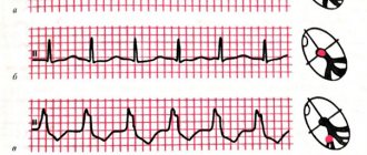
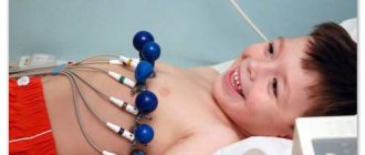
- ECG - the axis is deviated to the right, the right atrium is hypertrophied and dilated, rhythm disturbances in the form of paroxysmal tachycardia, atrial fibrillation and flutter, Wolff-Parkinson-White syndrome, blockade of the right branch of the Hiss pathway.
- FCG – systolic murmur over the projection of the right ventricle, 1 tone is weakened, 2 is bifurcated, 3 and 4 are high-amplitude.
- X-ray – the right atrium gives the contour of the heart the shape of a ball, the pulmonary fields are transparent.
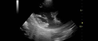
- Ultrasound of the heart - the valve leaflets are displaced downwards, close slowly, the right ventricle is poorly filled, blood is discharged through the atrial septum to the left. In the fetus, these abnormalities can be detected in approximately 65 - 70% of cases.
- CT and MRI make it possible to detail the degree of valve non-closure, the presence and intensity of blood shunting, and the size of the heart chambers.
- Ventriculography and probing of the cardiac cavities are used only when it is difficult to make a diagnosis.
Diagnostics
The key examination method in diagnosing an abnormality is an echocardiogram (ultrasound of the heart), usually transesophageal echocardiography gives the most accurate results. An echocardiogram can accurately assess the presence and extent of tricuspid valve abnormality and also identify most other congenital heart defects that may be present.
Adults and older children who receive initial evaluation for Ebstein's anomaly usually undergo an exercise test to assess their physical performance, blood oxygenation during exercise, and the response of heart rate and blood pressure to exercise. These measurements are useful in assessing the overall severity of a heart condition and the need and urgency of surgical treatment.
It is also important to evaluate people who have the abnormality for the presence of cardiac arrhythmia. In addition to annual electrocardiogram (ECG) and ambulatory ECG monitoring, most of these people should be evaluated by an electrophysiologist after they are diagnosed to assess the likelihood of developing potentially dangerous arrhythmias.
How does the disease manifest?
The anomaly manifests itself from the moment of birth; asymptomatic forms of the defect do not occur. The disease has 4 degrees of severity, each of which is characterized by its own manifestations - from subtle to pronounced.
Reasons why a defect may not be noticed or detected untimely:
- The first degree of severity of the disease, when the defect is barely pronounced and almost does not appear;
- Failure to register a pregnant woman;
- Low social literacy, lack of parental attention to external signs of illness in the child.
Symptoms in newborns
The defect manifests itself in the first hours of life . Since the fetal lungs do not participate in gas exchange, the baby normally has a purplish-bluish skin tone immediately after birth. If the baby's breathing is not difficult, an anomaly can be suspected by the persistent bluishness of the skin, especially the earlobes, lips, and fingertips.
Gradually, breathing becomes frequent, the child cannot suckle, as he begins to breathe through his mouth. During inhalation, the chest noticeably increases in volume.
Signs in children
Diagnosis occurs, as a rule, in the fifth or sixth year of life , which is associated with increased development of the body during this age period.
Symptoms: low body weight, weakness, shortness of breath during exercise, tendency to respiratory diseases, fainting. Children often breathe through their mouths, which leads to difficulty eating. Shortness of breath and bluish skin during physical activity intensify. Children complain of palpitations and chest pain.
On examination, the center of the chest has a visible pulsating protrusion formed by the enlarged right half of the heart, the nail phalanges are enlarged like “drumsticks”, the fingers are tight and cold, the nails are flat (symptom of “watch glasses”).
Manifestations in adults
In adults (not older than 30-40 years), the disease manifests itself as low body weight, asthenic physique, bluish skin, shortness of breath, and poor tolerance to physical activity. Patients feel interruptions in the functioning of the heart, complain of chest pain, swelling of the veins of the neck, visible pulsation in the xiphoid process, are exhausted, and are constantly cold.
Untreated adults are characterized by attacks of palpitations ; in particularly severe cases, the disease may have a single manifestation - sudden cardiac death.
Treatment of Ebstein's anomaly
In general, if the abnormality causes significant symptoms, treatment is surgical repair.
Surgery in newborns with severe Ebstein's anomaly is usually delayed as long as possible due to the high risk of repair of this problem in infants. These children are usually treated aggressively in the intensive care unit in an attempt to delay surgery until they are older. If possible, surgery is postponed for at least several months.
In older children and adults newly diagnosed with Ebstein's anomaly, surgical treatment is strongly recommended as soon as any symptoms appear. However, if they have significant heart failure, an attempt is made to stabilize them with drug therapy before surgery.
The surgical procedures used to correct the abnormality can be quite complex, and the specific surgical procedures that are performed vary from person to person, depending on the condition of the tricuspid valve, the presence or absence of additional congenital heart defects, and whether or not severe heart failure and the age of the patient.
Classification
The classification includes 4 types of disease. I form of Ebstein's anomaly is characterized by the following symptoms:
- the posterior valve leaflet is completely absent or significantly displaced;
- the septal is completely absent or displaced from its normal location;
- the front one is present, but large and unnaturally mobile.
Type II involves the following changes: the anterior, posterior and septal valve leaflets are most often present, but are small in size and displaced higher from the normal location.
Type III is characterized by the presence of the following problems:
- the septal and posterior valve leaflets are displaced and undeveloped;
- the anterior one is present, but has short and merging chords.
IV – has features:
- the anterior leaflet is significantly deformed, shifted closer to the tract from the right ventricle;
- septal consists of fibrous tissue;
- the posterior one is undeveloped or absent altogether.
Another congenital heart defect is no less dangerous - Fallot's triad. Familiarize yourself with its symptoms so that you can consult a doctor in time if necessary. Rarely, newborns are diagnosed with tetralogy of Fallot. Find out how it differs from the triad and other heart defects.
Forecast
The prognosis of Ebstein's anomaly depends on the severity of the tricuspid valve problem and the presence or absence of other congenital heart problems. Babies with the disease who are born critically have a high risk of mortality—more than 30 percent die before they are discharged from the hospital.
The risk of premature death when the abnormality is diagnosed in later childhood or adulthood also depends on the severity of the condition. However, in recent decades, aggressive surgical treatment and prophylactic treatment of potential cardiac arrhythmias have significantly improved the prognosis of people with Ebstein's anomaly.
Dangerous complications
In the absence of surgical treatment, the following consequences are possible:
- rapidly progressing congestive heart failure;
- aneurysm of the right chambers of the heart with a high risk of rupture;
- thromboembolism of large vessels;
- severe rhythm disturbances, insensitive to drug therapy;
- septic endocarditis.
The prognosis for life depends entirely on timely diagnosis and treatment. A prenatally isolated congenital defect with a good prognosis for life is not at all a mandatory indication for termination of pregnancy: a surgical operation performed at the optimal time with minimal signs of cardiac failure is a guarantee of a long and happy life for the child.
For 90% of operated patients, the prognosis for recovery is favorable - a person can live a full life, but with mandatory monitoring by a cardiologist and limiting severe physical activity.
The presence of severe and dangerous complications sharply reduces the effectiveness of drug and surgical therapy for the disease.