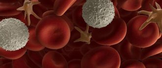
What is hemolytic anemia
Hemolytic anemia is anemia that occurs as a result of increased erythrodieresis, when the destruction of red blood cells prevails over their formation, which is due to their increased breakdown.
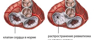
With hemolytic anemia, due to increased destruction of red blood cells, their life expectancy is shortened to 12–14 days. The incidence of hemolytic anemia is about 2 cases per 19 thousand population. The most commonly observed are Minkowski-Choffard anemia and elliptocytosis. Among other types of anemia, hemolytic ones occupy a share of 11%. The disease can begin in early childhood and show its first signs in adulthood.
In severe forms of hemolytic anemia, complications such as encephalopathy and biliary cirrhosis may occur.
Prognosis and prevention
When giving a prognosis for GA, specialists take into account the causes and clinical course of the disease. If it is possible to quickly resolve hereditary anemia, there is a good chance of stabilizing the patient’s well-being. With homozygous variants of hemoglobinopathies, the situation is more complicated: with severe forms, the risk of developing dangerous conditions remains.
With acquired types of GA, a dubious prognosis is often given, although the possibility of a long and quiet course of the disease or even spontaneous recovery cannot be ruled out.
Prevention of pathology consists of timely identification of a predisposition to hemolysis and its complications, taking appropriate measures, as well as eliminating external factors that contribute to exacerbations.
Causes of hemolytic anemia
Hemolytic anemia can develop at any age and in any population group. The main cause of hemolytic anemia is the destruction of red blood cells.
The destruction of red blood cells can be caused by blood diseases and exposure to poisons. Also, some diseases serve as a trigger for the destruction of red blood cells:
- hepatitis;
- Epstein-Barr disease;
- typhoid fever;
- sickle cell anemia;
- leukemia;
- lymphoma;
- various tumors;
- Wiskott Aldridge syndrome;
- lupus erythematosus;
- infection with Escherichia coli and streptococcus.
It happens that the use of certain medications causes hemolytic anemia, usually this is:
- penicillin;
- painkillers;
- acetaminophen.
Mechanism of development of hemolytic anemia
The disease is based on a genetic defect in the red blood cell membrane protein. The existing membrane anomaly leads to the penetration of excess sodium ions into the erythrocyte and increased accumulation of water in it, resulting in the formation of spherical erythrocytes (spherocytes). Spherocytes, unlike biconcave normal erythrocytes, do not have the ability to deform in narrow areas of the blood flow, for example, when passing into the sinuses of the spleen.
This leads to a slowdown in the movement of red blood cells in the sinuses of the spleen, the detachment of part of the surface of the red blood cell with the formation of microspherocytes (hence the name of the disease - “microspherocytosis”) and their gradual death. Destroyed red blood cells are absorbed by macrophages of the spleen. Constant hemolysis of red blood cells in the spleen leads to hyperplasia of its pulp cells and enlargement of the organ.
Due to the increased breakdown of red blood cells in the serum, the content of free bilirubin increases. Bilirubin entering the intestines in increased quantities is excreted from the body in the urine and mainly in feces in the form of stercobilin. The daily release of stercobilin in hereditary microspherocytosis exceeds the norm by 10-20 times. The consequence of increased release of bilirubin into bile is bile pleiochromia and the formation of pigment stones in the gallbladder and ducts.
Classification
In the general classification, diseases are divided into hereditary and acquired.
Hereditary
- Membranopathies. Pathologies such as microspherocytic, ovalocytic or acanthocytic are characterized by an altered shape of erythrocytes due to a violation of the protein and lipid structure of the membrane. Contains few proteins, being in pathological plasma, blood cells decrease in size, elongate, and acquire spherical outgrowths. A fragile membrane causes premature hemolysis.
- Enzymopenic anemia. The pathology is caused by a lack of red blood cell enzymes. The hemolytic condition is caused by the use of certain medications.
- Hemoglobinopathies. This group includes sickle cell anemia and thalassemia. The pathogenesis is based on disturbances in the structure of hemoglobin. At the same time, the shape of red blood cells and the internal structure change.
Purchased
- Immune hemolytic anemia. They develop as a result of a conflict between blood antibodies and native or donor-derived erythrocyte antibodies.
- Acquired membranopathies. Hemolysis is provoked by various physical and biochemical factors against the background of various diseases or surgical intervention.
- Toxic anemia. Hemolysis develops due to poisoning by chemicals that enter the body, including pathogenic microorganisms.
General classification
Symptoms of hemolytic anemia
Very often, a person who suffers from this disease for a long time may not feel symptoms at all. Outside periods of exacerbation, the disease may practically not make itself felt.
However, if the anemia worsens, the patient may feel:
- severe attacks of dizziness;
- weakness;
- a sharp increase in temperature.
One of the most striking signs of hemolytic anemia is jaundice of the skin . This phenomenon is caused by the excessive formation of bilirubin in the body, which under normal conditions is bound by the liver. If hemolytic anemia progresses, the liver does not have time to bind all the bilirubin, the excess of which manifests itself in the form of jaundice.
Another indisputable evidence of the presence of hemolytic anemia is bright red or dark brown feces , which contain hemoglobin in almost pure form.
Very often, people suffering from this disease complain of heaviness in the left side . This symptom is caused by a significant enlargement of the spleen, in which hemolysis (decomposition of red blood cells) occurs with increased intensity. In many cases, the spleen simply protrudes from under the costal arch, causing unbearable pain.
The most intense manifestation of symptoms is observed during periods of hemolytic crises, which can be provoked by:
- infection;
- hypothermia;
- pregnancy.
Treatment of hemolytic anemia
Hemolytic anemia is treated depending on its nature and cause. In many cases, a fairly effective method is splenectomy - removal of the spleen.
However, indications for this operation can only be frequent hemolytic crises, splenic infarctions, attacks of renal colic or severe anemia. In addition, to reduce hemolysis, special glucocorticoid hormonal drugs (Prednisolone) are prescribed, which, when taken correctly and in a properly calculated dosage, significantly reduce and sometimes completely stop the increased breakdown of red blood cells.
In general, the dosage of this drug is calculated at 1 mg/kg of human weight per day. As the patient's condition improves, the dosage is reduced to maintenance and is about 5-10 mg/day.
In some cases, immunosuppressants may be prescribed in combination with prednisolone. Treatment is a fairly lengthy process and may take 2-3 months until the increased breakdown of red blood cells stops completely.
Hemolytic anemia is treatable in many cases, but you need to understand that such a complex disease requires a lot of time to diagnose and treat. Therefore, it is very important to consult a doctor in time and undergo all the necessary examinations to identify the causes of the disease.
Complications
Chronic hemolysis causes increased release of bilirubin into the bile ducts, which leads to the formation of stones in the biliary ducts, gallbladder, and pancreas. Prolonged release of hemoglobin has been associated with pulmonary hypertension. Signs of complications: severe shortness of breath, chest pain, sometimes loss of consciousness. Further deterioration of the circulatory system in the lungs results in heart failure, swelling in the extremities, and abdominal dropsy.
During hemoglobinuric crises, the risk of severe thrombosis of the veins of the brain, heart and abdominal cavity develops. Blockage of blood vessels makes itself felt with aching and nagging pain. Such complications can be aggravated by organ infarctions. Portal vein thrombosis leads to splenic infarction, which further develops into portal hypertension and thrombophlebitic splenomegaly.
Other possible complications:
- inflammation of the retina (with possible progression to blindness);
- ataxic neuropathy;
- aplastic crises;
- encephalopathy;
- gangrene of fingers or toes;
- renal failure;
- anuria;
- nephrosis.
Prognosis for hemolytic anemia
The prognosis for hemolytic anemia is generally favorable: it is usually possible to completely stop or significantly reduce hemolysis.
Long-term results depend on the characteristics of the pathological process, pathogenetic therapy and active prevention. In some forms of acquired hemolytic anemia caused by exposure to chemical, physical and immunological factors, recovery is possible if the latter are eliminated and appropriate treatment is carried out. With congenital hemolytic anemia, complete cure does not occur, but in some cases it is possible to achieve long-term remission.
Clinical guidelines
The blood disease occurs as a result of increased destruction of erythroid cells. They can be:
- hereditary or acquired;
- with intravascular or intracellular type of hemolysis;
- associated with internal abnormalities of the red blood cells (RBCs) themselves or external influences.
The clinical picture is dominated by jaundice and hemorrhagic syndrome. In the blood test, the concentration of Hb, the content of erythrocytes and platelets are reduced. Anisocytosis, poikilocytosis, microspherocytosis are expressed, “styloid”, “helmet-shaped” cells and RBC fragments are detected.
In a biochemical blood test, there is an increase in LDH, indirect bilirubin, and a possible decrease in serum iron due to its loss in the urine. In the urine - hemosiderinuria, less often - hemoglobinuria.
The direct Coombs test is negative. The differential diagnostic criterion is a combination of anemia, red blood cell fragmentation, leukocytosis and thrombocytopenia.
The clinical picture of hemolytic anemia in children is varied - from asymptomatic forms to life-threatening. Anemia is characterized by a triad of symptoms: jaundice, splenomegaly, anemia. Delayed physical development and abnormalities of the skull and facial skeleton often occur. Blood tests reveal a decrease in Hb concentration, reticulocytosis, microspherocytosis, and a shift of the Price-Jones curve to the left. The osmotic resistance of red blood cells is reduced. The course of moderate and severe forms of the disease is sometimes accompanied by exacerbations.
- An exacerbation occurs spontaneously or against the background of an infection: jaundice appears or intensifies, the size of the spleen increases, and it becomes painful. In the blood, the concentration of Hb and the content of red blood cells decrease, reticulocytosis increases, and the concentration of indirect bilirubin and LDH increases. The first crisis, which occurs atypically and is rarely diagnosed, can develop during the newborn period.
- Aplastic crisis in hemolytic anemia is usually provoked by a parvovirus infection (parvovirus B19), which leaves lasting immunity, so such a crisis develops in patients once in a lifetime. The concentration of Hb and the content of RBC, and sometimes leukocytes and platelets, quickly decrease. The bilirubin content and the number of reticulocytes do not increase. The condition of patients is often severe due to developing hypoxemia and hypoxia. Relatively often, patients with hereditary spherocytosis develop cholelithiasis.
Diagnosis of hemolytic anemia
Determining the form of hemolytic anemia based on an analysis of the causes, symptoms and objective data is the responsibility of a hematologist. During the initial conversation, family history, frequency and severity of hemolytic crises are clarified. During the examination, the color of the skin, sclera and visible mucous membranes is assessed, and the abdomen is palpated to assess the size of the liver and spleen. Spleno- and hepatomegaly are confirmed by ultrasound of the liver and spleen.
Changes in the hemogram are characterized by normo- or hypochromic anemia, leukopenia, thrombocytopenia, reticulocytosis, and accelerated ESR. In autoimmune hemolytic anemia, a positive Coombs test is of great diagnostic importance. Biochemical blood samples reveal hyperbilirubinemia (increased indirect bilirubin fraction) and increased lactate dehydrogenase activity.
Urine examination reveals proteinuria, urobilinuria, hemosiderinuria, hemoglobinuria. The coprogram has an increased content of stercobilin. Examination of bone marrow puncture reveals hyperplasia of the erythroid lineage.
In the process of differential diagnosis, hepatitis, liver cirrhosis, portal hypertension, hepatolienal syndrome, and hemoblastosis are excluded.
Diagnostics
Doctors recommend that, even in the absence of symptoms, you undergo a general blood test, which helps to identify a number of diseases, including anemia. And then, with the help of additional tests, determine the type and cause of the disease.
First stage
Hemolysis of erythrocytes is divided into intracellular and intravascular. It is extremely important to determine what type of disease is progressing so that the doctor understands in which direction to look for the cause of the destruction of red blood cells.
Intracellular hemolytic anemia is examined using the following laboratory tests:
- hemoglobinemia – helps determine the level of free hemoglobin in the blood;
- hemoglobinuria - determines the level of unchanged hemoglobin in the urine;
- hemosiderinuria - indicates the presence of an oxidation product in the kidneys - hemosiderin.
Intravascular anemia is detected by the following laboratory tests:
- general blood test - determines a decrease in the number of red blood cells or hemoglobin;
- peripheral blood smear - shows abnormalities in the structure of red blood cells;
- biochemical analysis - helps monitor the increase in total bilirubin.
Second phase
Determining the cause of the disease can take a huge amount of time, so the doctor should collect an anamnesis - find out the places where the patient has been, in what conditions he lives, where he works and what symptoms he observed. If this information is missing, then a number of tests are prescribed:
- Circulating immune complexes.
- Hemoglobin electrophoresis is carried out to exclude qualitative and quantitative hemoglobinopathies.
- Direct and indirect Coombs test - a reaction to determine antibodies to red blood cells.
- Osmotic resistance of erythrocytes - with the congenital form of anemia, a decrease in osmotic resistance can be observed. With thalassemia, on the contrary, you can see an increase in the indicator.
- Studying the activity of erythrocyte enzymes helps to find the enzymes you are looking for. Quantitative determination of enzymes helps to monitor their decline in relation to normal values.
- Hem's test and Hartman's test determine the lifespan of red blood cells. Hartmann's test is considered positive if more than 4% of red blood cells are destroyed, and Hem's - more than 5%.
- The study of a “thick drop” of blood identifies malaria pathogens, the life cycle of which is associated with the destruction of red blood cells.
- Heinz body test - helps to detect insoluble hemoglobin in a blood smear. The test is performed to confirm G-6-FDG deficiency.
- Red blood cell sickling test - determines the change in the shape of red blood cells as the partial pressure of oxygen in the blood decreases. If the corpuscles take the shape of a sickle, then the diagnosis is considered confirmed.
- Bacteriological blood culture - determines the types of infectious agents circulating in the blood that bind to the bodies and cause their destruction.
- Myelogram is the result of a brain puncture. Allows you to detect malignant diseases. It also determines the proliferation of the erythroid germ, which indicates a high rate of compensatory production of red blood cells.
Classification of hemolytic anemia
Hemolytic anemias are classified into congenital and acquired. As a result of inheritance of genetic defects of functionally defective red blood cells, a child is born with congenital hemolytic anemia. Acquired hemolytic anemia is caused by the effect of destructive factors on red blood cells.
| Congenital hemolytic anemias | Acquired hemolytic anemia |
|
|
Autoimmune hemolytic anemia
Autoimmune hemolytic anemia is considered as a pathological process, which is based on the production of antibodies to the antigenic structure of one’s own red blood cells. For some reason, the immune system mistakes its own antigen for a foreign one and begins to fight it.
There are two types of autoimmune hemolytic anemia:
- Symptomatic autoimmune hemolytic anemia, which develops against the background of another pathology:
- hemoblastoses,
- chronic hepatitis,
- neoplasms,
- systemic lupus erythematosus,
- rheumatoid arthritis,
- lymphogranulomatosis.
- An idiopathic variant of autoimmune hemolytic anemia, provoked by various factors:
- infection,
- taking medications,
- injury,
- pregnancy,
- childbirth.
Hemolytic anemia during pregnancy
This form of anemia develops extremely rarely during pregnancy.
With moderate anemia, the course of pregnancy is usually not disturbed, but observation by a hematologist is necessary until delivery. If the disease occurs with frequent hemolytic crises, the doctor may consider it necessary to perform a splenectomy (removal of the spleen), after which a significant improvement in the condition occurs, and the remainder of pregnancy and childbirth proceed normally.
Women suffering from hereditary forms of hemolytic anemia need to prepare for motherhood, since pregnancy can occur with severe complications. Due to the fact that the condition of pregnant women with exacerbation of anemia very quickly deteriorates, splenectomy is also indicated for them. After surgery, the patients' condition improves, and in most cases the pregnancy ends in normal birth. In women who have undergone splenectomy before pregnancy, the disease usually does not worsen during the gestational period, and childbirth proceeds without complications.
Treatment
Treatment of hemolytic conditions depends on their causes and degree of development. Hereditary types, due to their etiology, cannot be completely cured. Therapy is mainly aimed at normalizing hematological parameters, relieving symptoms and preventing exacerbation of the disease.
One of the first roles in treatment is given to blood transfusion. Thus, there is a significant replenishment of the circulatory system with healthy red blood cells. Blood transfusion is mandatory in critical conditions.
Blood transfusion
Oxygen therapy is also important for stopping hemolytic crises. Artificial saturation of the lungs with oxygen prevents the development of life-threatening processes.
For membranopathies associated with extravascular hemolysis and other complications, surgery to remove the spleen is indicated. Splenectomy ensures the cessation of hemolysis and normalization of blood composition. Despite the fact that the red blood cells retain an abnormal shape with reduced osmotic resistance, the severity of the disease is noticeably reduced.
Against GA against the background of autoimmune diseases, treatment is carried out using glucocorticosteroid hormones (prednisolone), adjusting the dosage depending on the patient’s well-being and tests. Potassium-containing and alkaline drugs are prescribed simultaneously with corticosteroids. As an alternative to steroid therapy, modern drugs from the group of multiclonal antibodies (Rituximab) are used.
To suppress overactive antibodies, immunosuppressive therapy is used. In order to cleanse the blood of pathological elements of the immune system, the method of plasmapheresis is used, as a result of which the patient, after taking blood, returns his own purified or donor plasma with his own cellular material.
Another direction in the treatment of HA is to cleanse the body of excess iron.
To do this, the patient is regularly injected intravenously with complex agents that can combine with free iron, after which they are easily excreted through the excretory systems. The intensity of therapy is determined in accordance with the dynamics of the patient's condition. In case of recurring crises, the dosage of drugs is increased.
Prednisolone
Hemolytic anemia in children
Hemolytic anemia in children is the second most common disease belonging to the group of anemias. This anemia occurs because red blood cells, which are produced in normal quantities, are destroyed too quickly. Their lifespan is only a few weeks.
The causes of anemia in children are usually hereditary. There are two types of this type of anemia, depending on how the disease was inherited. As everyone probably remembers from the school course of general biology, two types of genes are responsible for all traits in the body - dominant or primary, and recessive or secondary.
If the dominant gene is diseased and the recessive gene is healthy, the disease will be dominant. Well, if a disease is caused by a combination of two recessive diseased genes, the disease is considered recessive.
The recessive type of hemolytic anemia in children is much more severe than the dominant one. However, fortunately, among hemolytic anemias, the most common dominant type of disease is called Minkowski-Choffard anemia. With this type of anemia, the destruction of red blood cells occurs in the spleen, where, in fact, it should occur. But with non-spherocytic hemolytic anemia, which is inherited in a recessive manner, red blood cells are destroyed everywhere - in the liver, in the bone marrow, in the spleen.
Hemolytic anemia in children has the following symptoms:
- Constant pallor of the skin at the onset of the disease.
- As the disease progresses, the yellowness of the skin becomes more and more pronounced.
- Periodic increase in body temperature up to 30 degrees.
Based on data from laboratory blood tests and well-being, doctors choose tactics on how to treat anemia in children. Most often, a blood transfusion is given to the child to alleviate the condition. And after stabilization of the condition, doctors often recommend a surgical method for treating hemolytic anemia - splenectomy. Splenectomy is the removal of the spleen, which is responsible for the destruction of red blood cells.
After this operation, a complete clinical cure occurs for a sick child with any degree of severity of dominant hemolytic anemia. However, a person still has a genetic defect, and in the future there is a very high probability of transmitting this disease by inheritance.
But the situation in the case of non-spherocytic forms of hemolytic anemia is much more complicated. Due to the fact that red blood cells are destroyed in many organs, not just the spleen, its removal has only a small effect, or is even downright useless.
Symptoms
In general, symptoms common to all types include:
- pale skin is a consequence of lack of oxygen in the tissues;
- fatigue syndrome - oxygen deficiency in the brain tissue causes a decrease in muscle tone and physical activity; often the symptoms are supplemented by dizziness and fainting;
- shortness of breath is another evidence of hypoxia that occurs during physical work or in conditions with rarefied air;
- jaundice - an excessive increase in the content of bilirubin in the blood due to hemolysis is manifested by typical jaundice symptoms: yellowing of the skin and whites of the eyes, darkening of urine, lightening of stool;
- splenomegaly - enlargement of the spleen due to blockage of blood vessels by pathological red blood cells, a drop in hemoglobin levels, and deposition of iron-containing compounds in the tissues; a complex of such problems can cause her to have a heart attack.
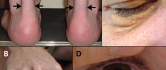
Young children with any form of HA may experience delayed growth and development. With hereditary diseases, congenital anomalies such as a tower skull, cervical ribs, polydactyly, strabismus are noted - such symptoms constitute the so-called hemolytic constitution. The disturbance in the structure of the skeleton is explained by a compensatory mechanism, within which, with the massive death of red blood cells, the processes of hematopoiesis in the red bone marrow are enhanced. As a result, the hematopoietic tissue of the bones grows, leading to their deformation.
Jaundice in a newborn